Avicenna J Dent Res. 17(2):118-123.
doi: 10.34172/ajdr.2178
Original Article
Evaluating the Remineralizing Potential of Carbonated Hydroxyapatite on Enamel After At-Home Bleaching: An In Vitro Study
S. Swathi Priyadharshini Conceptualization, Data curation, Investigation, Methodology, Validation, Writing – original draft, Writing – review & editing, 1, 2 
Syed Ashiyana Writing – original draft, Writing – review & editing, 2
Chinnasamy Ragavendran Formal analysis, Methodology, 1, *
Anand Sherwood Data curation, Formal analysis, Investigation, Supervision, Validation, 2
Author information:
1Department of Cariology, Saveetha Dental College and Hospitals, Saveetha Institute of Medical and Technical Science, (SIMATS), Saveetha University, Chennai, Tamil Nadu 600077, India
2Department of Conservative Dentistry and Enodontics, C.S.I. College of Dental Sciences and Research, Madurai, Tamil Nadu 625001, India
Abstract
Background: Tooth bleaching may result in enamel demineralization and increased surface roughness. This study aimed to investigate the effects of at-home bleaching on enamel surface morphology and assess the potential of carbonated hydroxyapatite (CHA) as a remineralizing agent compared to casein-phosphopeptide-amorphous calcium phosphate nanocomplex (CPP-ACP).
Methods: Extracted premolars were divided into four groups. Group 1 had no treatment, while group 2 was treated with 22% carbamide peroxide. In addition, group 3 was treated with 22% carbamide peroxide followed by remineralization with CPP-ACP, and group 4 was treated with 22% carbamide peroxide followed by remineralization with CHA. After bleaching with 22% carbamide peroxide, remineralization agents were applied for seven days. Scanning electron microscopy (SEM) and energy-dispersive X-ray spectroscopy were used to evaluate surface morphology and elemental composition.
Results: There was a statistically significant reduction in calcium and phosphorus levels but an increase in carbon content in Groups 2, 3, and 4 compared to Group 1 (P=0.000). Group 4 (bleached and CHA-treated) had the highest carbon content (40.2±0.9 wt (%)). SEM analysis revealed a smooth enamel surface in group 1, while group 2 (bleached) displayed increased surface irregularities. Groups 3 and 4, treated with CPP-ACP and CHA, respectively, confirmed significant mineral deposition on enamel surfaces.
Conclusion: Overall, at-home bleaching with 22% carbamide peroxide caused enamel damage and mineral loss. Both CPP-ACP and CHA demonstrated potential for remineralization following bleaching.
Keywords: Tooth bleaching, Casein phosphopeptide-amorphous calcium phosphate, Tooth remineralization
Copyright and License Information
© 2025 The Author(s); Published by Hamadan University of Medical Sciences.
This is an open-access article distributed under the terms of the Creative Commons Attribution License (
https://creativecommons.org/licenses/by/4.0), which permits unrestricted use, distribution, and reproduction in any medium provided the original work is properly cited.
Please cite this article as follows: Priyadharshini SS, Ashiyana S, Ragavendran C, Sherwood A. Evaluating the Remineralizing Potential of Carbonated Hydroxyapatite on Enamel After At-Home Bleaching: An In Vitro Study. Avicenna J Dent Res. 2025;17(2):118-123. doi:10.34172/ajdr.2178
Background
Dental discolorations can be due to various factors and may require different treatment durations (1). At-home bleaching involves using a custom-fitted tray with a bleaching gel every day for a specified period under the supervision of a dentist (2).
Numerous techniques are available for bleaching vital teeth that rely on the direct application of hydrogen peroxide or its precursor, carbamide peroxide (3). The success of this technique depends on the peroxides’ ability to penetrate or diffuse into the enamel and dentin (4). Tooth whiteners are categorized into three classes based on the delivered peroxide concentration, including over-the-counter (up to 10% carbamide peroxide), dentist-dispensed (up to 20% carbamide peroxide), and in-office (up to 45% carbamide peroxide or 40% hydrogen peroxide) gels (5).
Bleaching agents can drastically alter the morphology, composition, hardness, and even the fracture toughness of tooth surfaces (6). To lessen the negative effects of bleaching treatments, agents that decrease sensitivity and strengthen tooth enamel are professionally applied before, during, or after the procedure (7). Clinical trials have demonstrated the incidence of tooth sensitivity even when desensitizing and remineralizing products are applied, which may be attributed to structural changes in enamel and dentin (8).
Casein-phosphopeptide-amorphous calcium phosphate nanocomplex (CPP-ACP), a milk protein extract, has been extensively used due to its proven anticaries effects. Some tooth-whitening products incorporate amorphous calcium phosphate to reduce sensitivity, promote enamel remineralization after bleaching, and enhance tooth luster (1). By sealing dentinal tubules, CPP-ACP facilitates enamel surface remineralization, thus decreasing hypersensitivity. The efficacy of CPP-ACP relies on its ability to integrate nanocomplexes onto tooth surfaces and within dental plaque, thereby creating a reservoir of calcium and phosphate ions (9).
In dentistry, biomimetic approaches involve using bioinspired materials to replicate natural remineralization processes in damaged or diseased dental hard tissues (10). Hydroxyapatite, the main inorganic component of human bones and teeth, has been extensively studied in regenerative medicine since the mid-1900s due to its potential in various biomedical applications. It is rich in calcium and phosphate, which play a crucial role in promoting the remineralization of enamel that has been weakened by demineralization (11). The substitution of carbonate anions (CO₃2-) for hydroxyl or phosphate groups in the hydroxyapatite crystal structure creates unique physicochemical properties and structural complexities. Carbonated hydroxyapatite (CHA), a biomimetic material, has shown potential for remineralizing dental tissues. The incorporation of carbonate typically reduces crystallinity and increases solubility, enhancing biocompatibility, biomimetic mineralization, and resorption (12).
While previous studies have explored how at-home bleaching affects enamel surface morphology (13), the efficacy of CHA as a remineralizing agent in this context remains largely unexplored, warranting further investigation.
Accordingly, this research seeks to assess the effects of carbamide peroxide bleaching on enamel microstructure and mineral content, as well as the potential remineralizing effects of CHA on bleached enamel.
Methods
To ensure adherence to ethical guidelines, the study protocol was reviewed and granted authorization by an Institutional Review Board (with reference number SRB/SDC/PhD/ENDO-2309/23/TH-081). CHA was synthesized through a precipitation technique, following a previously established protocol from our prior research. The process utilized calcium nitrate tetrahydrate and diammonium hydrogen phosphate as sources of calcium and phosphate, respectively, in a 1:0.6 M molar ratio. Sodium hydrogen carbonate was employed to supply carbonate ions. The mixture containing carbonate and phosphate was slowly added to the calcium ion solution, which resulted in the formation of precipitates. The resulting material was subsequently dried and then ground into a fine powder. The detailed characterization of this CHA has already been reported in our earlier publication (14).
Sample Preparation
This study adopted the methodological framework outlined by Melo et al (15). A total of 45 premolars extracted exclusively for orthodontic purposes were utilized in the study. The specimens were carefully inspected under 10x magnification, with those showing fractures, decay, or innate enamel defects being excluded. The teeth were stored in distilled water at 25°C until study initiation. To obtain standardized test specimens, the teeth underwent a three-step sectioning process using a laboratory trimmer (Marathon, Korea) equipped with a diamond disc (Marathon, Korea) operating at 400 rpm. First, horizontal sectioning was performed at the cementoenamel junction. Subsequently, a mesiodistal cut separated the mesial and distal surfaces of the crown. Finally, a buccolingual cut isolated the buccal surface from the palatal/lingual surface, resulting in 40 samples measuring 3 × 3 × 3 mm ( ± 0.25 mm). Post-sectioning, the samples were cleaned ultrasonically in water and dried, and their dentin margins were coated with nail varnish to prevent bleaching agent leakage. The sample size calculation, performed using G*Power software, determined that 40 samples were required, with 10 samples in each of the 4 groups. This calculation was based on an alpha level of 0.05, a statistical power (β) of 0.9, and an effect size of 0.47. The samples were randomly assigned to four groups via simple randomization using a random number generator. Group 1 received no treatment, and group 2 was treated only with the bleaching agent. Group 3 received the bleaching protocol followed by CPP-ACP, and group 4 received the bleaching protocol followed by CHA.
Bleaching and Remineralization Procedure
Before applying the bleaching agent, the samples were dried using sterile gauze. The bleaching agent, a 22% carbamide peroxide in a neutral pH gel (FGM Whiteness Perfect Home Bleach, FGM Productos Odontologicos Ltd. a, Joinville, SC, Brazil) of approximately 0.25 g, was applied to labial tooth surfaces using applicator tips. The entire enamel surface was covered to ensure even distribution. The agent remained on the surface for 4 hours daily over 14 days. During and after treatment, specimens in groups 2, 3, and 4 were stored in artificial saliva (ICPA Wet Mouth, ICPA Health Products Ltd., Adarsh Industrial Estate, Andheri, Mumbai). Post-bleaching, group 3 was treated with approximately 0.5 g of 10% CPP-ACP (Tooth Mousse, GC, Tokyo, Japan), a commercial remineralizing agent. Group 4 received a 0.1 M CHA treatment using a homogenous suspension of CHA powder and distilled water. These agents were carefully applied for 3 minutes daily with a microbrush for 7 days. After each application, the samples were dried using sterile gauze and then immersed in regularly refreshed artificial saliva (ICPA Wet Mouth, ICPA Health Products Ltd., Adarsh Industrial Estate, Andheri, Mumbai). Table 1 details the composition of materials used in this study.
Table 1.
Composition of Materials Used in the Study
|
Material
|
Composition
|
| Whiteness Perfect bleaching gel (FGM Productos Odontologicos Ltd. a, Joinville, SC, Brazil) |
22% carbamide peroxide, Carbopol, potassium hydroxide, sodium fluoride, glycerol, and deionized water |
| Tooth Mousse (“GC”, MI Paste, GC Corp., TOKYO, Japan) |
Pure water, glycerol, Recaldent (CPP-ACP), D-sorbitol, CMC-Na, propylene glycol, silicon dioxide, titanium dioxide, xylitol, phosphoric acid, flavoring, zinc oxide, guar gum, propyl p-hydroxybenzoate, and butyl p-hydroxybenzoate |
| Artificial saliva (Wet Mouth, ICPA Health Products Ltd., Adarsh Industrial Estate, Andheri, Mumbai) |
Water, glycerin, sorbitol, propylene glycol, PEG 40 HCO, poloxamer, sodium benzoate, sodium CMC, flavour, cetylpyridinium chloride, parabens, xylitol, xanthan gum, disodium hydrogen phosphate, and sodium dihydrogen phosphate |
Micromorphological and Microanalytical Investigation Using Scanning Electron Microscopy-Energy-Dispersive X-Ray Spectroscopy
The samples’ surface morphology was evaluated using scanning electron microscopy (SEM). For SEM analysis, all samples were secured onto aluminum stubs. Subsequently, a uniform palladium-gold sputter coating was applied for two minutes. SEM imaging (TESCAN VEGA 3, Czech Republic) was performed at magnifications ranging from 5000 × to 10000 × .
Quantitative EDX (D-12489, BRUKER Nano, GmbH, Germany) point analysis, in conjunction with SEM analysis, was conducted on the enamel surface to quantify the elemental concentrations (in weight percent) of calcium and phosphorus. EDX is frequently combined with SEM to enable elemental analysis with high spatial resolution. The method involves directing high-energy electrons onto a sample, resulting in the emission of characteristic X-rays. These X-rays are detected and analyzed to identify the elemental composition of the sample (16).
Statistical Analysis
Statistical analysis involved calculating means and standard deviations (SDs) for the weight percentages of calcium, phosphorus, and carbon using SPSS software (version 20.0, SPSS Inc., Chicago, IL, USA). A one-way analysis of variance was employed to evaluate and compare the mean weight percentages of calcium, phosphorus, and carbon across the groups. Eventually, pairwise comparisons between groups were conducted using Tukey’s post-hoc testing, with statistical significance determined at P < 0.05.
Results
The average calcium, phosphorus, and carbon levels were assessed in all four groups, the results of which are presented in Table 2. Group 1 (control) exhibited mean calcium and phosphorus levels of 31.5 ± 0.7 wt (%) and 14.0 ± 0.6 wt (%), respectively. There was a statistically significant decrease in calcium and phosphorus values in all the other groups compared to sound enamel in group 1 (Table 2 and Figure 1). However, a statistically significant increase was found in the levels of carbon in all the groups except group 1 (P = 0.000). Group 4 (bleached and CHA-treated) had the highest carbon content with a mean of 40.2 ± 0.9 wt (%), followed by groups 2 (bleached) and 3 (bleached and CHA-treated) (P = 0.000). Post-hoc multiple comparisons of calcium, phosphorus, and carbon levels among different groups are provided in Table 3.
Table 2.
Elemental Analysis Data (wt%) for Each Group as Means ± SD
|
EDX Elemental Analysis of Enamel Surfaces
|
|
Groups
|
Mean Calcium
|
Mean Phosphorus
|
Mean Carbon
|
| 1 |
31.5 ± 0.7 |
14.2 ± 0.6 |
0 |
| 2 |
25.6 ± 1.0 |
9.4 ± 0.8 |
9.5 ± 0.7 |
| 3 |
26.5 ± 0.7 |
12.2 ± 0.6 |
6.4 ± 0.7 |
| 4 |
18.7 ± 0.2 |
7.8 ± 0.2 |
40.2 ± 0.9 |
Note. SD: Standard deviation; EDX: Energy-dispersive X-ray spectroscopy.
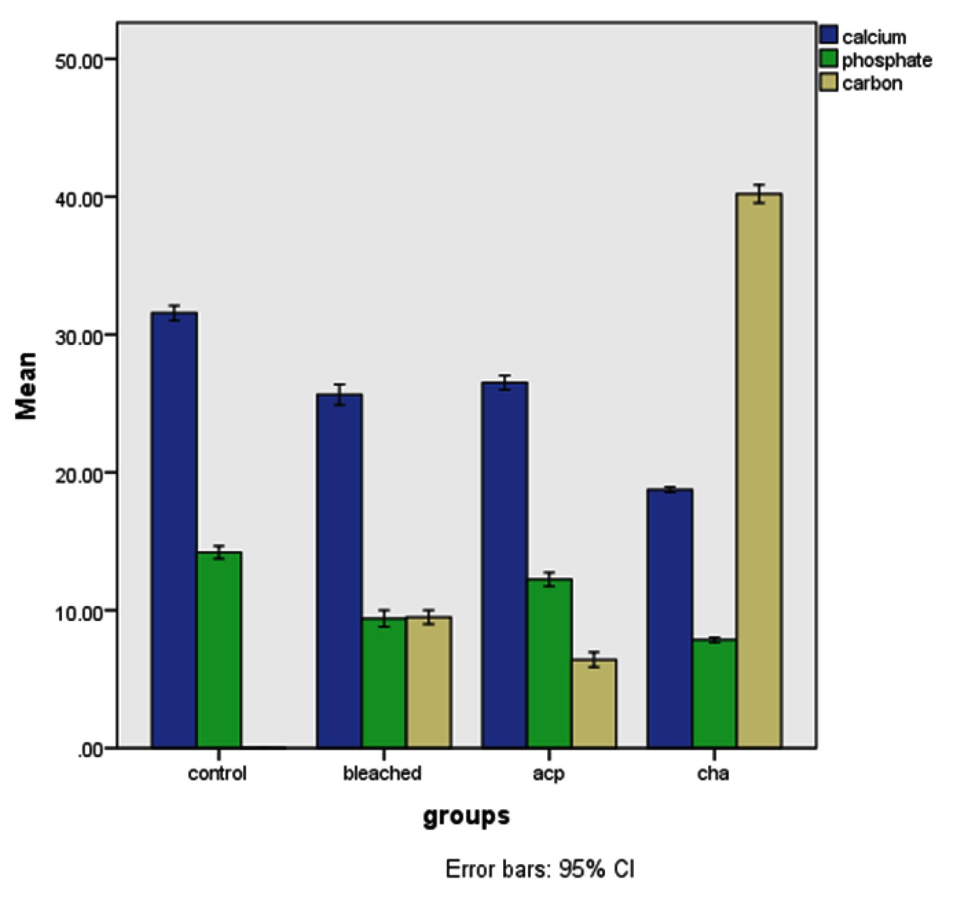
Figure 1.
Mean Values of Calcium, Phosphorus, and Carbon Across the Four Groups. Note. CI: Confidence interval; ACP: Amorphous calcium phosphate; ChA: Carbonated hydroxyapatite. *Significant increase in the levels of calcium and carbon among the groups
.
Mean Values of Calcium, Phosphorus, and Carbon Across the Four Groups. Note. CI: Confidence interval; ACP: Amorphous calcium phosphate; ChA: Carbonated hydroxyapatite. *Significant increase in the levels of calcium and carbon among the groups
Table 3.
Post-hoc Multiple Comparisons of Calcium, Phosphorus, and Carbon Levels Among Different Groups
|
Intergroup Comparison
|
Post-hoc Multiple Comparisons (
P
<0.05)
|
|
Calcium
|
Phosphorus
|
Carbon
|
| 1 |
2 |
0.00 |
0.00 |
0.00 |
| 1 |
3 |
0.00 |
0.00 |
0.00 |
| 1 |
4 |
0.00 |
0.00 |
0.00 |
| 2 |
3 |
0.06 |
0.00 |
0.00 |
| 2 |
4 |
0.00 |
0.00 |
0.00 |
| 3 |
4 |
0.00 |
0.00 |
0.00 |
The SEM analysis of group 1 demonstrated enamel with a smooth and uniform surface topography (Figure 2A). Group 2 revealed a significant increase in surface irregularities following the bleaching procedure (Figure 2B). Compared to the other groups, SEM analysis showed a significant increase in the amount of superficial mineral deposits on the surfaces in groups 3 and 4. Figures 2C and 2D illustrate mineral distribution on bleached enamel remineralized with CPP-ACP and CHA.
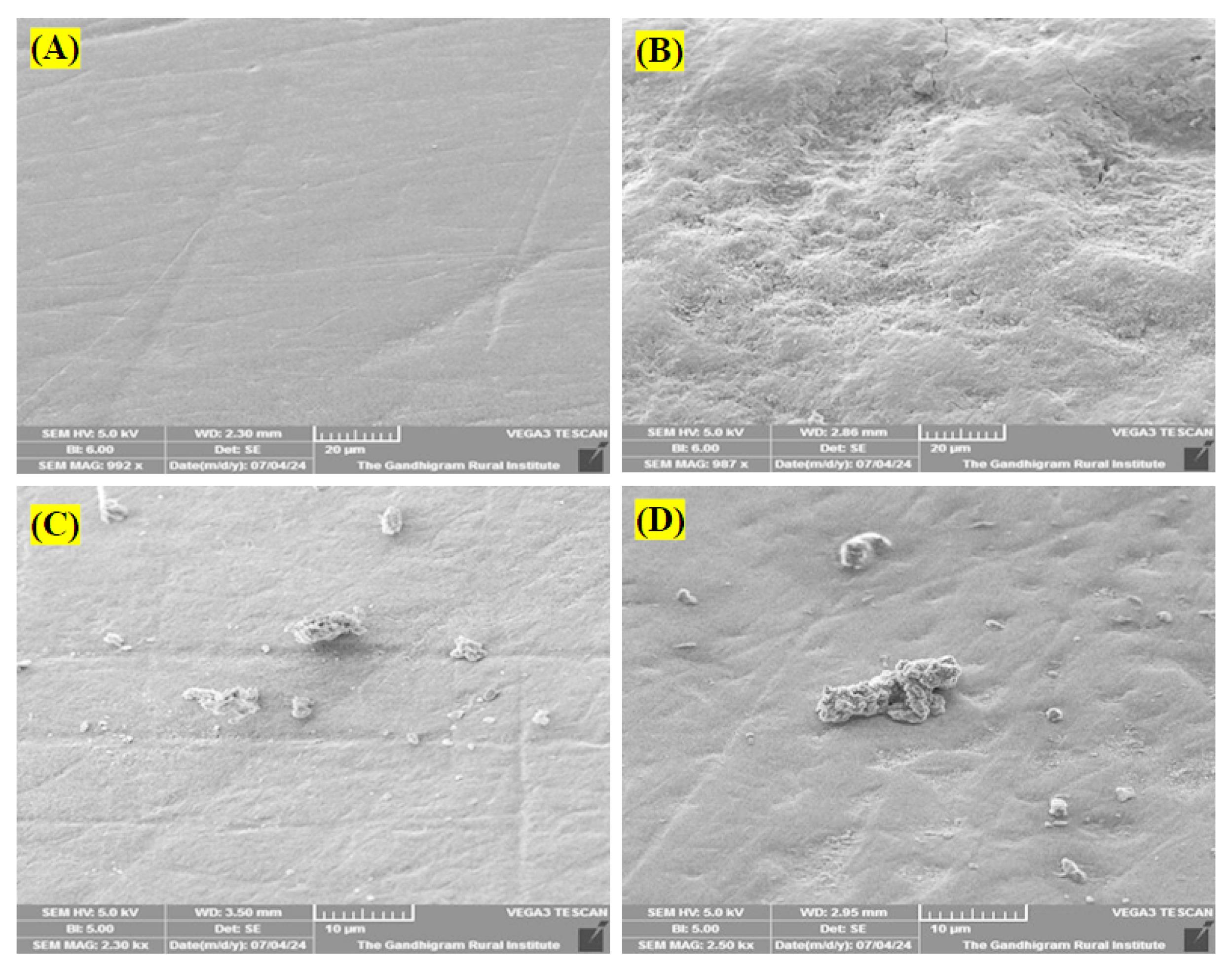
Figure 2.
(A) SEM Image of Intact Enamel (Group 1), (B) SEM Image of Enamel Exposed to Carbamide Peroxide (Group 2), (C) SEM Image of Bleached Enamel Remineralized With CPP-ACP (Group 3), and (D) SEM Image of Bleached Enamel Remineralized With CHA (Group 4). Note. SEM: Scanning electron microscopy; CPP: Casein-phosphopeptide; ACP: Amorphous calcium phosphate nanocomplex. (A) A smooth surface morphology can be observed. (B) The bleaching agent caused porosities, depressions, and superficial alterations
.
(A) SEM Image of Intact Enamel (Group 1), (B) SEM Image of Enamel Exposed to Carbamide Peroxide (Group 2), (C) SEM Image of Bleached Enamel Remineralized With CPP-ACP (Group 3), and (D) SEM Image of Bleached Enamel Remineralized With CHA (Group 4). Note. SEM: Scanning electron microscopy; CPP: Casein-phosphopeptide; ACP: Amorphous calcium phosphate nanocomplex. (A) A smooth surface morphology can be observed. (B) The bleaching agent caused porosities, depressions, and superficial alterations
Discussion
This study evaluated the remineralization potential of CHA on enamel that had undergone bleaching. The applied CHA was synthesized through precipitation and characterized as b-type CHA (14). The study employed 22% carbamide peroxide, a common at-home dental bleaching agent, which breaks down into hydrogen peroxide and urea (17). Vital tooth bleaching with carbamide peroxide often results in dentin hypersensitivity, possibly due to the diffusion of breakdown products through enamel and dentin to the pulp (18). As enamel lacks cells that may generate an extracellular matrix, apatitic crystals cannot spontaneously re-deposit in enamel and dentine after being disintegrated or abraded (19). Applying remineralizing agents post-bleaching has been proposed to mitigate the negative effects of the process (20).
CPP-ACP was chosen as a comparison due to its extensive research history and proven positive outcomes. The study utilized SEM and EDX for high-resolution surface morphology imaging and quantitative elemental analysis, respectively, providing a comprehensive assessment of morphological and compositional changes in the enamel.
Impact of Bleaching on Enamel
Surface analysis revealed damaged enamel areas in group 2 (Figure 2A), which underwent bleaching only, supporting the concept that bleaching can demineralize enamel and potentially increase susceptibility to erosion and caries. Groups treated with remineralizing agents, such as CPP-ACP and CHA, demonstrated evidence of surface remineralization, indicated by crystal deposition around enamel prisms (Figures 2C and 2D). This suggests that while these agents may not fully reverse mineral loss from bleaching, they may offer some protective benefits. EDX analysis confirmed a statistically significant reduction in calcium and phosphorus levels in groups 2, 3, and 4 compared to group 1, likely due to the demineralizing effect of hydrogen peroxide. Group 4, treated with CHA, exhibited lower calcium levels and higher carbon content than the other groups, yet represented tooth remineralization under SEM. This could highlight the successful incorporation of carbon into the hydroxyapatite structure by CHA, potentially affecting overall mineral content measurement, with remineralization possibly resulting from increased carbonate deposition on the bleached enamel. This framework can subsequently facilitate the deposition of minerals, such as calcium and phosphate, enhancing remineralization. Additional research into the precise composition of the remineralized layer would be valuable.
The significant decrease of calcium in all the groups, except in group 1, was due to the loss of calcium and phosphorus content due to bleaching. Ben-Amar et al utilized SEM to assess the impact of Opalescence home bleaching (10% carbamide peroxide) on enamel surface topography (21). Their findings revealed that bleaching caused superficial enamel defects (pitting), attributed to the presence of highly reactive hydroxyl radicals in the bleaching agent. These radicals facilitate the removal of organic components from the enamel, potentially altering its mechanical properties, including abrasion resistance.
The findings of the study are consistent with those of Melo et al (15), indicating that CPP-ACP and hydroxyapatite showed a more pronounced accumulation of surface deposits compared to formulations containing 8% arginine or fluoride. The present study supports the findings of Godinho et al (22), demonstrating a decrease in enamel calcium and phosphorus content following bleaching procedures. They examined the effectiveness of post-bleaching remineralization strategies using casein-phosphopeptide-amorphous calcium phosphate fluoride paste and xylitol-coated calcium phosphate fluoride varnish and reported that both agents successfully restored enamel surface morphology, with fluoride varnish underlining superior restorative effects.
Remineralization Potential of Carbonated Hydroxyapatite and Casein-Phosphopeptide-Amorphous Calcium Phosphate
Casein phosphopeptides, derived from the proteolytic breakdown of milk proteins, such as αS1-, αS2-, and β-casein, contain a key cluster sequence, Ser(P)-Ser(P)-Ser(P)-Glu-Glu, which helps stabilize the nanoclusters of ACP (23). ACP maintains a supersaturated state of calcium and phosphate ions on the enamel surface, thereby facilitating remineralization. The synergistic interaction between casein phosphopeptide and calcium phosphate components within ACP is believed to support this mechanism of action.
Nagi et al (20) investigated the impact of different bleaching methods on enamel erosion. They treated enamel with 25% hydrogen peroxide, followed by an application of ACP gel, and used Nite White ACP, an at-home bleaching product with pre-incorporated ACP. Enamel treated with hydrogen peroxide and then ACP gel represented a relatively smooth surface, with minimal pores and no visible etching. In contrast, enamel bleached with Nite White ACP displayed more significant morphological changes when subjected to erosion.
Group 4 showed a substantial increase in carbon content compared to other groups. The incorporation of carbonate ions into the hydroxyapatite structure leads to the formation of point defects and vacancies in the Ca-sublattice and OH-sublattice, as well as microstrains in CHA nanocrystals. This increased defect concentration enhances the bioactivity of CHA samples (24). This could explain why CHA was able to remineralize enamel despite having lower calcium levels. Research has demonstrated that CHA can promote the deposition of a biomimetic apatite coating on enamel and dentine surfaces, potentially aiding in the remineralization process after teeth have undergone bleaching treatments (19).
Consistent with the findings of Rimondini et al (25), CHA displayed promising potential for dentin remineralization. They reported the material’s effectiveness in remineralizing dentin surfaces etched by orthophosphoric acid. They further concluded that CHA rapidly decreased exposed dentinal tubules, leading to mineralized tissue regeneration within hours. Its biomimetic properties allow it to adhere to tooth surfaces and promote mineral deposition, crucial for restoring bleaching-compromised tooth structure. The incorporation of ions, such as carbonate, zinc, fluoride, magnesium, and strontium, into hydroxyapatite has been shown to influence the formation, growth, alignment, and dissolution properties of the crystals (26).
Contemporary dentistry must adapt to evolving lifestyles and address challenges such as demineralization, which can result in significant oral health issues, including sensitivity, occlusal alterations, and potential tooth loss (27). This research underscores the potential adverse effects of at-home bleaching on enamel mineral content and suggests that both CPP-ACP and CHA may provide remineralizing benefits post-bleaching. Additional studies quantifying mineral deposition and composition would be beneficial. More research is needed to fully understand these agents’ effectiveness in restoring mineral loss and the specific mechanisms of CHA’s carbon incorporation.
Limitations of the Study
This study has some limitations, including the need for validation through larger sample sizes and extended observation periods to confirm the long-term efficacy of CHA and other remineralizing agents. In addition, the in vitro nature of the study limited its ability to fully replicate the oral cavity’s complexities, necessitating in vivo investigations to corroborate findings in a more clinically relevant context. Individual variations in enamel composition and response to bleaching and remineralizing agents were not considered, potentially affecting the generalizability of our results.
Conclusion
It can be concluded that 22% carbamide peroxide caused morphological alterations in enamel and decreased its calcium and phosphorus content. Nonetheless, the post-bleaching application of the investigated remineralizing agents demonstrably restored the surface topography of the enamel and replenished its mineral content.
Competing Interests
Nil.
Funding
Nil.
References
- Alqahtani MQ. Tooth-bleaching procedures and their controversial effects: a literature review. Saudi Dent J 2014; 26(2):33-46. doi: 10.1016/j.sdentj.2014.02.002 [Crossref] [ Google Scholar]
- de Sousa Barros Júnior E, Ribeiro ME, Lima RR, da Silva E Souza Júnior MH, Loretto SC. Excessive dental bleaching with 22% carbamide peroxide combined with erosive and abrasive challenges: new insights into the morphology and surface properties of enamel. Materials (Basel) 2022; 15(21):7496. doi: 10.3390/ma15217496 [Crossref] [ Google Scholar]
- Féliz-Matos L, Hernández LM, Abreu N. Dental bleaching techniques; hydrogen-carbamide peroxides and light sources for activation, an update Mini review article. Open Dent J 2014; 8:264-8. doi: 10.2174/1874210601408010264 [Crossref] [ Google Scholar]
- Minoux M, Serfaty R. Vital tooth bleaching: biologic adverse effects-a review. Quintessence Int 2008; 39(8):645-59. [ Google Scholar]
- Cvikl B, Lussi A, Moritz A, Flury S. Enamel surface changes after exposure to bleaching gels containing carbamide peroxide or hydrogen peroxide. Oper Dent 2016; 41(1):E39-47. doi: 10.2341/15-010-l [Crossref] [ Google Scholar]
- Heshmat H, Hoorizad Ganjkar M, Miri Y, Kharrazi Fard MJ. The effect of two remineralizing agents and natural saliva on bleached enamel hardness. Dent Res J (Isfahan) 2016; 13(1):52-7. doi: 10.4103/1735-3327.174713 [Crossref] [ Google Scholar]
- Pintado-Palomino K, Tirapelli C. The effect of home-use and in-office bleaching treatments combined with experimental desensitizing agents on enamel and dentin. Eur J Dent 2015; 9(1):66-73. doi: 10.4103/1305-7456.149645 [Crossref] [ Google Scholar]
- Reis A, Tay LY, Herrera DR, Kossatz S, Loguercio AD. Clinical effects of prolonged application time of an in-office bleaching gel. Oper Dent 2011; 36(6):590-6. doi: 10.2341/10-173-c [Crossref] [ Google Scholar]
- Rajendran R, Antony SD, Ashik PM, Bharath S, Thomas AJ, Heboyan A. Remineralization potential of strontium-doped nano-hydroxyapatite dentifrice and casein phosphopeptide-amorphous calcium phosphate cream on white spot lesions in enamel following orthodontic debonding - a randomized controlled trial. SAGE Open Med 2024; 12:20503121231221634. doi: 10.1177/20503121231221634 [Crossref] [ Google Scholar]
- Kranz S, Heyder M, Mueller S, Guellmar A, Krafft C, Nietzsche S. Remineralization of artificially demineralized human enamel and dentin samples by zinc-carbonate hydroxyapatite nanocrystals. Materials (Basel) 2022; 15(20):7173. doi: 10.3390/ma15207173 [Crossref] [ Google Scholar]
- Kengadaran S, Sakthi S, Anusha D, Arumugham IM, Kumar RP. Effect of nano-hydroxyapatite crystal incorporated herbal dentifrice on remineralization of incipient caries lesion-a pilot study. J Pharm Res Int 2020; 32(20):13-9. doi: 10.9734/JPRI/2020/v32i2030724 [Crossref] [ Google Scholar]
- Adekanmi DG, Garcia CR, Lopez-Badillo CM. Carbonate hydroxyapatite-a multifunctional bioceramics with non-medical applications. Eng Chem 2024; 7:1-24. doi: 10.4028/p-518pjS [Crossref] [ Google Scholar]
- Karimi Z, Saoui H, Sakout M, Abdallaoui F. Effect of vital bleaching on micromorphology of enamel surface: an in vitro study. Prim Dent J 2021; 10(1):126-31. doi: 10.1177/2050168420980966 [Crossref] [ Google Scholar]
- Priyadharshini SS, Ragavendran C, Sherwood A, Ramya JR, Krithikadatta J. Evaluation of mineral induction ability and cytotoxicity of carbonated hydroxyapatite for pulp tissue regeneration: an in vitro study. Restor Dent Endod 2024; 49(4):e40. doi: 10.5395/rde.2024.49.e40 [Crossref] [ Google Scholar]
- Melo M, Fioresta R, Sanz JL, Pecci-Lloret MP, Llena C. Effect of highly concentrated bleaching gels on enamel microhardness and superficial morphology, and the recovery action of four remineralizing agents. BMC Oral Health 2022; 22(1):645. doi: 10.1186/s12903-022-02693-2 [Crossref] [ Google Scholar]
- Tromp RM. Energy-dispersive X-ray spectroscopy in a low energy electron microscope. Ultramicroscopy 2024; 259:113935. doi: 10.1016/j.ultramic.2024.113935 [Crossref] [ Google Scholar]
- D'Arce MB, Lima DA, Aguiar FH, Bertoldo CE, Ambrosano GM, Lovadino JR. Effectiveness of dental bleaching in depth after using different bleaching agents. J Clin Exp Dent 2013; 5(2):e100-7. doi: 10.4317/jced.51063 [Crossref] [ Google Scholar]
- Haywood VB. Dentine hypersensitivity: bleaching and restorative considerations for successful management. Int Dent J 2002; 52(S5P2):376-84. doi: 10.1002/j.1875-595X.2002.tb00937.x [Crossref] [ Google Scholar]
- Roveri N, Foresti E, Lelli M, Lesci IG. Recent advancements in preventing teeth health hazard: the daily use of hydroxyapatite instead of fluoride. Recent Pat Biomed Eng 2009; 2(3):197-215. doi: 10.2174/1874764710902030197 [Crossref] [ Google Scholar]
- Nagi SM, Nabil SH, Zaazou MH. Elemental and morphological analysis of enamel following the application of two bleaching systems with amorphous calcium phosphate: effect on enamel erosion susceptibility. Bull Natl Res Cent 2021; 45(1):68. doi: 10.1186/s42269-021-00527-9 [Crossref] [ Google Scholar]
- Ben-Amar A, Liberman R, Gorfil C, Bernstein Y. Effect of mouthguard bleaching on enamel surface. Am J Dent 1995; 8(1):29-32. [ Google Scholar]
- Godinho M, de Noronha de Ataide I, Lambor R, Fernandes M. Influence of two remineralizing agents on bleached enamel surface morphology and mineral composition - an in vitro study. Indian J Dent Res 2022; 33(2):188-92. doi: 10.4103/ijdr.ijdr_896_21 [Crossref] [ Google Scholar]
- Sionov RV, Tsavdaridou D, Aqawi M, Zaks B, Steinberg D, Shalish M. Tooth mousse containing casein phosphopeptide-amorphous calcium phosphate prevents biofilm formation of Streptococcus mutans. BMC Oral Health 2021; 21(1):136. doi: 10.1186/s12903-021-01502-6 [Crossref] [ Google Scholar]
- Kovaleva ES, Shabanov MP, Putlayev VI, Filippov YY, Tretyakov YD, Ivanov VK. Carbonated hydroxyapatite nanopowders for preparation of bioresorbable materials. Materwiss Werksttech 2008; 39(11):822-9. doi: 10.1002/mawe.200800383 [Crossref] [ Google Scholar]
- Rimondini L, Palazzo B, Iafisco M, Canegallo L, Demarosi F, Merlo M. The remineralizing effect of carbonate-hydroxyapatite nanocrystals on dentine. Mater Sci Forum 2007; 539-543:602-5. doi: 10.4028/www.scientific.net/MSF.539-543.602 [Crossref] [ Google Scholar]
- Ionescu AC, Degli Esposti L, Iafisco M, Brambilla E. Dental tissue remineralization by bioactive calcium phosphate nanoparticles formulations. Sci Rep 2022; 12(1):5994. doi: 10.1038/s41598-022-09787-5 [Crossref] [ Google Scholar]
- Yuwanati M, Chitra S. Investigating the effect of bioactive glasses on enamel remineralization through morphological and elemental analysis. Biomed Biotechnol Res 2023; 7(2):181-6. doi: 10.4103/bbrj.bbrj_24_23 [Crossref] [ Google Scholar]