Avicenna J Dent Res. 17(3):178-191.
doi: 10.34172/ajdr.2127
Review Article
Impact of Self-Etch Adhesives Increased Application Time on Dental Bond Strength: A Systematic Review and Meta-Analysis
Azam Valian Conceptualization, Data curation, Investigation, Methodology, Resources, Supervision, Validation, Visualization, Writing – original draft, Writing – review & editing, 1 
Amir Ghasemi Conceptualization, Data curation, Investigation, Validation, Writing – review & editing, 2
Shahin Kasraei Conceptualization, Data curation, Investigation, Validation, Writing – review & editing, 1
Kiana Shakeri Investigation, Writing – original draft, Writing – review & editing, 2
Farzaneh Sadeghi Mahounak Conceptualization, Data curation, Formal analysis, Investigation, Methodology, Project administration, Resources, Software, Supervision, Visualization, Writing – review & editing, 3, *
Elham Zanguei Data curation, Investigation, Writing – review & editing, 4
Author information:
1Department of Restorative Dentistry, School of Dentistry, Shahid Beheshti University of Medical Sciences, Tehran, Iran
2Department of Restorative Dentistry, School of Dentistry, Dental Research Center, Research Institute of Dental Sciences, Shahid Beheshti University of Medical Sciences, Tehran, Iran
3Department of Restorative Dentistry, School of Dentistry, Tehran University of Medical Sciences, Tehran, Iran
4Department of Restorative Dentistry, School of Dentistry, Birjand University of Medical Sciences, South Khoasan, Iran
Abstract
In this systematic review and meta-analysis, it was intended to determine whether increasing the application time of self-etch adhesives and universal adhesives could increase the bond strength between composite resin and enamel/dentin. Electronic databases, such as MEDLINE via PubMed, ISI (Web of Science), and Scopus, were thoroughly searched based on the PICOS strategy in order to gather in vitro articles in English with no publication year limit. Studies assessing the bond strength between enamel/dentin and composite resin were selected and further evaluated using self-etch or universal adhesives. Meta-analysis was performed using a random-effect model to determine the difference in the bond value between the specimens that underwent longer adhesive application and the control groups. Amongst 123 gathered studies, 59 were selected for full-text analysis, and 26 were chosen for inclusion in this review. Overall, 14 studies were included in the meta-analysis, and a random-model effect was used for all but one analysis dependent on the heterogeneity percentage. The meta-analysis demonstrated a significant increase in bond strength subsequent to increasing the application time of the aforementioned bonding systems to an extent. Doubling the application time increased the bond strength to both enamel and dentin. However, tripling the application time seemed to be only beneficial to bond strength between enamel and self-etch/universal adhesive systems. The one-component adhesive systems were more impacted by the increased application time than two-component systems. However, not enough data existed on the effect of the pH level of adhesive systems on bond strength value to draw a reach conclusion. Under the limitations of this review, it was concluded that increasing the application time of self-etch and universal adhesives to a certain extent could increase bond strength to both ground dentin and enamel under in vitro conditions.
Keywords: Self-etch adhesive, Universal adhesive, Bond strength, Application time
Copyright and License Information
© 2025 The Author(s); Published by Hamadan University of Medical Sciences.
This is an open-access article distributed under the terms of the Creative Commons Attribution License (
http://creativecommons.org/licenses/by/4.0), which permits unrestricted use, distribution, and reproduction in any medium provided the original work is properly cited.
Please cite this article as follows: Valian A, Ghasemi A, Kasraei S, Shakeri K, Sadeghi Mahounak F, Zanguei E. Impact of self-etch adhesives increased application time on dental bond strength: a systematic review and meta-analysis. Avicenna J Dent Res. 2025;17(3):178-191. doi:10.34172/ajdr.2127
Background
The longevity of restoration depends on many factors, one of which is the adequate adhesion of dental material to tooth structure (1,2). Utilizing adhesive systems in order to form a cohesive bond between the restorative materials and the hard dental tissue is a crucial step in restorative treatments. Without a proficient bonding system or the lack of adequate application of one, the restoration would be prone to microleakage and/or secondary caries (3-5).
Since the development of the first bonding system in the 1970s (6), different generations of bonding systems with diverse compositions and methods of application have emerged (7,8), including total etch/etch and rinse adhesives (TEA), self-etch adhesives (SEA), and universal/multi-mode adhesives (UA) (8,9).
While TEAs use phosphoric acid for complete removal of the smear layer, SEAs utilize acidic monomers to demineralize the smear layer and simultaneously prime the tooth structure (10-12). By eliminating the rinsing step and streamlining the process, SEAs offer advantages, such as reducing technique sensitivity, decreasing contamination risks, and minimizing the risk of postoperative sensitivity (6,13-15). Considering that UAs are also a modified generation of self-etching primers, they possess the main aforementioned advantages (16,17). Nevertheless, the SEAs have shortcomings, since combining diverse functional monomers with varying sizes, weights, and different solvents introduces new forms of technique sensitivity, potentially compromising bonding strength (11,18-21). Furthermore, there are several concerns about bonding to both dentin and enamel, since using acidic monomers instead of phosphoric acid seems to induce less surface roughness in dental tissue and consequently produce less bond strength (11,15,22).
Despite the advancements in the production of SEAs, pH adjustments, and incorporation of various acidic monomers, solvents, initiators, hydrophilic monomers, and crosslinkers, the bonding process remains a subject of controversy. Hence, there remains a need to optimize bonding efficacy by adjusting other clinical factors (23). These methods include but are not limited to reducing moisture by using a rubber dam (20), utilizing proteolytic agents (e.g., sodium hypochlorite) (24), applying multiple coats of adhesive (13,25), texturing the surface of sclerotic dentin using dental burs (26), employing phosphoric acid as a pre-treatment for self-adhesives/universal adhesives (19,27), or altering the application time.
Several studies have explored the impact of modifying the priming time of various adhesive systems. However, these investigations have yielded different outcomes, likely attributed to variations in the adhesive materials employed or whether they were bonding with dentin or enamel (13,15,19,20,28). While some findings stated that alterations in the manufacturer’s recommended priming time have no significant impact on bond strength (15,20), others suggested that increasing the application time may result in higher bond strength values (19,24-26).
In light of the aforementioned cases, examining different durations of primer application time may contribute valuable insights to our clinical and technical knowledge, which can result in further improving bond strength values in dental restorative treatments. To the best of our knowledge, no other systematic reviews have been performed based on this query to this extent. Accordingly, the objective of this review and meta-analysis is to investigate whether the adjustments in the conditioning time of SEAs and UAs can affect the bond strength between adhesive systems/resin composite and tooth structure. The null hypothesis posits that altering the application time does not induce a significant alteration in bond strength.
Materials and Methods
This systematic review was conducted based on the guidelines of the Preferred Reporting Items for Systematic Reviews and Meta-Analyses statement (29). The research question was as follows:
Does increasing the application time of self-etch adhesives improve the bond strength of composite resins to dentin and enamel?
Protocol and Registration
The systematic review protocol was registered in the International Prospective Register of Systematic Reviews database (under the registration number CRD42022304646), and the method section followed the described methodology of a study that was published in the Journal of Adhesive Dentistry (30).
Information Sources
The last literature search was independently performed by two reviewers on 24 March 2025 without the limitation of publication year. Three databases were investigated, including MEDLINE via PubMed, Web of Science, and Scopus. In addition, a manual search was conducted to find gray literature. The references of included articles were searched for additional papers, and the cited articles were also tracked using Scopus citation tools.
Search Strategy
The PICOS questions were termed as P-population: Self-etch and universal adhesive systems; I-intervention: Increased application time of aforementioned adhesives; C-control: Application time based on the manufacture’s instruction; O-outcome: Bond strength of composite to the enamel/dentin specimens; S-study designs: In vitro studies. The following search strategy was employed in the search query box of PubMed, Scopus, and Web of Science databases:
(((“self-etch adhesives”) OR (“universal adhesives”)) AND ((“application time”) OR (“conditioning time”))) AND (((((((((“bond strength”) OR (“shear bond strength”)) OR (“shear strength”)) OR (“micro shear bond strength”)) OR (”micro shear strength”)) OR (“tensile bond strength”)) OR (“tensile strength”)) OR (“micro tensile bond strength”)) OR (“micro tensile strength”)).
Study Selection and Eligibility Criteria
Two reviewers evaluated the titles/abstracts of all of the included studies individually. Articles related to self-etch and universal adhesive systems evaluating dentin and/or enamel bond strength to composite by using SEAs or UAs were chosen for analysis. In vitro studies published in English were subjected to further evaluation. Furthermore, articles whose titles/abstracts suggested to meet the inclusion criteria or articles whose titles/abstracts presented insufficient data in order to make a clear decision were selected for detailed full-text review. Two independent review authors conducted a thorough assessment of the full-text papers. Any disagreements in the eligibility of the included studies were resolved through discussion with a third reviewer. An overview of the aforementioned criteria is provided in Table 1. Studies with a proper control group that demonstrated a standardized method of measurement were further evaluated for use in meta-analysis.
Table 1.
Inclusion and Exclusion Criteria
|
Inclusion Criteria
|
Exclusion Criteria
|
-
Paper published in English
-
In vitro studies
-
Studies measuring the bond strength of composite resin to enamel/dentin by means of self-etch and universal adhesives in self-etch mode
-
Studies using the bonding agents commercially available and tested by shear/tensile mode on sound enamel/dentin of permanent teeth
|
-
Enamel/dentin samples, which were contaminated or not treated under clinical conditions before the application of the dentin-bonding agents
-
Studies without data analysis
-
Studies performed on deciduous teeth
-
Papers that did not provide sufficient raw data in the published articles, and attempts to access the data were unsuccessful
-
Studies that compared self-etch bonding systems and etch-and-rinse bonding systems
-
Studies measuring the bond strength to ceramics and other dental materials
-
Studies that measured bond strength to abnormal enamel/dentin (carious, demineralized, sclerotic, and the like)
|
Data Extraction and Collection
The required data were gathered using a form on Microsoft Excel software, and any disagreements concerning the selection of articles were resolved by consulting the third reviewer. The extracted information included demographic information (e.g., first author and year of publication), adhesive systems tested (commercial name and number of steps), source of samples and type of substrate (enamel/dentine), sample size, reported statistical parameters, failure mode, evaluated outcomes, means/standard deviations of bond strength, type of bond strength test (e.g., macro, micro, shear, and tensile), and composite (commercial name and type). In the case of missing data, the authors were contacted to provide unpublished information, and articles that lacked the needed information were excluded from the statistical analysis. Meta-analysis was performed based on the doubling or tripling of advocated primer application time and used substrate (enamel/dentin). Furthermore, the subgroup analysis was conducted according to the mode of adhesive application and their level of acidity (Table 2).
Table 2.
Features of the Included Studies
Author/
Publication Year
|
Composite Material
|
Adhesive System/Number of Steps
|
Source and Substrate
|
Specimen Number per Group
|
Bond Strength Test
|
Failure Mode
|
Increased Application Time
|
| Karadas 2021 (31) |
Filtek Ultimate |
All-Bond Universal
Scotchbond Universal
Tokuyama Universal |
Bovine incisor/enamel |
24 |
Micro-shear |
Adhesive |
Double |
| Ostby et al 2021 (32) |
--- |
Transbond Plus SEP/1 |
Human molars/enamel |
20 |
Shear |
Bracket-adhesive interface |
Triple |
| Karalar and Bayındır 2021 (27) |
Voco Grandio Universal Nanohybrid |
Single Bond Universal
Clearfil Universal Bond Quick
One coat 7 Universal |
Human molars/dentin |
5 |
Shear |
Adhesive |
Double-Triple |
| Burrer et al 2020 (33) |
Filtek supreme |
Scotchbond Universal |
Human molars/dentin |
10 |
Microtensile |
Adhesive |
Double |
| Ahmed et al 2019 (34) |
Clearfil AP-X |
Clearfil Universal Bond Quick
Scotchbond Universal
Clearfil SE Bond 2/2 |
Human molars/dentin |
40 |
Microtensile |
Adhesive |
Double |
| Zecin-Deren et al 2019 (35) |
Flow-Art |
Adper Easy One/1
Xeno V/1
Prime & Bond One Select/1
Single Bond Universal |
Human molars/dentin |
14 |
Shear |
--- |
Double-Triple |
| Saikaew et al 2016 (36) |
Clearfil AP-X |
G-Premio Bond
Clearfil Universal Bond
Scotchbond Universal Adhesive |
Human molars/dentin |
5 |
Microtensile |
Mixed-Adhesive |
Double |
| Pashaev et al 2017 (37) |
Filtek Ultimate Universal |
Single-Bond Universal
All-Bond Universal
Adper Easy One/1 |
Human molars/dentin |
15 |
Microtensile |
Adhesive |
Double |
| Huang et al 2017 (38) |
G-ænial Sculpt |
G-Premio Bond |
Human molars/dentin |
20 |
Microtensile |
Adhesive |
Double |
| Protásio et al 2016 (39) |
--- |
Transbond Plus Self-Etching Primer/1 |
Bovine incisor/ enamel |
15 |
Shear |
--- |
Double |
| Cardenas et al 2016 (25) |
Z350 |
Clearfil Universal
Scotchbond Universal
Futurabond U |
Human molars/enamel |
24 |
Microshear |
Adhesive |
Double |
| Saikaew et al 2018 (28) |
Clearfil AP-X |
Clearfil Universal
G-Premio Bond
Scotchbond Universal |
Human molars/dentin |
15 |
Microtensile |
Mixed |
Double |
| Amsler et al 2015 (20) |
Filtek Z250 |
Clearfil SE Bond/2
AdheSE/2
Xeno select/1
Scotchbond Universal |
Human molars/dentin |
15 |
Shear |
Cohesive- Adhesive |
Double |
| Tekçe et al 2015 (19) |
Filtek Z250 |
Clearfil S3 Bond/1
G-Aenial Bond/1 |
Human molars/dentin |
47-67 |
Microtensile |
Adhesive-Mixed |
Double |
| Mena-Serrano et al 2013 (26) |
Opallis |
Adper SE Bond/1
GO/2 |
Bovine incisor/dentin |
10-14 |
Microtensile |
Adhesive-Mixed |
|
| Kimmes et al 2010 (6) |
Z100 |
Peak SE /(2)
Adper Prompt L-Pop/(1)
Clearfil SE/(2)
Xeno V/(1)
AdheSE One Viva Pen/(1)
OptiBond All-In-One (1)
Clearfil S3/(1)
Xeno IV/(1) |
Human molar/dentin
/enamel |
10 |
Shear |
Adhesive |
Double-Triple |
| Osorio et al 2010 (40) |
Tetric Ceram |
Prompt-L-Pop/1 |
Human molars/dentin |
20 |
Microtensile |
Mixed |
Double |
| Tsuchiya et al 2010 (41) |
Estelite Quick, Clearfil AP-x, Venus, premise |
Bond Force/1
Clearfil Tri-S Bond/1
iBond Self-Etch/1
OptiBond All-in-One/1 |
Bovine incisor/enamel |
10 |
Shear |
Adhesive |
Double-Triple |
| Erhardt et al 2009 (13) |
Tetric Ceram |
Clearfil SE Bond/2
Resulcin AquaPrime/2
Etch & Prime/1
One-Up Bond F |
Human molars/dentin |
30 |
Microtensile |
Adhesive-Mixed |
Double |
| Barkmeier et al 2009 (42) |
Z100 |
Adper Prompt L-Pop/1
Clearfil SE Bond/2
Clearfil S3 Bond/1
Xeno IV/1 |
Human molars/enamel |
10 |
Shear |
Adhesive |
Triple |
| Iijima et al 2009 (43) |
--- |
Transbond Plus/2
Beauty Ortho Bond/2 |
Human premolars/enamel |
15 |
Shear |
---- |
Double |
| Britta et al 2009 (11) |
Rok—Lot |
Clearfil SE Bond/2
AdheSE/2
Futurabond NR/1
One Up Bond F Plus/1 |
Human molars/enamel |
16 |
Microtensile |
Mixed |
Double |
| Pivetta et al 2008 (15) |
Filtek flow |
Clearfil SE Bond/2
Adper Prompt L-Pop/1 |
Human molars/enamel |
6 |
Shear |
Mixed |
Double |
| Toledano et al 2007 (24) |
Arabesk |
Futurabond/1 |
Human molars-bovine incisor/dentin |
30 |
Microtensile |
Adhesive |
Double |
| Velasquez et al 2006 (14) |
Z100 |
Clearfil SE Bond/2
Xeno III/1
AdheSE/2 |
Human molars/enamel-dentin |
10 |
Shear |
--- |
Double-Triple |
| Perdigão et al 2006 (44) |
Filtek Z250 |
Adper prompt Lpop/1
adheSE/2
Clearfil SE Bond/2
Tyrian SPE unit-dose/One-Step Plus/2 |
Human molars/enamel |
6 |
Microtensile |
--- |
Double |
Risk of Bias Assessment
The risk of bias assessment was based on a protocol from a paper depicting Guidelines for Reporting Preclinical in Vitro Studies on Dental Materials (45). The assessment examined the description of several parameters in each section for evaluating the quality of the study, including (1) a structured summary (encompassing trial design, methods, results, and conclusions), (2) scientific background with an explanation of rationale, (2) specific objectives and/or hypotheses, and (3) the intervention per group. Other parameters were (4) completely defined, pre-specified primary and secondary measures of outcome, (5) sample size determination process, (6) method of generation for the random allocation sequence, and (7) method of implementation for the random allocation sequence (describing any steps taken to conceal the sequence until intervention was assigned). Moreover, several other parameters encompassed (8) the implementation of the generator of the random allocation sequence and enroller and assigner of teeth to the intervention, (9) identification of the blinded operator (if performed blind), and (10) statistical methods used to compare groups for primary and secondary outcomes. The remaining explained parameters were (11) precision of primary and secondary outcome, results for each group, and the estimated size of the effect and its precision, (12) trial limitations, addressing sources of potential bias and imprecision, (13) sources of funding, and (14) availability of full trial protocol (Table 3).
Table 3.
Risk of Bias Assessment
|
Articles
|
1
|
2a
|
2b
|
3
|
4
|
5
|
6
|
7
|
8
|
9
|
10
|
11
|
12
|
13
|
14
|
Overall Risk
|
| Karadas 2021 (31) |
Yes |
Yes |
Yes |
Yes |
Yes |
Yes |
Yes |
Yes |
Yes |
Yes |
Yes |
Yes |
No |
Yes |
No |
87% |
| Ostby et al 2021 (32) |
Yes |
Yes |
Yes |
Yes |
Yes |
Yes |
Yes |
Yes |
Yes |
Yes |
Yes |
Yes |
No |
Yes |
No |
87% |
| Karalar and Bayındır 2021 (27) |
No |
Yes |
Yes |
Yes |
Yes |
No |
No |
No |
No |
No |
Yes |
Yes |
Yes |
Yes |
No |
53% |
| Burrer et al 2020 (33) |
No |
Yes |
Yes |
Yes |
Yes |
No |
Yes |
No |
No |
No |
Yes |
Yes |
Yes |
Yes |
No |
60% |
| Ahmed et al 2019 (34) |
Yes |
Yes |
Yes |
Yes |
Yes |
No |
No |
No |
No |
No |
Yes |
Yes |
No |
Yes |
No |
53% |
| Zecin-Deren et al 2019 (35) |
Yes |
Yes |
Yes |
Yes |
Yes |
No |
No |
No |
No |
No |
Yes |
No |
No |
Yes |
No |
47% |
| Saikaew et al 2018 (28) |
Yes |
Yes |
Yes |
Yes |
Yes |
Yes |
Yes |
Yes |
Yes |
Yes |
Yes |
Yes |
Yes |
Yes |
Yes |
100% |
| Pashaev et al 2017 (37) |
Yes |
Yes |
Yes |
Yes |
Yes |
No |
No |
No |
No |
No |
Yes |
No |
No |
No |
No |
40% |
| Huang et al 2017 (38) |
Yes |
Yes |
Yes |
Yes |
Yes |
No |
No |
No |
No |
No |
Yes |
No |
No |
Yes |
No |
47% |
| Protásio et al 2016 (39) |
Yes |
Yes |
Yes |
Yes |
Yes |
Yes |
Yes |
Yes |
Yes |
Yes |
Yes |
Yes |
No |
Yes |
No |
87% |
| Cardenas et al 2016 (25) |
Yes |
Yes |
Yes |
Yes |
Yes |
Yes |
Yes |
Yes |
Yes |
Yes |
Yes |
Yes |
No |
Yes |
Yes |
93% |
| Saikaew et al 2016 (36) |
Yes |
Yes |
Yes |
Yes |
Yes |
No |
No |
No |
No |
No |
Yes |
No |
No |
Yes |
Yes |
53% |
| Amsler et al 2015 (20) |
Yes |
Yes |
Yes |
Yes |
Yes |
Yes |
Yes |
Yes |
Yes |
Yes |
Yes |
Yes |
Yes |
Yes |
No |
93% |
| Tekçe et al 2015 (19) |
Yes |
Yes |
Yes |
Yes |
Yes |
Yes |
Yes |
Yes |
Yes |
Yes |
Yes |
Yes |
No |
No |
No |
80% |
| Mena-Serrano et al 2013 (26) |
Yes |
Yes |
Yes |
Yes |
Yes |
No |
Yes |
No |
No |
No |
Yes |
Yes |
No |
No |
No |
53% |
| Kimmes et al 2010 (6) |
Yes |
Yes |
Yes |
Yes |
Yes |
Yes |
Yes |
Yes |
Yes |
Yes |
Yes |
Yes |
Yes |
Yes |
No |
93% |
| Osorio et al 2010 (40) |
Yes |
Yes |
Yes |
Yes |
Yes |
Yes |
Yes |
Yes |
Yes |
Yes |
Yes |
Yes |
Yes |
Yes |
No |
93% |
| Tsuchiya et al 2010 (41) |
Yes |
Yes |
Yes |
Yes |
Yes |
No |
Yes |
Yes |
Yes |
Yes |
Yes |
Yes |
Yes |
Yes |
No |
87% |
| Erhardt et al 2009 (13) |
Yes |
Yes |
Yes |
Yes |
Yes |
Yes |
Yes |
Yes |
Yes |
Yes |
Yes |
Yes |
Yes |
Yes |
No |
93% |
| Barkmeier et al 2009 (42) |
No |
Yes |
No |
Yes |
Yes |
No |
Yes |
Yes |
No |
No |
Yes |
Yes |
No |
No |
No |
47% |
| Iijima et al 2009 (43) |
Yes |
Yes |
Yes |
Yes |
Yes |
Yes |
Yes |
Yes |
Yes |
Yes |
Yes |
Yes |
No |
No |
No |
80% |
| Britta et al 2009 (11) |
Yes |
Yes |
Yes |
Yes |
Yes |
No |
Yes |
Yes |
Yes |
Yes |
Yes |
Yes |
No |
Yes |
No |
80% |
| Pivetta et al 2008 (15) |
Yes |
Yes |
Yes |
Yes |
Yes |
Yes |
Yes |
Yes |
Yes |
Yes |
Yes |
Yes |
Yes |
Yes |
Yes |
100% |
| Toledano et al 2007 (24) |
Yes |
Yes |
Yes |
Yes |
Yes |
Yes |
Yes |
Yes |
Yes |
Yes |
Yes |
Yes |
Yes |
Yes |
No |
93% |
| Velasquez et al 2006 (14) |
No |
Yes |
Yes |
Yes |
Yes |
No |
Yes |
No |
No |
No |
Yes |
Yes |
No |
No |
No |
47% |
| Perdigão et al 2006 (44) |
Yes |
Yes |
Yes |
Yes |
Yes |
Yes |
Yes |
Yes |
Yes |
Yes |
Yes |
Yes |
Yes |
No |
No |
87% |
Following the method from a previous systematic review (46), the articles were classified into low, high, and unclear risk groups; they had low risk of bias if all criteria were fulfilled, high risk of bias if one or more criteria were not fulfilled, and unclear risk of bias when insufficient data were available for classification as ‘high’ or ‘low’ risk.
Data Analysis
The meta-analysis was conducted using Stata/MP 17.0. Due to high levels of heterogeneity (I2 > 50%), a random-effects model was used, and the data regarding doubled and tripled conditioning time compared with the manufacturer’s recommended time were assessed accordingly. The high heterogeneity shown in the meta-analyses could be because of the lack of cohesiveness in protocols, different adhesive materials, sample sizes, and the like. Subgroup analysis was performed to evaluate the effect of adhesive acidity and application mode (one step/two steps). Each adhesive system/application time was considered an independent parameter in studies that involved various conditioning times or adhesives.
Results
Study Selection
There were no duplicates, and 123 potentially relevant studies were chosen, 59 of which underwent full-text analysis. Among them, 26 were eligible to be included in the systematic review, and 14 were considered for the meta-analysis (Figure 1).
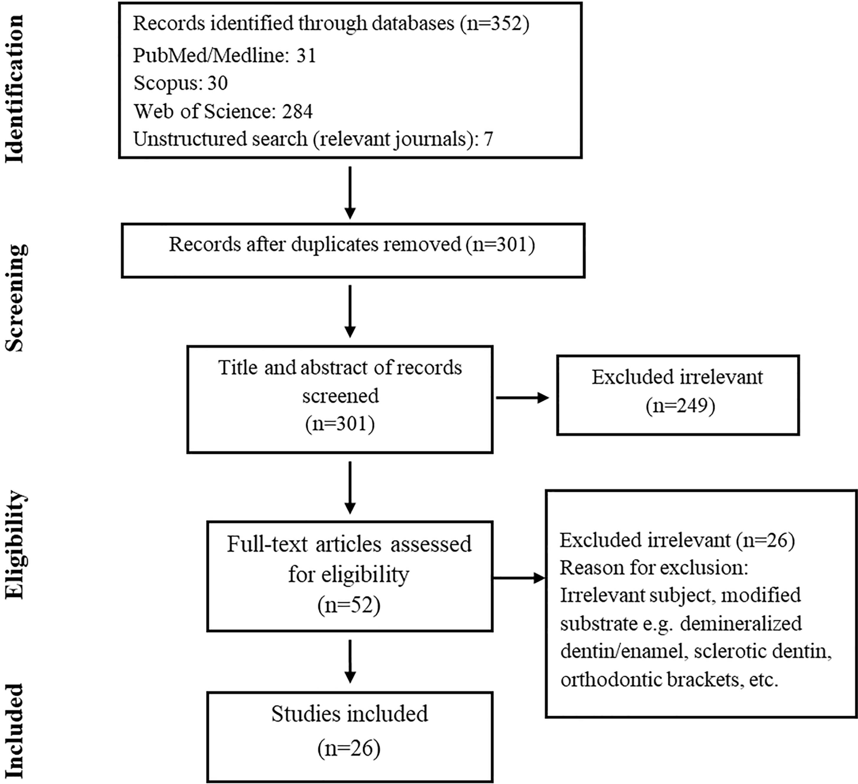
Figure 1.
Flowchart of Study Selection for Inclusion in the Systematic Review And Meta-analysis
.
Flowchart of Study Selection for Inclusion in the Systematic Review And Meta-analysis
Risk of Bias
The quality of the involved studies was assessed according to the modified CONSORT form, the results of which are presented in Table 3 (45). According to the 26 in vitro studies, the average score was 74%. All studies obtained a value greater than 47%, except for one that obtained 40% (37). Two studies showed low risk, reaching 100% (15,28), while others demonstrated moderate or high levels of bias. The least observed parameters were mention of the full trial protocol (Table 3). Concerning the overall risk of included articles, most studies represented moderate to high levels of risk of bias.
Qualitative Analysis
Overall, 26 studies were eligible for this review, all of which were in vitro studies and were performed on extracted teeth. Of this number, 4 (26,31,39,41) and 21 (6,11,13-15,19,20,24,25,27,28,32-38,40,42-44) studies used extracted bovine incisors and extracted human molars, respectively, and one study utilized both human extracted molars and bovine incisors (24). The minimum and maximum number of specimens were reported to be 5 and 34, and all studies employed either self-etch or universal adhesive systems. In all included studies, after the bonding procedure, the samples were stored in 37 ºC water for a period of time (minimum 24 hours and maximum 2 years). All the studies performed bond strength tests using a shear/tensile load at a crosshead speed of 0.5 or 1 mm/min, with the exception of one article which reported the crosshead speed of 2 mm/minute. The microtensile bond test was the most commonly used method (13 studies), followed by the shear bond test (11 studies) and micro-shear test (2 studies), respectively.
Meta-Analysis
Meta-analyses were performed using 14 studies that met the required criteria. The results were analyzed using the random-effects model in all but one analysis because I2 tests showed high heterogeneity (over 50%). In the general analysis of specimens undergoing doubled application time, ground dentin demonstrated higher bond strength compared to the recommended time by manufacturers (95% confidence interval [CI]: -1.509, -0.171, Z = 2.46, P = 0.014). Doubling the application time in one-component adhesives induced higher bond strength in ground dentin compared to the recommended time by manufacturers (95% CI: -1.624, -0.225, Z = 2.59, P = 0.010). However, by doubling the application time for two-component adhesives, the difference in bond strength was not significant (95% CI: -1.803, 2.088, Z = 0.14, P = 0.886, Figure 2). Taking into account the acidity of adhesives, doubling the application time of moderate adhesives seems to have increased the bond strength to the ground dentin (95% CI: -3.035, -0.381, Z = 2.52, P= 0.012), while mild (95% CI: -1.374, 0.244, Z = 1.37, P= 0.171) and strong (95% CI: -0.002, 1.053, Z = 1.95, P= 0.057) adhesives were not impacted as much (Figure 3). According to the overall analysis of adhesives concerning both aforementioned factors (i.e., the number of components and the level of acidity), one-component moderate adhesives seem to increase bond strength to ground dentine following the doubled application time (95% CI: -3.787, -0.734, Z = 2.90, P= 0.004). Conversely, other groups either lacked relevant information due to a lack of studies and experiments (i.e., two-component mild adhesives and two-component strong adhesives), or the available information indicated that their impact was insignificant (i.e., two-component moderate adhesives) (Figure 4).
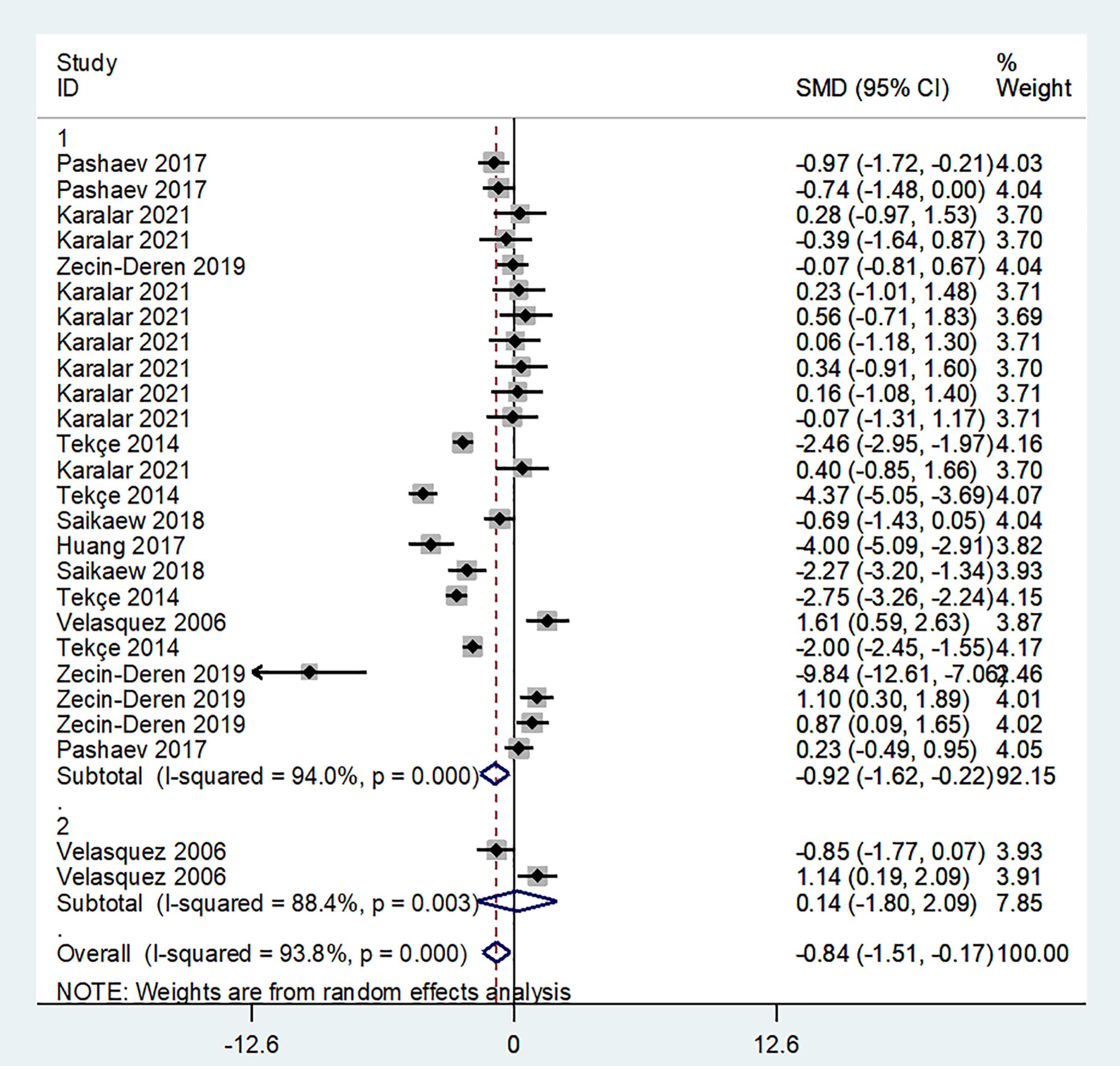
Figure 2.
Influence of Doubling the Application Time Compared With Manufacture Recommended Time on Bond Strength to Ground Dentin in One-Component and Two-Component Adhesives
.
Influence of Doubling the Application Time Compared With Manufacture Recommended Time on Bond Strength to Ground Dentin in One-Component and Two-Component Adhesives
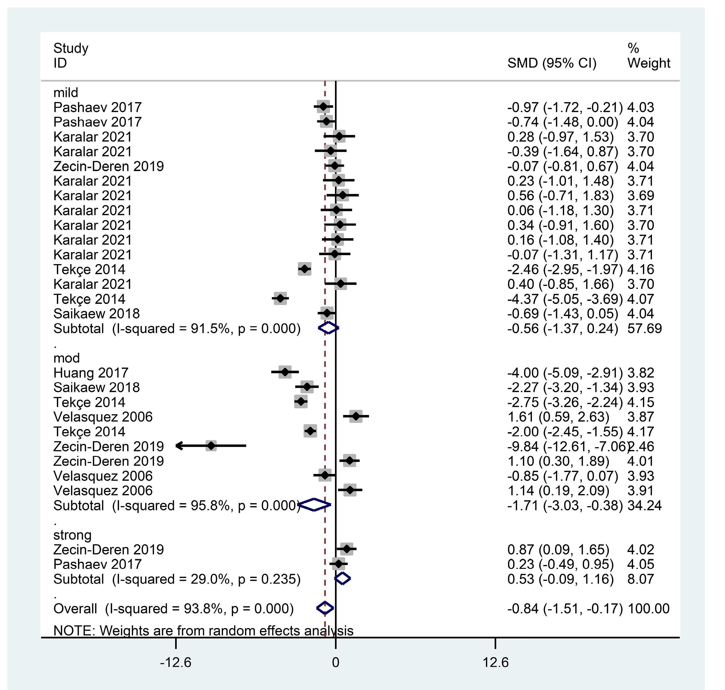
Figure 3.
Influence of Doubling the Application Time Compared With Manufacture Recommended Time on Bond Strength to Ground Dentin in Mild, Moderate and Strong Adhesives
.
Influence of Doubling the Application Time Compared With Manufacture Recommended Time on Bond Strength to Ground Dentin in Mild, Moderate and Strong Adhesives
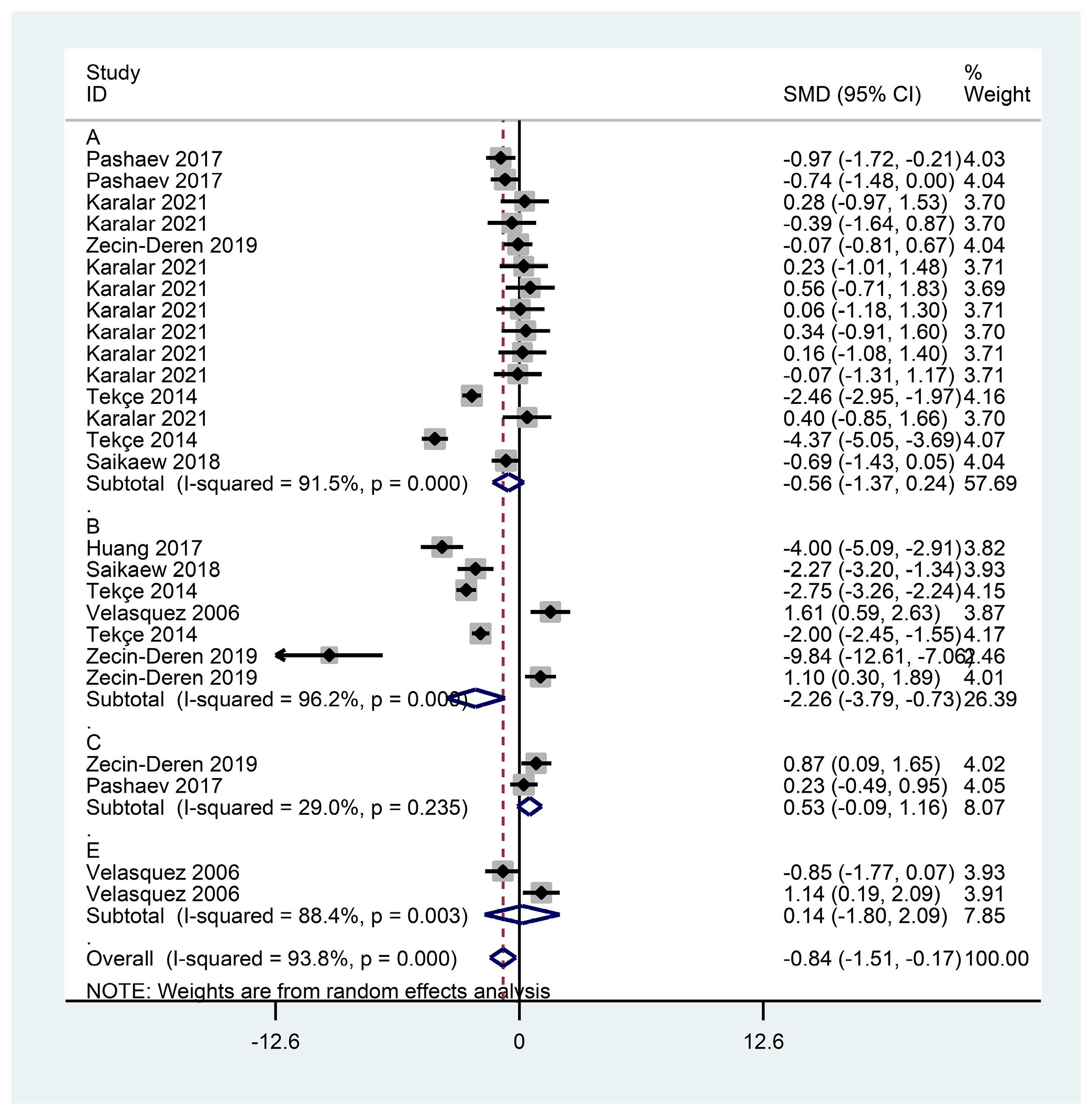
Figure 4.
Overall Analysis of the Influence of Doubling the Application Time Compared With Manufacture Recommended Time on Bond Strength to Ground Dentin
.
Overall Analysis of the Influence of Doubling the Application Time Compared With Manufacture Recommended Time on Bond Strength to Ground Dentin
In the general analysis of grounded enamel undergoing doubled application time, the specimen showed higher bond strength in comparison to the recommended time by manufacturers (95% CI: -1.287, -0.262, Z = 2.96, P= 0.003). Doubling the application time in one-component adhesives could induce higher bond strength in ground enamel compared to the recommended time by manufacturers (95% CI: -1.628, 0.453, Z = 3.47, P= 0.001). Contrarily, the difference in bond strength was not significant by doubling the application time for two-component adhesives (95% CI: -0.397, 0.600, Z = 0.40, P= 0.690, Figure 5). Based on the level of acidity, doubling the application time of mild adhesives increased the bond strength to the ground enamel (95% CI: -2.051, - 0.292, Z = 2.67, P= 0.009), whereas moderate adhesives (95% CI: -1.058, 0.049, Z = 1.79, P= 0.074) were not affected by doubled application time (Figure 6). Overall, considering both the number of components and level of acidity, the analysis reported a significant increase in bond strength following the duplication of application time for one-component mild (95% CI: -2.051, -0.292, Z = 2.67, P = 0.009) and one-component moderate (95% CI: -1.505, -0.553, Z = 4.24, P= 0.000) adhesives. The other subgroups of adhesive material faced the same limitations as the dentin group (Figure S1).
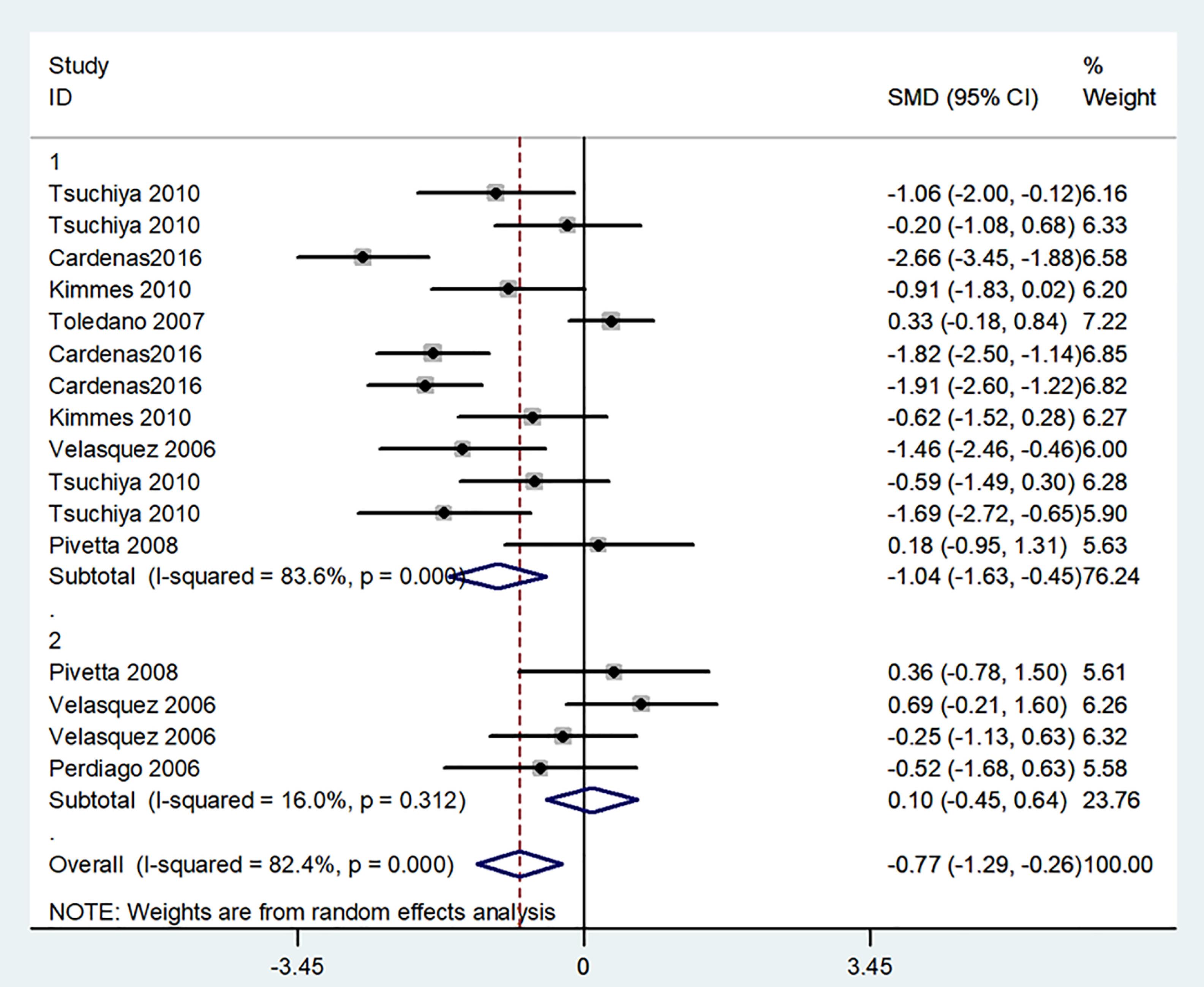
Figure 5.
Influence of Doubling the Application Time Compared With Manufacture Recommended Time on Bond Strength to Ground Enamel in One-Component and Two-Component Adhesives
.
Influence of Doubling the Application Time Compared With Manufacture Recommended Time on Bond Strength to Ground Enamel in One-Component and Two-Component Adhesives
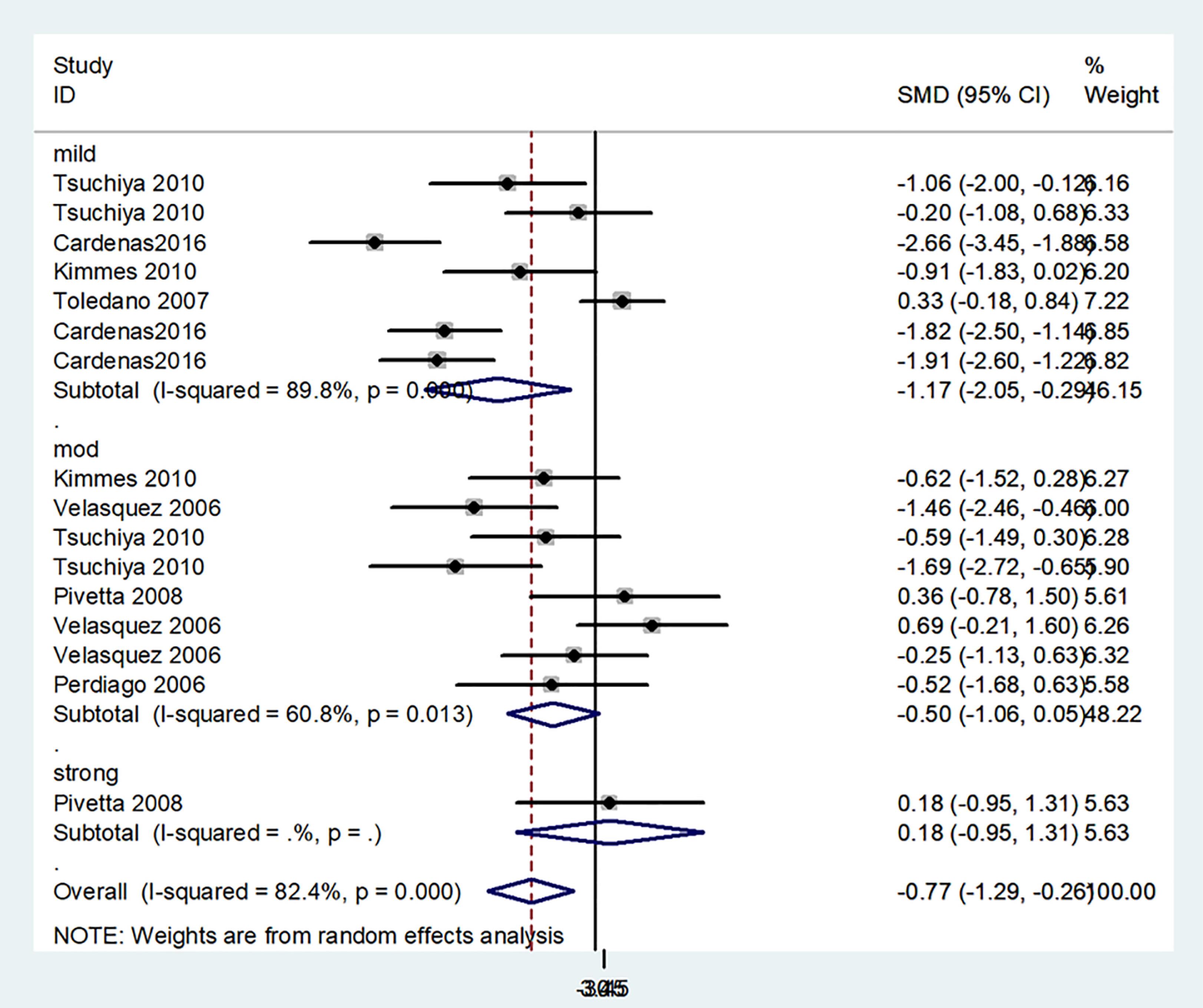
Figure 6.
Influence of Doubling the Application Time Compared With Manufacture Recommended Time on Bond Strength to Ground Enamel in Mild, Moderate and Strong Adhesives
.
Influence of Doubling the Application Time Compared With Manufacture Recommended Time on Bond Strength to Ground Enamel in Mild, Moderate and Strong Adhesives
In the general analysis of the specimen undergoing tripled application time, ground dentin displayed no significant increase in bond strength in any of the groups (95% CI: -0.779, -0.270, Z = 0.32, P= 0.750). Based on the reports from further specific analysis on one-component (95% CI: -0.927, 0.686, Z = 0.29, P= 0.774) and two-component (95% CI: -1.369, 0.895, Z = 041, P= 0.682) adhesives, tripling the application time in self-etch and universal adhesives could not induce increased bond strength in grounded dentin (Figure S2). The same results were obtained considering the level of acidity (95% CI: -0.811, 0.584, Z = 0.32, P= 0.750) (Figure S3) and thus factors revolving around adhesives and ground dentin (95% CI: -0.811, 0.584, Z = 0.32, P= 0.75, Figure S4).
In contrast, the general analysis of grounded enamel undergoing tripled application time revealed a significant increase in bond strength (95% CI: -0.779, -0.270, Z = 4.04, P= 0.000) compared to the manufacturer’s recommended time. Given that the subgroup analysis of one-component and two-component adhesives produced a low value of heterogeneity (I2 < 50%), the overall analysis used the fixed model instead of the random model for this specific analysis. Based on the reports from further specific analysis on one-component adhesives using a fixed model (I2 = 34.6%), tripling the application time induced a significant increase in bond strength to ground enamel (95% CI: -1.180, -0.518, Z = 5.02, P= 0.000). Nonetheless, using the fixed model on further analysis of two-component adhesives (I2 = 27.7%) did not illustrate any significant increase (95% CI: -0.454, 0.342, Z = 0.28, P= 0.782, Figure S5). Based on the level of acidity, tripling the application time of mild (95% CI: -1.159, - 0.238, Z = 2.97, P= 0.003) and moderate (95% CI: -0.976, -0.012, Z = 2.01, P= 0.045) adhesives could increase the bond strength to ground enamel. Not enough experimental data were available on strong adhesives (Figure S6). In general, considering the number of components and level of acidity, the analysis confirmed a significant increase in bond strength following the duplication of application time for one component mild (95% CI: -1.159, -0.238, Z = 2.97, P= 0.003) and one-component moderate (95% CI: -0.979, -0.012, Z = 2.01, P= 0.045) adhesives. Other subgroups lacked relevant information due to a lack of studies and experiments (i.e., one-component strong adhesives and two-component mild, moderate, and strong adhesives) (Figure S7).
The results of the evaluation of the diffusion pattern using the Egger method revealed that the bias was not significant in any of the meta analyses as follows:
(P= 0.075) for grounded dentin groups with the duplication of primer application time (Figure S8).
(P= 0.944) for grounded enamel groups with the duplication of primer application time (Figure S9).
(P= 0.054) for grounded dentin groups with the triplication of primer application time (Figure S10).
(P= 0.000) for grounded enamel groups with the triplication of primer application time (Figure S11).
Discussion
This systematic review and meta-analysis assessed the impact of increased application time on the bond strength between self-etch/universal adhesive systems and tooth structure. To the best of our knowledge, no other similar systematic review has been conducted so far. A thorough search of existing literature was performed in English, with no publication date restrictions. To conduct the meta-analysis, self-etch and universal adhesive systems were further categorized and independently analyzed based on their pH values (mild, moderate, and strong) and the number of involved components (one or two bottles). This approach was taken to accommodate potential variations in behavior depending on the adhesive system’s composition and how different numbers of steps in the clinical application process might influence the outcomes. Subsequently, a complete analysis was conducted, considering both acidity and the number of components.
The null hypothesis was rejected based on the existing data and the performed meta-analysis. Overall, the results indicated that doubling the application time of adhesives could increase the bond strength to dentin or enamel. While tripling the application time did not significantly affect the bond strength between dentin and adhesives, it could increase the bond strength between enamel and SEAs and UAs. However, there were several limitations due to the limited number of available studies in certain subgroups. For instance, there was a lack of data exploring whether tripling the application time in strong adhesive systems could enhance the bond strength to dentin. Similar limitations were observed for 2-component mild adhesives and 2-component strong adhesives.
In relation to doubling the application time of one-component self-etch and universal adhesives, a significant increase was observed in bond strength between these systems and dentin. Our findings align with those of other studies (19,24,47). Considering that the high content of the remaining solvent could compromise the mechanical properties and conversion degree of adhesive systems (48,49), it appears that prolonging the application time would allow for increased solvent evaporation and continued monomer diffusion into the dentin structure. This process could contribute to the formation of a stronger resin-dentin bond strength (37,50). However, the prolonged application time of self-etch and universal adhesive systems may not exhibit a significant increase in all cases, depending on the amount of prolongation (51). These data are in line with our findings considering the tripled application time of one component adhesive and resin-dentin bond strength.
Dentin is a hydrated hard dental tissue, and its composition and structural features are different from those in enamel. While hydroxyapatite minerals compose close to 90% wt of enamel, dentin has a higher organic content (52,53). Acidic monomers in self-adhesive bonding agents interact and partially dissolve the hydroxyapatite minerals in dentin structure, modifying the smear layer as opposed to dissolving it (35,51,54). The residual hydroxyapatite crystals along with smear debris remnants would become part of the final hybrid layer (50,54). Following this step, hydrophilic monomers present in the bond solution would infiltrate and interact with the exposed collagen fibrils (55). As longer exposure of the mineral content to acidic monomers would result in more demineralization, the hydrated collagen matrix might be able to partially maintain its structural height and volume. However, ongoing demineralization might compromise the mechanical support of the matrix (56) and thus likely affect the final bond value. Based on these facts and our statistical results, it is possible to assume that increasing the application time of self-etch adhesives over a certain limit could be destructive to resin-dentin bond strength.
With regard to doubling and tripling the application time of one component adhesive systems on enamel, our findings suggest a significant impact, which is consistent with the results of previous investigations (25,41). According to Cardenas et al (25), since the acidic monomers in self-etch and universal adhesives are not strong enough to create a retentive etching pattern, the prolonged application time might improve the interaction of acidic monomers with the enamel, thereby creating a more retentive pattern and increasing the resin impregnation into the dental tissue. However, the effect of prolonged application time on resin-enamel bond strength has not been substantial in all studies. As reported by Kimmes et al (6), while the enamel bond strength increased slightly with the extended treatment time, it did not induce a significant increase in bond strength compared to the recommended treatment time. Nonetheless, this result might be due to different experimental conditions and materials used. Additionally, because of enamel and dentin chemical and structural differences (57,58), utilizing the same adhesive system might result in different outcomes for dentin and enamel, as certain adhesive systems might yield higher bond strength values for dentin compared to enamel, or conversely (23,59-61).
Recent studies have confirmed the correlation between the pH value of the applied universal adhesives and their bonding performances, highlighting the importance of this knowledge for clinical practitioners (62,63). Self-etch and universal adhesive systems can be classified into ultra-mild (pH > 2.5), mild (2.5 > pH > 2), moderate (1 < pH < 2), and strong (pH < 1) categories based on the pH value (11,15,53). Self-etch adhesives employ acidic monomers for conditioning tooth structures, in contrast to traditional phosphoric acid etching in total-etch methods. However, while strong self-etch adhesives with lower pH values could create demineralization patterns in enamel/dentin similar to that of total-etch systems (64), mild self-etch adhesives did not generate an equivalent level of porosity in enamel surfaces as achieved through phosphoric acid etching. Furthermore, the collateral collapse of collagen fibers in dentin followed by strong adhesive conditioning could result in weaker bond strength to dentin (65,66), leaving the possibility that the resulting bond strength from self-etch adhesives might be related to other bonding factors rather than only the level of pH (15,27,33,55). According to Oliveira et al (56), the bonding mechanism of self-etch adhesives to dentin is largely dependent on the formation of a “hybrid layer”, which is created following the infiltration of resinous monomers in demineralized dentin and the creation of a molecular interaction between the resin and the fibers of collagen (67,68). However, in some self-etch adhesives, the presence of other functional monomers could contribute to the bond strength as well. For example, the ionic interaction of the functional monomer 10-methacryloyloxydecyl dihydrogen phosphate with calcium from the remaining hydroxyapatite around the partially exposed collagen seems to have had a positive impact on the resin-dentin bond value in certain adhesives (13,15,20,21,33).
According to our statistical findings, doubling and tripling the application time of mild one-component self-etch and universal adhesives led to an increase in bond strength to the enamel. However, such variations in the application time had no significant impact on bond strength to dentin. Moreover, the duplication of the application time of one-component moderate adhesive resulted in increased adhesive-dentin bond strength, while tripling the application time produced higher bond strength to the enamel. Nevertheless, it is important to note that there is a limited body of research exploring the relationship between the prolonged application time and the acidity levels of self-etch and universal adhesives and how these factors influence bond strength to dentin and enamel. Consequently, our findings in this specific matter may not offer conclusive clinical guidance due to the current gaps in knowledge.
Most articles stored the samples in water for 24 hours before bond strength tests. However, samples in one study did not undergo water storage (44). The speed per minute of the machine testing the bond strength varied in different machines (0.5, 1, and 2 mm/s) and different bond tests (shear, micro-shear, tensile, and microtensile) used for experimenting. Therefore, the high heterogeneity shown in the meta-analyses could be because of the lack of cohesiveness in protocols, different adhesive materials, sample sizes, and the like. Additionally, the risk of bias was considered medium and high in most studies. Given the identified risk of bias and the observed high heterogeneity, there is an imperative need for standardized experimental methods for further investigations.
Considering the current findings and acknowledged limitations, the prolonged application time of one-component self-etch and universal adhesives could enhance the bond strength of both dentin and enamel. Nevertheless, dental practitioners should interpret these results with caution for clinical purposes. Moreover, further in vitro and clinical investigation is needed to confirm the impact of increased application time of different adhesive systems and provide evidence-based recommendations for clinical practice.
Conclusion
Based on the gathered data and performed analysis, duplicating the application time of self-etch and universal adhesives could increase the bond strength to ground dentine/enamel under in vitro conditions. However, while tripling the application time of self-etch and universal adhesives could increase the bond strength to ground enamel, it did not have the same impact on ground dentin. Furthermore, the overall increased application time of one-component adhesives could result in better bond strength compared to two-component adhesives. Considering the diverse variables affecting the bond strength value and the present limitations in current literature related to this matter, no coherent conclusion could be reached considering the effect of the number of components or the pH level of adhesives on bond strength value.
Acknowledgements
The author would like to thank Dr. Alireza Akbarzadeh Baghaban, Ph.D. in Biostatistics, for assistance with data analysis.
Competing Interests
The authors declared that there is no conflict of interests regarding the publication of this paper.
Data Availability Statement
The data are available upon request from the authors.
Declaration of Generative Artificial Intelligence in Scientific Writing
The authors declare the use of generative artificial intelligence in scientific writing upon submission of the paper.
Ethical Approval
Not applicable.
Funding
This research received no financial support.
Supplementary Files
Supplementary file 1 contains Figures S1-S11.
(pdf)
References
- de Amoêdo Campos Velo MM, Coelho LV, Basting RT, do Amaral FL, França FM. Longevity of restorations in direct composite resin: literature review. RGO Rev Gaúch Odontol 2016; 64(3):320-6. doi: 10.1590/1981-8637201600030000123109 [Crossref] [ Google Scholar]
- Manhart J, García-Godoy F, Hickel R. Direct posterior restorations: clinical results and new developments. Dent Clin North Am 2002; 46(2):303-39. doi: 10.1016/s0011-8532(01)00010-6 [Crossref] [ Google Scholar]
- Dionysopoulos D, Gerasimidou O, Papadopoulos C. Current modifications of dental adhesive systems for composite resin restorations: a review in literature. J Adhes Sci Technol 2022; 36(5):453-68. doi: 10.1080/01694243.2021.1924499 [Crossref] [ Google Scholar]
- Abou Neel EA, Young AM. Antibacterial adhesives for bone and tooth repair. In: Zhou Y, Breyen MD, eds. Joining and Assembly of Medical Materials and Devices. Amsterdam: Woodhead Publishing; 2013. p. 491-513. doi: 10.1533/9780857096425.4.491.
- Rathi SD, Nikhade P, Chandak M, Motwani N, Rathi C, Chandak M. Microleakage in composite resin restoration-a review article. J Evol Med Dent Sci 2020; 9(12):1006-11. doi: 10.14260/jemds/2020/216 [Crossref] [ Google Scholar]
- Kimmes NS, Barkmeier WW, Erickson RL, Latta MA. Adhesive bond strengths to enamel and dentin using recommended and extended treatment times. Oper Dent 2010; 35(1):112-9. doi: 10.2341/09-081-l [Crossref] [ Google Scholar]
- Sofan E, Sofan A, Palaia G, Tenore G, Romeo U, Migliau G. Classification review of dental adhesive systems: from the IV generation to the universal type. Ann Stomatol (Roma) 2017; 8(1):1-17. doi: 10.11138/ads/2017.8.1.001 [Crossref] [ Google Scholar]
- Arandi NZ. The classification and selection of adhesive agents; an overview for the general dentist. Clin Cosmet Investig Dent 2023; 15:165-80. doi: 10.2147/ccide.S425024 [Crossref] [ Google Scholar]
- Chen H, Feng S, Jin Y, Hou Y, Zhu S. Comparison of bond strength of universal adhesives using different etching modes: a systematic review and meta-analysis. Dent Mater J 2022; 41(1):1-10. doi: 10.4012/dmj.2021-111 [Crossref] [ Google Scholar]
- Saikaew P, Sattabanasuk V, Harnirattisai C, Chowdhury A, Carvalho R, Sano H. Role of the smear layer in adhesive dentistry and the clinical applications to improve bonding performance. Jpn Dent Sci Rev 2022; 58:59-66. doi: 10.1016/j.jdsr.2021.12.001 [Crossref] [ Google Scholar]
- Britta LC, Martins M, França FM. Influence of different primer application times on bond strength of self-etching adhesive systems to unground enamel. Oper Dent 2009; 34(1):43-50. doi: 10.2341/08-35 [Crossref] [ Google Scholar]
- Eliades G, Vougiouklakis G, Palaghias G. Heterogeneous distribution of single-bottle adhesive monomers in the resin-dentin interdiffusion zone. Dent Mater 2001; 17(4):277-83. doi: 10.1016/s0109-5641(00)00082-8 [Crossref] [ Google Scholar]
- Erhardt MC, Osorio R, Pisani-Proenca J, Aguilera FS, Osorio E, Breschi L. Effect of double layering and prolonged application time on MTBS of water/ethanol-based self-etch adhesives to dentin. Oper Dent 2009; 34(5):571-7. doi: 10.2341/08-060-l [Crossref] [ Google Scholar]
- Velasquez LM, Sergent RS, Burgess JO, Mercante DE. Effect of placement agitation and placement time on the shear bond strength of 3 self-etching adhesives. Oper Dent 2006; 31(4):426-30. doi: 10.2341/05-52 [Crossref] [ Google Scholar]
- Pivetta MR, Moura SK, Barroso LP, Lascala AC, Reis A, Loguercio AD. Bond strength and etching pattern of adhesive systems to enamel: effects of conditioning time and enamel preparation. J Esthet Restor Dent 2008; 20(5):322-35. doi: 10.1111/j.1708-8240.2008.00202.x [Crossref] [ Google Scholar]
- Bahrololumi N, Beglou A, Najafi-Abrandabadi A, Sadr A, Sheikh-Al-Eslamian SM, Ghasemi A. Effect of water storage on ultimate tensile strength and mass changes of universal adhesives. J Clin Exp Dent 2017; 9(1):e78-83. doi: 10.4317/jced.53048 [Crossref] [ Google Scholar]
- Chen C, Niu LN, Xie H, Zhang ZY, Zhou LQ, Jiao K. Bonding of universal adhesives to dentine--old wine in new bottles?. J Dent 2015; 43(5):525-36. doi: 10.1016/j.jdent.2015.03.004 [Crossref] [ Google Scholar]
- Pheerarangsikul N, Wayakanon P, Wayakanon K. Effects of various functional monomers on adhesion between immediate dentin sealing and resin cement. Oper Dent 2022; 47(5):562-73. doi: 10.2341/21-057-l [Crossref] [ Google Scholar]
- Tekçe N, Demirci M, Tuncer S, Uysal Ö. Effect of different application techniques of all-in-one adhesives on microtensile bond strength to sound and caries-affected dentin. J Adhes 2015; 91(4):245-61. doi: 10.1080/00218464.2014.885842 [Crossref] [ Google Scholar]
- Amsler F, Peutzfeldt A, Lussi A, Flury S. Bond strength of resin composite to dentin with different adhesive systems: influence of relative humidity and application time. J Adhes Dent 2015; 17(3):249-56. doi: 10.3290/j.jad.a34400 [Crossref] [ Google Scholar]
- Mine A, De Munck J, Cardoso MV, Van Landuyt KL, Poitevin A, Van Ende A. Dentin-smear remains at self-etch adhesive interface. Dent Mater 2014; 30(10):1147-53. doi: 10.1016/j.dental.2014.07.006 [Crossref] [ Google Scholar]
- Ahn J, Jung KH, Son SA, Hur B, Kwon YH, Park JK. Effect of additional etching and ethanol-wet bonding on the dentin bond strength of one-step self-etch adhesives. Restor Dent Endod 2015; 40(1):68-74. doi: 10.5395/rde.2015.40.1.68 [Crossref] [ Google Scholar]
- Ozer F, Blatz MB. Self-etch and etch-and-rinse adhesive systems in clinical dentistry. Compend Contin Educ Dent 2013; 34(1):12-8. [ Google Scholar]
- Toledano M, Proença JP, Erhardt MC, Osorio E, Aguilera FS, Osorio R. Increases in dentin-bond strength if doubling application time of an acetone-containing one-step adhesive. Oper Dent 2007; 32(2):133-7. doi: 10.2341/06-32 [Crossref] [ Google Scholar]
- Cardenas AM, Siqueira F, Rocha J, Szesz AL, Anwar M, El-Askary F. Influence of conditioning time of universal adhesives on adhesive properties and enamel-etching pattern. Oper Dent 2016; 41(5):481-90. doi: 10.2341/15-213-l [Crossref] [ Google Scholar]
- Mena-Serrano AP, Garcia EJ, Perez MM, Martins GC, Grande RH, Loguercio AD. Effect of the application time of phosphoric acid and self-etch adhesive systems to sclerotic dentin. J Appl Oral Sci 2013; 21(2):196-202. doi: 10.1590/1678-7757201302136 [Crossref] [ Google Scholar]
- Karalar B, Bayındır YZ. Effects of adhesive application time and air blowing pressure on dentin bond strength of self-etch mode universal adhesives. J Adhes Sci Technol 2022; 36(3):287-301. doi: 10.1080/01694243.2021.1917905 [Crossref] [ Google Scholar]
- Saikaew P, Matsumoto M, Chowdhury A, Carvalho RM, Sano H. Does shortened application time affect long-term bond strength of universal adhesives to dentin?. Oper Dent 2018; 43(5):549-58. doi: 10.2341/17-205-l [Crossref] [ Google Scholar]
- Moher D, Liberati A, Tetzlaff J, Altman DG. Preferred reporting items for systematic reviews and meta-analyses: the PRISMA statement. Int J Surg 2010; 8(5):336-41. doi: 10.1016/j.ijsu.2010.02.007 [Crossref] [ Google Scholar]
- Dreweck FD, Zarpellon D, Wambier LM, Loguercio AD, Reis A, Gomes OM. Is there evidence that three-step etch-and-rinse adhesives have better retention rates than one-step self-etch adhesives in noncarious cervical lesions? A systematic review and meta-analysis. J Adhes Dent 2021; 23(3):187-200. doi: 10.3290/j.jad.b1367811 [Crossref] [ Google Scholar]
- Karadas M. Influence of reduced application time on bonding durability of universal adhesives to demineralized enamel. Clin Oral Investig 2021; 25(12):6843-55. doi: 10.1007/s00784-021-03972-y [Crossref] [ Google Scholar]
- Ostby AW, Bishara SE, Laffoon J, Warren JJ. Influence of self-etchant application time on bracket shear bond strength. Angle Orthod 2007; 77(5):885-9. doi: 10.2319/092006-383 [Crossref] [ Google Scholar]
- Burrer P, Dang H, Par M, Attin T, Tauböck TT. Effect of over-etching and prolonged application time of a universal adhesive on dentin bond strength. Polymers (Basel) 2020; 12(12):2902. doi: 10.3390/polym12122902 [Crossref] [ Google Scholar]
- Ahmed MH, Yoshihara K, Mercelis B, Van Landuyt K, Peumans M, Van Meerbeek B. Quick bonding using a universal adhesive. Clin Oral Investig 2020; 24(8):2837-51. doi: 10.1007/s00784-019-03149-8 [Crossref] [ Google Scholar]
- Zecin-Deren A, Lukomska-Szymanska M, Szczesio-Wlodarczyk A, Piwonski I, Sokolowski J, Lapinska B. The influence of application protocol of simplified and universal adhesives on the dentin bonding performance. Appl Sci 2019; 10(1):124. doi: 10.3390/app10010124 [Crossref] [ Google Scholar]
- Saikaew P, Chowdhury AF, Fukuyama M, Kakuda S, Carvalho RM, Sano H. The effect of dentine surface preparation and reduced application time of adhesive on bonding strength. J Dent 2016; 47:63-70. doi: 10.1016/j.jdent.2016.02.001 [Crossref] [ Google Scholar]
- Pashaev D, Demirci M, Tekçe N, Tuncer S, Baydemir C. The effect of double-coating and times on the immediate and 6-month dentin bonding of universal adhesives. Biomed Mater Eng 2017; 28(2):169-85. doi: 10.3233/bme-171665 [Crossref] [ Google Scholar]
- Huang XQ, Pucci CR, Luo T, Breschi L, Pashley DH, Niu LN. No-waiting dentine self-etch concept-Merit or hype. J Dent 2017; 62:54-63. doi: 10.1016/j.jdent.2017.05.007 [Crossref] [ Google Scholar]
- Protásio MF, Frota PH, Costa JF, Carneiro KK, Bauer J. Effects of application mode of self-etching primer on shear bond strength of orthodontic brackets. Rev Port Estomatol Med Dent Cir Maxilofac 2016; 57(1):9-13. doi: 10.1016/j.rpemd.2016.01.002 [Crossref] [ Google Scholar]
- Osorio R, Osorio E, Aguilera FS, Tay FR, Pinto A, Toledano M. Influence of application parameters on bond strength of an “all in one” water-based self-etching primer/adhesive after 6 and 12 months of water aging. Odontology 2010; 98(2):117-25. doi: 10.1007/s10266-010-0126-3 [Crossref] [ Google Scholar]
- Tsuchiya H, Tsubota K, Iwasa M, Ando S, Miyazaki M, Platt JA. Influence of adhesive application time on enamel bond strength of single-step self-etch adhesive systems. Oper Dent 2010; 35(1):77-83. doi: 10.2341/09-064-l [Crossref] [ Google Scholar]
- Barkmeier WW, Erickson RL, Kimmes NS, Latta MA, Wilwerding TM. Effect of enamel etching time on roughness and bond strength. Oper Dent 2009; 34(2):217-22. doi: 10.2341/08-72 [Crossref] [ Google Scholar]
- Iijima M, Ito S, Yuasa T, Muguruma T, Saito T, Mizoguchi I. Effects of application time and agitation for bonding orthodontic brackets with two self-etching primer systems. Dent Mater J 2009; 28(1):89-95. doi: 10.4012/dmj.28.89 [Crossref] [ Google Scholar]
- Perdigão J, Gomes G, Lopes MM. Influence of conditioning time on enamel adhesion. Quintessence Int 2006; 37(1):35-41. [ Google Scholar]
- Faggion CM Jr. Guidelines for reporting pre-clinical in vitro studies on dental materials. J Evid Based Dent Pract 2012; 12(4):182-9. doi: 10.1016/j.jebdp.2012.10.001 [Crossref] [ Google Scholar]
- Fortea L, Sanz-Serrano D, Luz LB, Bardini G, Mercade M. Update on chelating agents in endodontic treatment: a systematic review. J Clin Exp Dent 2024; 16(4):e516-38. doi: 10.4317/jced.60989 [Crossref] [ Google Scholar]
- Reis A, de Carvalho Cardoso P, Vieira LC, Baratieri LN, Grande RH, Loguercio AD. Effect of prolonged application times on the durability of resin-dentin bonds. Dent Mater 2008; 24(5):639-44. doi: 10.1016/j.dental.2007.06.027 [Crossref] [ Google Scholar]
- Reis A, Grandi V, Carlotto L, Bortoli G, Patzlaff R, de Lourdes Rodrigues Accorinte M. Effect of smear layer thickness and acidity of self-etching solutions on early and long-term bond strength to dentin. J Dent 2005; 33(7):549-59. doi: 10.1016/j.jdent.2004.12.003 [Crossref] [ Google Scholar]
- Ye Q, Spencer P, Wang Y, Misra A. Relationship of solvent to the photopolymerization process, properties, and structure in model dentin adhesives. J Biomed Mater Res A 2007; 80(2):342-50. doi: 10.1002/jbm.a.30890 [Crossref] [ Google Scholar]
- Hosaka K, Prasansuttiporn T, Thanatvarakorn O, Kunawarote S, Takahashi M, Foxton RM. Smear layer-deproteinization: improving the adhesion of self-etch adhesive systems to caries-affected dentin. Curr Oral Health Rep 2018; 5(3):169-77. doi: 10.1007/s40496-018-0185-z [Crossref] [ Google Scholar]
- Sabatini C, Wu Z. Effect of desensitizing agents on the bond strength of mild and strong self-etching adhesives. Oper Dent 2015; 40(5):548-57. doi: 10.2341/14-190-l [Crossref] [ Google Scholar]
- Shahmoradi M, Bertassoni LE, Elfallah HM, Swain M. Fundamental structure and properties of enamel, dentin and cementum. In: Ben-Nissan B, ed. Advances in Calcium Phosphate Biomaterials. Berlin, Heidelberg: Springer; 2014. p. 511-47. doi: 10.1007/978-3-642-53980-0_17.
- de Oliveira FG, Machado LS, Sundfeld-Neto D, Giannini M, Briso AL, Dos Santos PH. Two-year clinical evaluation of a nanofilled etch-and-rinse and a self-etch adhesive system containing MDPB and fluoride in non-carious cervical lesions. Compend Contin Educ Dent 2017; 38(3):e1-4. [ Google Scholar]
- Ferreira JC, Pires PT, de Melo PR, Silva MJ. Etch-and-rinse and self-etch adhesives behavior on dentin. In: Rudawska A, ed. Adhesives-Applications and Properties. IntechOpen; 2016. doi: 10.5772/64856.
- Bin Hasan M. Self-etching adhesive systems in operative dentistry: a literature review. Res Rev J Dent Sci 2017; 5(2):23-32. [ Google Scholar]
- Oliveira SS, Marshall SJ, Hilton JF, Marshall GW. Etching kinetics of a self-etching primer. Biomaterials 2002; 23(20):4105-12. doi: 10.1016/s0142-9612(02)00149-7 [Crossref] [ Google Scholar]
- Van Meerbeek B, Conn LJ Jr, Duke ES, Eick JD, Robinson SJ, Guerrero D. Bonding to enamel and dentin. In: Fundamentals of Operative Dentistry: A Contemporary Approach. 3rd ed. Chicago: Quintessence; 2006. p. 183-260.
- Van Meerbeek B, De Munck J, Yoshida Y, Inoue S, Vargas M, Vijay P. Buonocore memorial lecture Adhesion to enamel and dentin: current status and future challenges. Oper Dent 2003; 28(3):215-35. [ Google Scholar]
- Elshehawy T. Influence of different universal adhesives on fracture resistance of endodontically treated teeth. Egypt Dent J 2021; 67(3):2805-15. doi: 10.21608/edj.2021.74983.1617 [Crossref] [ Google Scholar]
- Blatz MB. Bonding protocols for improved long-term clinical success. Compend Contin Educ Dent 2014; 35(4):276-7. [ Google Scholar]
- Sato T, Takagaki T, Hatayama T, Nikaido T, Tagami J. Update on enamel bonding strategies. Front Dent Med 2021; 2:666379. doi: 10.3389/fdmed.2021.666379 [Crossref] [ Google Scholar]
- Cuevas-Suárez CE, de Oliveira da Rosa WL, Lund RG, da Silva AF, Piva E. Bonding performance of universal adhesives: an updated systematic review and meta-analysis. J Adhes Dent 2019; 21(1):7-26. doi: 10.3290/j.jad.a41975 [Crossref] [ Google Scholar]
- Chasqueira AF, Arantes-Oliveira S, Portugal J. Bonding performance of simplified dental adhesives with three application protocols: an 18-month in vitro study. J Adhes Dent 2020; 22(3):255-64. doi: 10.3290/j.jad.a44549 [Crossref] [ Google Scholar]
- Pashley DH, Tay FR. Aggressiveness of contemporary self-etching adhesives Part II: etching effects on unground enamel. Dent Mater 2001; 17(5):430-44. doi: 10.1016/s0109-5641(00)00104-4 [Crossref] [ Google Scholar]
- Inoue S, Vargas MA, Abe Y, Yoshida Y, Lambrechts P, Vanherle G. Microtensile bond strength of eleven contemporary adhesives to dentin. J Adhes Dent 2001; 3(3):237-45. [ Google Scholar]
- De Munck J, Van Meerbeek B, Satoshi I, Vargas M, Yoshida Y, Armstrong S. Microtensile bond strengths of one- and two-step self-etch adhesives to bur-cut enamel and dentin. Am J Dent 2003; 16(6):414-20. [ Google Scholar]
- Betancourt DE, Baldion PA, Castellanos JE. Resin-dentin bonding interface: mechanisms of degradation and strategies for stabilization of the hybrid layer. Int J Biomater 2019; 2019:5268342. doi: 10.1155/2019/5268342 [Crossref] [ Google Scholar]
- Nakabayashi N, Nakamura M, Yasuda N. Hybrid layer as a dentin-bonding mechanism. J Esthet Dent 1991; 3(4):133-8. doi: 10.1111/j.1708-8240.1991.tb00985.x [Crossref] [ Google Scholar]