Avicenna J Dent Res. 17(1):35-40.
doi: 10.34172/ajdr.1873
Review Article
Assessing the Prognostic Reliability of Autophagy-Related Biomarkers in Oral Squamous Cell Carcinoma: A Systematic Review
Nishanthi Raja Conceptualization, Data curation, Funding acquisition, Visualization, Writing – original draft, Writing – review & editing, 1, * 
Anuradha Ganesan Investigation, Supervision, Validation, 1
KV Swathi Formal analysis, Project administration, 1
Krithika Chandrasekar Lakshmi Methodology, 1
Author information:
1SRM Dental College, Ramapuram, India
Abstract
Background: Oral squamous cell carcinoma (OSCC) is one of the most common cancer types worldwide. Due to the limited availability of biomarkers and therapeutic targets, OSCC is the major leading cause of cancer death. Although many studies have shown the role of the autophagy-related biomarker in cell survival and progression of several cancers, it is unclear whether the autophagy-related biomarker could be a marker in tumorigenesis and prognosis in OSCC. The aim of this review was to evaluate the available evidence about the possibly significant role of autophagy-related genes (ATG) in tumorigenesis and prognosis in OSCC.
Methods: A systematic review was conducted according to the Preferred Reporting Items for Systematic Reviews and Meta-analysis statement, and the PICOS question was, "Whether autophagy genes can be a marker in tumorigenesis and prognosis in oral cancer." A search strategy was elaborated to retrieve studies (2018-2023) from various databases, such as PubMed, Google Scholar, Cochrane Library, and Web of Science. The risk of bias was assessed using the QUADAS tool in Cochrane Rev-Man software 5.4.
Results: Based on the inclusion and exclusion criteria, three out of 178 studies found through the search were included in this systematic review. The majority of the studies accurately demonstrated features of the tumor with a worse prognosis in OSCC that were associated with an autophagy-related biomarker.
Conclusion: According to the review, investigations indicate that biomarkers related to autophagy can be used to predict the diagnosis and prognosis of OSCC.
Keywords: Oral squamous cell carcinoma, Autophagy, Biomarker, Prognosis, Survival rate
Copyright and License Information
© 2025 The Author(s); Published by Hamadan University of Medical Sciences.
This is an open-access article distributed under the terms of the Creative Commons Attribution License (
https://creativecommons.org/licenses/by/4.0), which permits unrestricted use, distribution, and reproduction in any medium provided the original work is properly cited.
Please cite this article as follows: Raja N, Ganesan A, Swathi KV, Lakshmi KC. Assessing the prognostic reliability of autophagy-related biomarkers in oral squamous cell carcinoma: a systematic review. Avicenna J Dent Res. 2025;17(1):35-40. doi:10.34172/ajdr.1873
Background
Cancer is a global health problem and the leading cause of death worldwide. Of all types of cancer, oral cancer ranks ninth in terms of incidence and is a major cause of cancer-related mortality. In 2018, there were roughly 350 000 new cases of oral cancer, and 170 000 fatalities were related to the disease. In India and Southeast Asia, oral cancer accounts for approximately 40% of all cancer cases, while in Western nations, it accounts for only 4% (1). The important risk factors for oral squamous cell carcinoma (OSCC) include smoking, chewing tobacco, alcohol use, poor oral hygiene, and genetic alterations(2). India has witnessed a surging trend in late diagnosis, where around 70% of cases are diagnosed only in the advanced stages, thus lowering the five-year survival rate to 20% (3). The potential of OSCC to spread and interfere with upper gastrointestinal and respiratory tract functioning also results in a low survival rate and a high deformity rate (4). Despite developments in modern medicine and diagnostic technology, the five-year survival rate of many countries across the globe is below 50%. The fact that head and neck cancers are diverse tumors makes it challenging to plan a good treatment strategy in many cases (5). Preventive measures are essential since oral cancer has a poor prognosis. OSCC has a poor prognosis due to its vast genetic diversity and variability. The identification and prediction of malignant progression at an earlier stage of OSCC will be possible with the improved comprehension of processes leading to carcinogenesis. Different types of oral cancers vary greatly in their biological behavior, clinical course, and treatment responsiveness (6).
Eukaryotic cells are exposed to a wide range of physical, chemical, and biological stimuli during their lifetimes, which may cause an imbalance in homeostasis. Cells possess an array of internal defensive mechanisms to fight and adapt to such stress (7). Autophagy is an adaptive process that keeps cells alive and intact. Double-membrane structures called autophagosomes are found intracellularly and hold parts of the cytoplasm and proteins. In addition, these structures are brought to the lysosomes, where their contents are degraded (8). Genes known as autophagy-related genes (ATG) systematically regulate autophagy. Many of the ATGs, including ULK1, ATG5, Beclin-1, and ATG12, play important roles in autophagosome formation. Certain stimuli, such as hypoxia, starvation, genotoxicity, oxidative stress, protein accumulation, and pathogens, can trigger autophagy to maintain cellular homeostasis (9). Autophagy is dysregulated in a variety of clinical conditions, such as infection, aging, neurological disorders, and cancer. Early in the development of cancer, autophagy in cancer cells may restrict tumor growth by degrading harmful chemicals and halting the spread of damage, including alterations to DNA (10). On the other hand, autophagy is a promoter in the tumor process, particularly at advanced stages of tumor development, because it can sustain tumor viability under adverse situations. Hence, autophagy in cancer cells is referred to as a “double-edged sword”. Apart from its function in developing tumors, autophagy also plays a crucial part in resistance to many types of therapies, presenting a serious obstacle to effective treatment (11). According to previous studies, Cancer Genome Atlas (TCGA) HNSCC cohort analysis revealed a potential correlation between reduced overall survival (OS) and greater autophagic activity. Specifically, the high mRNA expression of ATG5 or Beclin-1 (BECN1) is linked to a lower chance of surviving (12). As a result, research on autophagy has gained popularity in the field of OSCC. Thus, this study seeks to evaluate the existing evidence confirming that ATGs could be a marker in tumorigenesis and prognosis in OSCC.
Methods
Study Design
The Preferred Reporting Items for Systematic Reviews and Meta-analysis (PRISMA) checklist was used as the reporting guide for conducting this systematic review.
Research Question and Outcome
The acronym PICOS was utilized to structure the research question as follows:
Can autophagy genes be a marker in tumorigenesis and prognosis in oral cancer?
P (patients) = Individuals diagnosed with oral cancer
I (Intervention) = Autophagy-related biomarker
C (comparator) = No autophagy-related biomarker
O (outcomes) = Prognosis of the tumor
S (study design) = Studies on humans
Search Strategy
A search of many databases, including PubMed, PubMed Central, ScienceDirect, and the Cochrane Library, was conducted electronically by two independent investigators. A tailored keyword search was performed on each database. The MeSH/keywords included (Mouth Neoplasm) OR (Neoplasms, Oral) OR (Cancer of Mouth) OR (Mouth Cancers) OR (Mouth Cancer) OR (Autophagocytosis biomarker) OR (Reticulophagy biomarker) OR (ER Phagy biomarker) OR (Nucleophagy biomarker) OR (Ribophagy biomarker) AND (Lipophagy biomarker) AND (Rate, Survival) OR (Survival Rates) OR (Mean Survival Time) OR (Time, Mean Survival) OR (Cumulative Survival Rate).
Inclusion and Exclusion Criteria
The studies that met eligibility requirements had to assess the prognostic validity of a biomarker associated with autophagy in oral cancer. Studies that were selected for review included articles that were published in English and provided sufficient information to enable prognosis and the survival rate estimate, research published in the last five years (2018–2023), and studies of different designs (e.g., cohort, case-control, cross-sectional, and prospective/retrospective studies). On the other hand, articles published before January 2018, publications without primary results, review articles, letters to the editor, studies conducted on cell lines and animals, and non-accessible full-text versions were excluded from the review.
Results
Data Extraction
The research involved participants with OSCC. Study characteristics included the name of the author, year of publication, study design, sample size, participant baseline demographics (age and gender), sample distribution, sample collection methodology, autophagy biomarker studied, autophagy biomarker evaluation methodology, and clinical outcomes such as OS, disease-free survival, and progression-free survival. The computer program Review Manager 5.4.1 (RevMan) was used for evaluation.
Literature Evaluation
Overall, 178 studies were found in the search. After screening 59 records for titles and abstracts, 119 articles were eliminated, along with 17 studies that failed to meet the inclusion criteria. Five studies were chosen for the assessment of the entire text. Following the entire manuscript evaluation, unpublished articles were eliminated, and three papers were added to the systematic review. Figure 1 illustrates the PRISMA flowchart related to selection processes.
Three studies were included and extensively reviewed in this systematic review (Table 1). Figure2 shows the distribution of samples in the included studies.
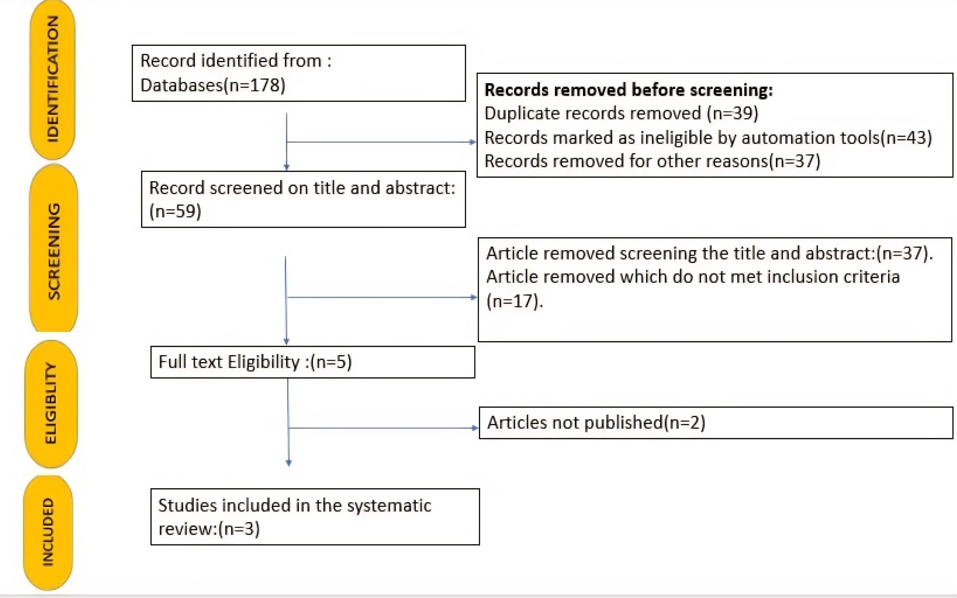
Figure 1.
PRISMA Flow Diagram. Note. PRISMA: The Preferred Reporting Items for Systematic Reviews and Meta-analysis
.
PRISMA Flow Diagram. Note. PRISMA: The Preferred Reporting Items for Systematic Reviews and Meta-analysis
Table 1.
Characteristics of the Included Studies
|
Author, Year of Publication, and Journal
|
Sample Distribution
|
Methodology Sample Collection
|
Evaluation of Inflammatory Biomarkers
|
Results
|
Inferences
|
| Liu et al (13) 2018, J Clin Med |
Group 1: Patients included 181 BMSCC, 244 TSCC, and 73 LSCC patients.
Group 2: Corresponding tumor-adjacent normal tissues at three subsites of oral SCC |
Tissue biopsy-IHC |
MAP1LC3B and SQSTM1 |
MAP1LC3B was associated with a poor prognosis only in TSCC. SQSTM1 was associated with poor differentiation in three subsites, while the association with lymph node invasion was only observed in BMSCC. |
Co-expression of higher MAP1LC3B and
SQSTM1 demonstrated a significantly worse
disease-specific survival (DSS) and disease-free survival (DFS) in patients with BMSCC and LSCC. |
| Wang et al (14) Int J Clin Exp Pathol.2018 |
Group 1: 186 patients with OSCC
Group 2: Corresponding adjacent normal tissues in all 186 patients |
Tissue biopsy |
ALDH1, Beclin1, and p16 |
Positive rates of VM, ALDH1, and Beclin1 were significantly higher, while levels of p16 were significantly lower in OSCC than in normal oral tissues. |
The expression of ALDH1, Beclin1, and p16 represents promising markers for metastasis
and prognosis, and potential therapeutic targets for OSCC. |
| Liu et al (15), 2019,Cancers |
Group 1: Tissue microarray comprising specimens from 428 OSCC patients, including 179 BMSCC and 249 TSCC patients.
Group 2: Corresponding tumor-adjacent normal tissues at two subsites of OSCC |
Tissue biopsy -IHC |
ATG4B and phosphorylated ATG4B proteins |
High co-expression levels of ATG4B and phospho-Ser383/392-ATG4B were associated with poor DFS only in TSCC patients, whereas they had no significant association with DSS in BMSCC and TSCC patients. |
ATG4B might be a biomarker for
the diagnosis/prognosis of OSCC
and a potential therapeutic target for OSCC patients. |
Note. SCC: Squamous cell carcinoma; OSCC: Oral squamous cell carcinoma; BMSCC: Buccal mucosal squamous cell carcinoma; TSCC: Tongue squamous cell carcinoma; LSCC: Lung squamous cell carcinoma; IHC: Immunohistochemistry; VM: Vasculogenic mimicry.

Figure 2.
Sample Distribution of the Included Studies
.
Sample Distribution of the Included Studies
Quality Assessment of Studies
According to selection criteria, the search method produced three articles, whose quality was then evaluated using the QUADAS tool 2. Four categories comprise the quality assessment of research on diagnostic accuracy, including patient sampling, index test, reference standard, and flow and timing. There are two to four questions in each of these domains, and the responses are “yes”, “no”, or “unclear”. The above data were entered into the Review Manager 5.4.1 software, which produced a color-coded chart showing the danger of bias and applicability issues.
Risk of Bias and Applicability Concerns
Studies performed by Liu et al (13) and Liu et al (15) had a high risk of bias, whereas those conducted by Wang et al (14) had a low risk of bias (Figure 3A-B).
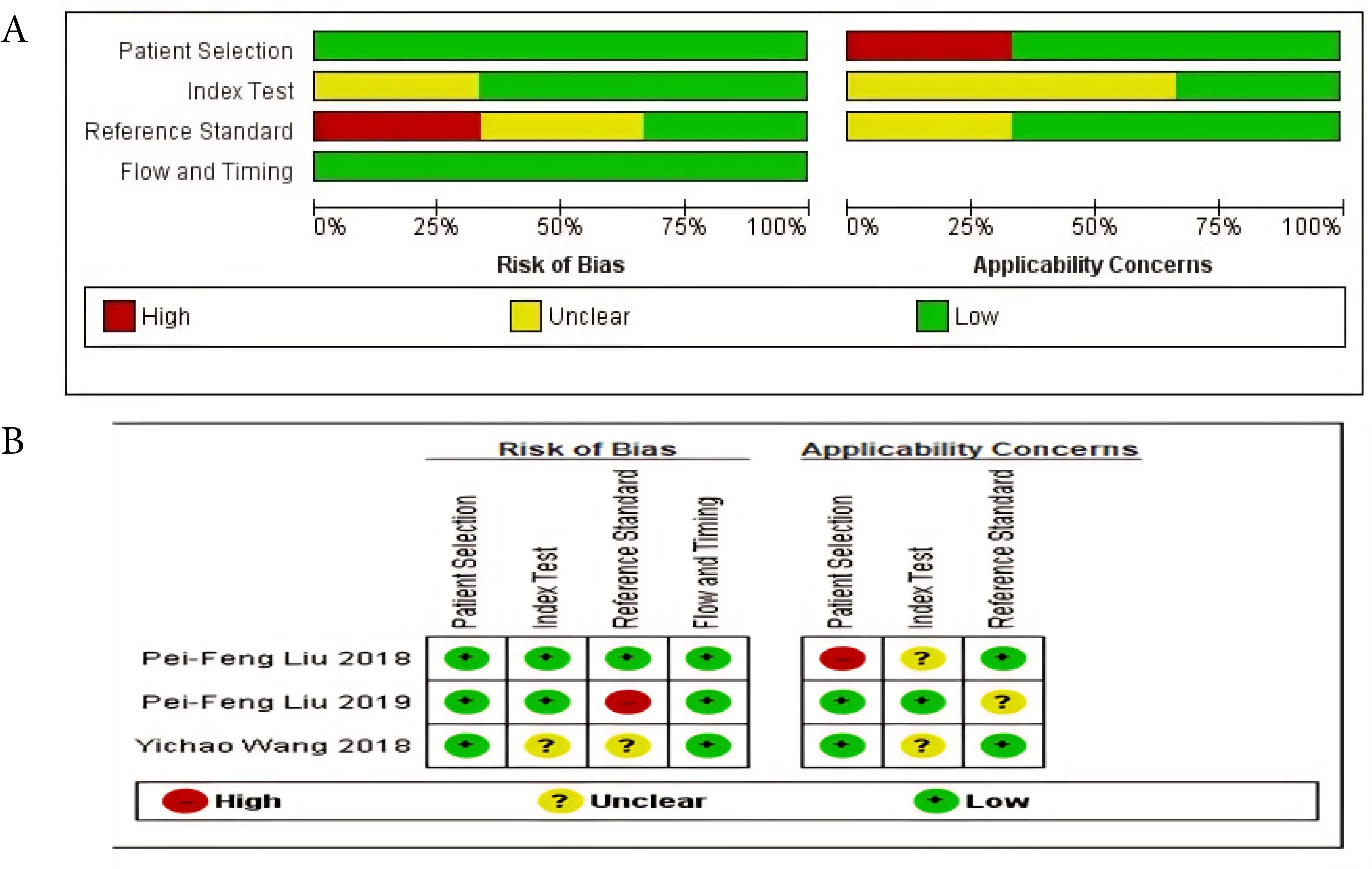
Figure 3.
(A-B) Risk of Bias and Applicability Concern Graph: Review Authors’ Judgements About Each Domain Presented as Percentages Across the Included Studies
.
(A-B) Risk of Bias and Applicability Concern Graph: Review Authors’ Judgements About Each Domain Presented as Percentages Across the Included Studies
Discussion
Over 275 000 individuals worldwide are affected by OSCC, the most prevalent type of oral cancer. Due to the aggressive nature of OSCC, most patients expire within three to five years after being diagnosed (16). Even though OSCC is caused by aberrant migration and development of oral epithelial cells, the carcinoma microenvironment is formed by immune cells and fibroblasts (17). The significance of studying the tumor microenvironment has increased over the last ten years. Autophagy is a new cellular microenvironment in OSCC, which is present in all living things from yeasts to mammals and encourages the breakdown of intracellular waste products such as macromolecules and organelles in order to preserve cell homeostasis (18). All cells have a basic autophagic tone, but in response to a specific stress, autophagy is upregulated to repair the damage. When there is no stress, autophagy operates at low levels to break down damaged cell components and recycle nutrients to keep the cell in an energetic condition (19). Various studies proved that there is a connection between autophagy and certain cancer hallmarks. For example, although autophagy and apoptosis are typically considered antagonistic processes, they can cooperate to induce cell death in specific biological contexts. ATGs are the proteins involved in autophagy (20). ATG8, sometimes referred to as MAP1LC3 (or simply LC3), is a crucial component of the autophagy process. ATG proteins have a pivotal role in the various phases of autophagosome production and are necessary for the biogenesis of the autophagosome. Autophagy has been demonstrated to be triggered by tumor-suppressor genes that activate anti-tumor signalling pathways, including PTEN, TSC1/2, LKB1, and p53. The role of autophagy in OSCC highlights the potential for autophagy gene-targeted treatments to regulate this challenging disease (21). Hence, the researchers of this study have systematically reviewed the current literature to verify the existing evidence about the ATG’s possible role in the progression and prognosis of OSCC. Liu et al (13) evaluated the role of the autophagy marker microtubule-associated protein light chain 3B (MAP1LC3B) and the adaptor sequestosome 1 (SQSTM1) in OSCC, which are widely used proteins for evaluating autophagy in tumor tissues. They used the tissue microarray method to study 498 OSCC patients. According to this study, in three subsites (BMSCC, TSCC, and LSCC), the expression levels of SQSTM1 and MAP1LC3B were higher in tumor tissues than in nearby normal tissues. They also discovered that silencing MAP1LC3B and SQSTM1 reduced autophagy, cell proliferation, and invasion and made BMSCC cells more susceptible to paclitaxel treatment. High expression of MAP1LC3B and SQSTM1 was further associated with poor survival, with shorter disease-specific survival and disease-free survival, especially in BMSCC and LSCC patients but not in those with TSCC patients. The results revealed that MAP1LC3B and SQSTM1 may influence autophagy for tumor development. In BMSCC cells, the suppression of these genes decreased autophagy, cell proliferation, and chemoresistance in a manner similar to treatment with an autophagy inhibitor. The results further demonstrated that autophagy may stimulate tumor growth at specific oral cancer subsites, especially in BMSCC. Yichao Wang et al examined the metastatic and prognostic significance of p16, ALDH1, Beclin1, and vasculogenic mimicry (VM) in OSCC. VM, ALDH1, Beclin1, and p16 were identified using immunohistochemical and histochemical staining in 186 complete OSCC specimens. Additionally, the results showed that p16 levels were significantly lower in OSCC than in normal oral tissues and that positive rates of VM, ALDH1, and Beclin1 were significantly higher. These findings are related to tumor grade, primary tumor (pT), lymph node metastasis (LNM), and tumor-node-metastasis (TNM) stages and represent an inverse relationship with patients’ OS time. The primary tumor, LNM, TNM stage, and tumor grade were all strongly correlated with Beclin1 expression. Based on the findings, it was determined that Beclin1 overexpression should be significant for OSCC invasion, metastasis, and prognosis (14). Liu et al analyzed the clinical significance of ATG4B and phospho-Ser383/392-ATG4B for OSCC, namely, in buccal mucosal SCC (BMSCC) and tongue SCC (TSCC). Using a tissue microarray that included samples from 428 OSCC patients (179 BMSCC and 249 TSCC patients), they discovered that the tumor tissues of BMSCC and TSCC had higher amounts of the ATG4B protein than the surrounding normal tissues. Patients with OSCC who had high protein levels of ATG4B had considerably lower disease-specific survival, especially if their tumors were at an advanced stage. Their findings indicated that ATG4B might be a useful biomarker for TSCC diagnosis or prognosis. However, ATG4B knockdown dramatically reduced the proliferation, migration, and invasion of oral cancer cells, indicating that ATG4B may be a target for future oral cancer treatments (15).
Conclusion
This review unraveled the relationship between autophagy and OSCC and analyzed the prognostic reliability of ATGs in OSCC. Comprehensive studies on OSCC explored the exact roles of autophagy, and studies with long-term follow-ups from OSCC initiation to tumor formation and malignant progression evaluated the prognostic reliability of autophagy-related markers in OSCC. The role of autophagy in the progression of oral cancers still requires extensive research. Further research in this field would bring forth several new methods of cancer prevention and treatment.
Limitations and Future Scope
One of the study’s limitations was the small sample size, which was due to a limited number of studies available in the literature. Future modifications could include studies that identify novel target autophagy-related biomarkers with long-term follow-ups and assess the prognostic value and five-year survival rate in OSCC.
Competing Interests
None declared.
Consent to Participate
Not applicable (participants were not included).
Data Availability Statement
The datasets generated and/or analyzed during the current study are available from the corresponding author upon reasonable request.
Ethical Approval
Not applicable.
Funding
This study was self-funded by the authors and received no external financial support from any funding organization.
References
- Ho AS, Kim S, Tighiouart M, Gudino C, Mita A, Scher KS. Metastatic lymph node burden and survival in oral cavity cancer. J Clin Oncol 2017; 35(31):3601-9. doi: 10.1200/jco.2016.71.1176 [Crossref] [ Google Scholar]
- Ganesan A, Kumar NG. Assessment of lipid peroxides in multiple biofluids of leukoplakia and oral squamous cell carcinoma patients-a clinico- biochemical study. J Clin Diagn Res 2014; 8(8):ZC55-8. doi: 10.7860/jcdr/2014/10200.4768 [Crossref] [ Google Scholar]
- Kim K, Lee DJ. The updated AJCC/TNM staging system (8th edition) for oral tongue cancer. Transl Cancer Res 2019; 8(Suppl 2):S164-6. doi: 10.21037/tcr.2019.01.02 [Crossref] [ Google Scholar]
- Warnakulasuriya S, Fennell N, Diz P, Seoane J, Rapidis A. An appraisal of oral cancer and pre-cancer screening programmes in Europe: a systematic review. J Oral Pathol Med 2015; 44(8):559-70. doi: 10.1111/jop.12267 [Crossref] [ Google Scholar]
- Abd El-Aziz YS, Leck LYW, Jansson PJ, Sahni S. Emerging role of autophagy in the development and progression of oral squamous cell carcinoma. Cancers (Basel) 2021; 13(24):6152. doi: 10.3390/cancers13246152 [Crossref] [ Google Scholar]
- Chen G, Liu H, Zhang Y, Liang J, Zhu Y, Zhang M. Silencing PFKP inhibits starvation-induced autophagy, glycolysis, and epithelial mesenchymal transition in oral squamous cell carcinoma. Exp Cell Res 2018; 370(1):46-57. doi: 10.1016/j.yexcr.2018.06.007 [Crossref] [ Google Scholar]
- Lin CW, Chin HK, Lee SL, Chiu CF, Chung JG, Lin ZY. Ursolic acid induces apoptosis and autophagy in oral cancer cells. Environ Toxicol 2019; 34(9):983-91. doi: 10.1002/tox.22769 [Crossref] [ Google Scholar]
- Xu Z, Huang CM, Shao Z, Zhao XP, Wang M, Yan TL. Autophagy induced by areca nut extract contributes to decreasing cisplatin toxicity in oral squamous cell carcinoma cells: roles of reactive oxygen species/AMPK signaling. Int J Mol Sci 2017; 18(3):524. doi: 10.3390/ijms18030524 [Crossref] [ Google Scholar]
- Yang C, Mei H, Peng L, Jiang F, Xie B, Li J. Prognostic correlation of an autophagy-related gene signature in patients with head and neck squamous cell carcinoma. Comput Math Methods Med 2020; 2020:7397132. doi: 10.1155/2020/7397132 [Crossref] [ Google Scholar]
- Yao Y, Chen S, Lu N, Yin Y, Liu Z. LncRNA JPX overexpressed in oral squamous cell carcinoma drives malignancy via miR-944/CDH2 axis. Oral Dis 2021; 27(4):924-33. doi: 10.1111/odi.13626 [Crossref] [ Google Scholar]
- Tang JY, Fang YY, Hsi E, Huang YC, Hsu NC, Yang WC. Immunopositivity of Beclin-1 and ATG5 as indicators of survival and disease recurrence in oral squamous cell carcinoma. Anticancer Res 2013; 33(12):5611-6. [ Google Scholar]
- Mizushima N, Sugita H, Yoshimori T, Ohsumi Y. A new protein conjugation system in human The counterpart of the yeast Apg12p conjugation system essential for autophagy. J Biol Chem 1998; 273(51):33889-92. doi: 10.1074/jbc.273.51.33889 [Crossref] [ Google Scholar]
- Liu PF, Chang HW, Cheng JS, Lee HP, Yen CY, Tsai WL. Map1lc3b and Sqstm1 modulated autophagy for tumorigenesis and prognosis in certain subsites of oral squamous cell carcinoma. J Clin Med 2018; 7(12):478. doi: 10.3390/jcm7120478 [Crossref] [ Google Scholar]
- Wang Y, Wang X, Zhang Y, Yu L, Zhu B, Wu S. Vasculogenic mimicry and expression of ALDH1, Beclin1, and p16 correlate with metastasis and prognosis in oral squamous cell carcinoma. Int J Clin Exp Pathol 2018; 11(3):1599-609. [ Google Scholar]
- Liu PF, Chen HC, Cheng JS, Tsai WL, Lee HP, Wang SC. Association of ATG4B and phosphorylated ATG4B proteins with tumorigenesis and prognosis in oral squamous cell carcinoma. Cancers (Basel) 2019; 11(12):1854. doi: 10.3390/cancers11121854 [Crossref] [ Google Scholar]
- Roma-Rodrigues C, Mendes R, Baptista PV, Fernandes AR. Targeting tumor microenvironment for cancer therapy. Int J Mol Sci 2019; 20(4):840. doi: 10.3390/ijms20040840 [Crossref] [ Google Scholar]
- Ribeiro Franco PI, Rodrigues AP, de Menezes LB, Pacheco Miguel M. Tumor microenvironment components: allies of cancer progression. Pathol Res Pract 2020; 216(1):152729. doi: 10.1016/j.prp.2019.152729 [Crossref] [ Google Scholar]
- Ramesh N, Krithika CL, Kannan A, Anuradha G, Aniyan YK. Expression of sirtuin 3 in oral cancer and oral leukoplakia: a cross-sectional observational study. J Int Oral Health 2022; 14(3):290-7. doi: 10.4103/jioh.jioh_9_22 [Crossref] [ Google Scholar]
- Saravana Murthy P, Kannan A, Ganesan A, Lakshmi KC, Aniyan Kumbalaparambil Y. Evaluating the expression of microRNA-15a-5p and YAP1 gene in oral squamous cell carcinoma in comparison with normal tissue: a cross-sectional study. J Oral Pathol Med 2023; 52(7):593-600. doi: 10.1111/jop.13451 [Crossref] [ Google Scholar]
- Huang T, Song X, Yang Y, Wan X, Alvarez AA, Sastry N. Autophagy and Hallmarks of Cancer. Crit Rev Oncog 2018; 23(5-6):247-67. doi: 10.1615/CritRevOncog.2018027913 [Crossref] [ Google Scholar]
- Choi AM, Ryter SW, Levine B. Autophagy in human health and disease. N Engl J Med 2013; 368(7):651-62. doi: 10.1056/NEJMra1205406 [Crossref] [ Google Scholar]