Avicenna J Dent Res. 17(1):8-13.
doi: 10.34172/ajdr.1804
Original Article
Reliability Assessment of Cone-Beam Computed Tomography Images of Maxillary and Frontal Sinuses for Age Estimation and Sex Determination: A Cross-sectional Study
Abbas Shokri Conceptualization, Methodology, Supervision, Visualization, 1 
Behrooz Khodabandehloo Investigation, Project administration, Resources, Writing – original draft, Writing – review & editing, 2, *
Leila Tapak Validation, 3
Fatemeh Hafezmaleki Data curation, 1
Hossein Hosseini Zarch Data curation, 4
Author information:
1Department of Oral and Maxillofacial Radiology, Dental Implant Research Center, School of Dentistry,Hamadan University of Medical Sciences,Hamadan,Iran
2Department of Orthodontics, School of Dentistry, Hamadan University of Medical Sciences, Hamadan, Iran
3Department of Biostatistics and Epidemiology, School of Public Health and Modeling of Communicable Diseases Research Center, Hamedan University of Medical Sciences, Hamadan, Iran
4Department of Oral and Maxillofacial Radiology, School of Dentistry, Mashhad University of Medical Sciences, Mashhad, Iran
Abstract
Background: Sex determination and age estimation in forensic medicine are important for identification. In this study, the volume and dimensions of the maxillary and frontal sinuses (FS) derived from cone-beam computed tomography (CBCT) images were measured, and their reliability in gender identification and age estimation underwent evaluation.
Methods: The CBCT of 240 patients, including 112 males and 128 females, was performed at 18–51 years old. The images were converted to DICOM format and entered the semi-automated segmentation software ITK-SNAP 3.6.0 beta that measured the volume, length, width, and height of the FS, right maxillary sinus (RMS), and left maxillary sinus (LMS). Discriminant analysis was used to assess the reliability of the dimensions and volume of frontal and maxillary sinuses for determining gender and age estimation. In addition, the Pearson correlation coefficient was utilized to evaluate the relationship between quantitative and age variables.
Results: The dimension and volume of FS, RMS, and LMS were significantly higher in males than in females (P<0.05). By measuring the volume and dimensions of these sinuses, it is possible to correctly identify individuals with an overall accuracy of 80%. These parameters do not significantly differ at different ages.
Conclusion: The volume and dimensions of FS, RMS, and LMS derived from CBCT images have a high ability to determine gender. Therefore, these variables can be employed to identify the gender of cadavers that are not known in forensic medicine. Using these parameters cannot estimate the age with a high confidence level.
Keywords: Skeletal age measurement, Gender identity, Frontal sinus, Maxillary sinus, Cone-beam computed tomography
Copyright and License Information
© 2025 The Author(s); Published by Hamadan University of Medical Sciences.
This is an open-access article distributed under the terms of the Creative Commons Attribution License (
https://creativecommons.org/licenses/by/4.0), which permits unrestricted use, distribution, and reproduction in any medium provided the original work is properly cited.
Please cite this article as follows: Shokri A, Khodabandehloo B, Tapak L, Hafezmaleki F, Hosseini Zarch H. Reliability assessment of cone-beam computed tomography images of maxillary and frontal sinuses for age estimation and sex determination: a cross-sectional study. Avicenna J Dent Res. 2025;17(1):8-13. doi:10.34172/ajdr.1804
Background
Today, identity recognition (including age and gender) is important in forensic medicine. To find out a person’s identity, various methods (e.g., clinical examination, radiography, and other paraclinical methods) are used in forensic medicine (1,2). However, in some accidents, such as burns and severe trauma, it is impossible to use these methods. In such cases, it is necessary to utilize structures such as paranasal sinuses, such as maxillary and frontal sinuses (FS), that are resistant to environmental trauma (3,4). Paranasal sinuses are structures that are covered by respiratory mucus. The function of paranasal sinuses includes reducing the effect of damage to the cranial area, lightening the skull, and balancing the sound (5). There are anatomical differences between men and women (6,7), so this difference can also be investigated in the structure of paranasal sinuses. There is no consensus on the relationship between the volume and dimensions of sinuses and age in various studies (8,9). Studying more about this issue can provide useful data for forensic medicine (to be more accurate in age estimation). Cone-beam computed tomography (CBCT) is an imaging method that has been developed recently. This imaging method can measure skull and facial structures with high dimensional accuracy (10). Our goal is to investigate the reliability of CBCT images of FS, right maxillary sinus (RMS), and left maxillary sinus (LMS) in estimating age and determining gender.
Materials and Methods
The archive of CBCT images prepared in a private center was used in this study. The number of images was 240, including 128 women and 112 men, and the range of age was 18–51 years, with an average of 34.5 years. These images include FS, RMS, and LMS. To comply with ethical considerations, patient information is kept confidential with the researcher. The patient information registration system has confirmed the chronological age of all samples. The inclusion criteria included CBCT scans without any imaging errors/distortions. Patients with a history of trauma and fractures in the jaw and face, orthognathic surgery, congenital craniofacial disorders, severe facial asymmetry, extracted molar teeth in the maxilla, and aplasia or evidence of pathology sinuses, such as sinusitis, mucosal thickening, and odontogenic cyst, were excluded from the study. CBCT was prepared by the “Planmeca ProMax” machine (Helsinki, Finland). The exposure parameters were 84 kVp, 12 mA, a time of 12 seconds, and a field of view (FOV of 12 × 12 inch). The images were saved in DICOM format. Then, these data were entered into the ITK-SNAP beta software 3.6.0 (Tandon School of Engineering, North Carolina, United States). The dimensions of FS, RMS, and LMS were measured as follows:
-
Length: The greatest distance of the most anterior from the most posterior point of the sinus in the axial section
-
Width: The greatest distance between the innermost and outermost point of the sinus in the axial section
-
Height: The greatest distance of the highest from the lowest point of the sinus in the coronal section
The dimensions of FS, RMS, and LMS are shown in Figure 1. The measurement of the dimensions of sinuses is depicted in axial, coronal, and sagittal dimensions. To estimate the volume of sinuses, these structures were segmented into three coronal, sagittal, and axial dimensions and colored in three dimensions by the tools in the software.
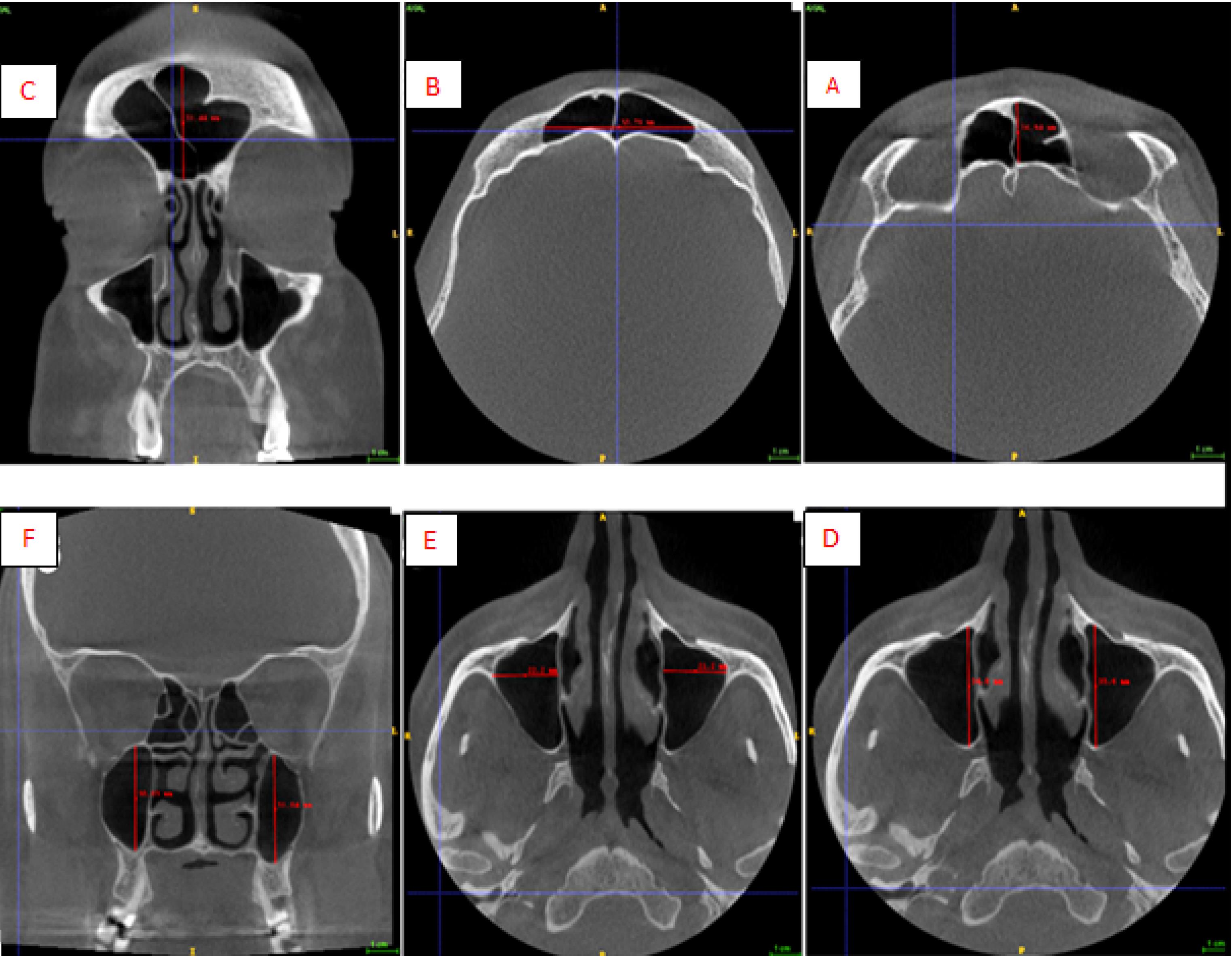
Figure 1.
Dimensions of the Maxillary and Frontal Sinuses: (A) Length, (B) Width, (C) Height of Frontal Sinus, (D) Length, (E) Width, and (F) Height of Maxillary Sinuses
.
Dimensions of the Maxillary and Frontal Sinuses: (A) Length, (B) Width, (C) Height of Frontal Sinus, (D) Length, (E) Width, and (F) Height of Maxillary Sinuses
Then, the volume of these structures is reported by the software. Painted sinuses to measure their volume are displayed in Figure 2. The measurement of the volume of the sinuses is shown in axial, coronal, and sagittal dimensions.
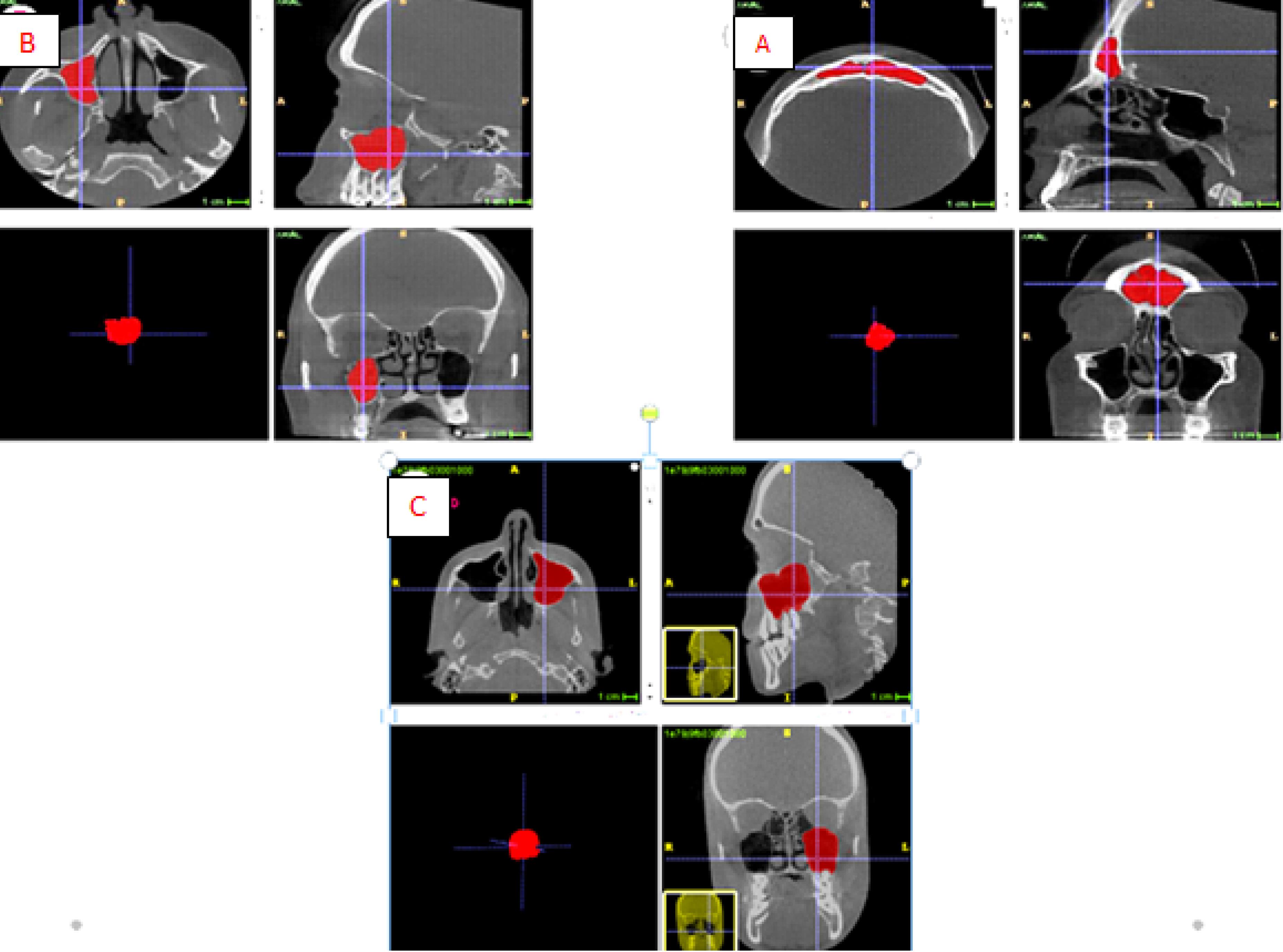
Figure 2.
(A) Frontal Sinus Volume, (B) Right Maxillary Sinus Volume, and (C) Left Maxillary Sinus Volume
.
(A) Frontal Sinus Volume, (B) Right Maxillary Sinus Volume, and (C) Left Maxillary Sinus Volume
The structures were measured by two researchers, and each researcher measured them twice with an interval of two weeks. In the current research, in addition to providing descriptive statistics such as means and standard deviations (SDs) and frequency tables, an independent t-test for two groups (gender) and variance analysis (age groups) were used to compare the average of the studied variables as a single variable. Therefore, the multivariate method of discriminant analysis was utilized to determine gender and age. This analysis examined the ability to detect each of the variables separately and together in the model. The data were analyzed using SPSS software, version 24. An independent t-test was employed to compare the mean sizes of the desired variables in two gender groups. In addition, Pearson’s correlation coefficient was used to evaluate the relationship between quantitative and age variables. Audit analysis was utilized to make an equation to determine gender and estimate age. Intraclass correlation was also employed to investigate the agreement between the two observers.
Results
two observers each observed the samples twice, and then the percentage of similarity between each observer and the two observers was evaluated. All agreements were equal to or greater than 0.85.
The means ± SDs and the significance level of each measured parameter in both genders for each of the sinuses are presented in Tables 1 and 2. The means ± SDs and significance level of each parameter measured in three age ranges for each of the sinuses are provided in Tables 3 and 4. All parameters estimated in FS, RMS, and LMS were significantly different between genders.
Table 1.
Descriptive Analysis of Maxillary Sinuses Measurements in Males and Females
|
Parameters
|
Side
|
Gender
|
Mean±SD
|
P
Value
|
| Length (mm) |
Right |
Male |
38.31 ± 3.59 |
< 0.05* |
| Left |
Female |
36.25 ± 3.62 |
| Right |
Male |
38.23 ± 4.21 |
< 0.05* |
| Left |
Female |
36.62 ± 3.52 |
| Width (mm) |
Right |
Male |
26.96 ± 4.62 |
< 0.05* |
| Left |
Female |
25.90 ± 4.73 |
| Right |
Male |
25.98 ± 4.38 |
< 0.05* |
| Left |
Female |
24.35 ± 4.12 |
| Height (mm) |
Right |
Male |
37.98 ± 5.67 |
< 0.05* |
| Left |
Female |
33.64 ± 5.92 |
| Right |
Male |
38.09 ± 5.37 |
< 0.05* |
| Left |
Female |
33.62 ± 5.09 |
| Volume (mm3) |
Right |
Male |
16386 ± 510 |
< 0.05* |
| Left |
Female |
13520 ± 463 |
| Right |
Male |
16386 ± 510 |
< 0.05* |
| Left |
Female |
13520 ± 463 |
SD: Standard deviation.
Note. *P value < 0.05 is considered significant (independent t test).
Table 2.
Descriptive Analysis of Frontal Sinus Measurements in Males and Females
|
Parameters
|
Gender
|
Mean±SD
|
P
Value
|
| Length (mm) |
Male |
16.08 ± 4.04 |
< 0.05* |
| Female |
13.71 ± 2.89 |
| Width (mm) |
Male |
56.35 ± 14.09 |
< 0.05* |
| Female |
49.51 ± 12.42 |
| Height (mm) |
Male |
29.54 ± 6.26 |
< 0.05* |
| Female |
26.20 ± 6.85 |
| Volume (mm3) |
Male |
10108 ± 556 |
< 0.05* |
| Female |
6192 ± 608 |
SD: Standard deviation.
* P value < 0.05 is considered significant (independent t test).
Table 3.
Descriptive Analysis of Maxillary Sinuses Measurements in Three Age Ranges
|
Parameters
|
Side
|
Age
|
Mean±SD
|
P
Value
|
| Length (mm) |
Right |
< 30 |
36.59 ± 3.54 |
0.11 |
| 30-40 |
36.83 ± 3.66 |
| > 40 |
36.32 ± 3.96 |
| Left |
< 30 |
37.67 ± 3.33 |
0.09 |
| 30-40 |
37.65 ± 4.08 |
| > 40 |
36.29 ± 4.11 |
| Width (mm) |
Right |
< 30 |
25.78 ± 3.83 |
0.49 |
| 30-40 |
26.26 ± 5.04 |
| > 40 |
25.35 ± 5.05 |
| Left |
< 30 |
25.08 ± 3.85 |
0.26 |
| 30-40 |
25.43 ± 4.57 |
| > 40 |
24.26 ± 4.08 |
| Height (mm) |
Right |
< 30 |
36.23 ± 5091 |
0.61 |
| 30-40 |
36.24 ± 6.73 |
| > 40 |
36.41 ± 4.43 |
| Left |
< 30 |
35.90 ± 5.87 |
0.06 |
| 30-40 |
35.73 ± 5.87 |
| > 40 |
35.17 ± 4.64 |
| Volume (mm3) |
Right |
< 30 |
15447 ± 535 |
0.10 |
| 30-40 |
14940 ± 507 |
| > 40 |
13418 ± 432 |
| Left |
< 30 |
14832 ± 504 |
0.32 |
| 30-40 |
15334 ± 533 |
| > 40 |
14031 ± 415 |
Table 4.
Descriptive Analysis of Frontal Sinus Measurements in Three Age Ranges
|
Parameters
|
Age
|
Mean±SD
|
P
Value
|
| Length (mm) |
< 30 |
37.67 ± 3.33 |
0.09 |
| 30-40 |
37.65 ± 4.08 |
| > 40 |
36.29 ± 4.11 |
| Width (mm) |
< 30 |
25.08 ± 3.85 |
0.26 |
| 30-40 |
25.43 ± 4.57 |
| > 40 |
24.26 ± 4.07 |
| Height (mm) |
< 30 |
35.90 ± 5.87 |
0.06 |
| 30-40 |
35.73 ± 5.88 |
| > 40 |
35.17 ± 4.64 |
| Volume (mm3) |
< 30 |
14832 ± 504 |
0.32 |
| 30-40 |
15334 ± 533 |
| > 40 |
14031 ± 415 |
The results (Table 1) demonstrated that all measured parameters of RMS and LMS were significantly higher in men than in women.
Based on the data in Table 2, all measured parameters of FS were significantly higher in men than in women.
The results (Table 3) showed that there was no significant difference in the three age groups in terms of all measured parameters of RMS and LMS.
Based on the results (Table 4), there was no significant difference in the three age groups regarding all the measured parameters of FS.
The following formula is a function to determine gender using all desired variables. The cut-point of this function is 3.21. Figures above and below this value indicate male and female genders, respectively.
Di = -2.617– 0.004 length (FS) – 0.054 width (FS) – 0.001 height (FS) + 0.000315 volume (FS) + 0.055 length (RMS) – 0.011 width (RMS) + 0.015 height (RMS) – 0.000033 volume (RMS) – 0.081 length (LMS) – 0.029 width (LMS) + 0.137 height (LMS) + 0.000011 volume (LMS).
Using all the variables, the classification accuracy was 78% for females and 82% for males, with an overall accuracy of 80%. In age estimation, discriminant analysis was utilized to evaluate the volume and dimensions of FS, RMS, and LMS. According to this function, none of the parameters had a significant difference in the three age ranges, so the age of people cannot be correctly recognized by using these parameters.
Discussion
Estimating the age and the gender of corpses whose identity is unknown is highly important in forensic medicine (11). Many previous studies have shown that maxillary and FSs have anatomical variations between genders (12-14). In the present study, the volume and dimensions of the frontal and maxillary sinuses in women were less than in men, and these indicators are highly predictive in determining gender. The results of this study are in line with those of the study of Michel et al (15) and Hamed et al (16). Saccucci et al (17) reported that there was no significant difference in the volume of the maxillary sinus between genders. One of the main reasons for the difference between the results of the above-mentioned study and those of the current study is the significant difference in the number of statistical samples in their study; the number of statistical samples was less. The loss of the maxillary molar teeth can be effective in changing the volume of the maxillary sinus (18,19). In the current study, people who had lost their first and second maxillary molar teeth for any reason were excluded from the study, while this issue was not mentioned in the study of Saccucci et al (17).
In this study, in addition to measuring the dimensions of sinuses, their volume was also measured both in the FS and in the maxillary sinus. The volume parameter compared to other parameters showed the greatest difference in both genders. In the study of Bengi et al, it was shown that measuring the sinus volume along with its dimensions can significantly increase the accuracy of gender determination (11).
The results of our study indicated that it is possible to estimate the gender of people with 80% accuracy by using the mentioned parameters. The accuracy of gender determination in this study was higher than that in other studies (13,20-22). Probably, the increase in the parameters measured in this study has increased the accuracy of gender determination (21). The difference in the rate of accuracy in previous studies can be due to diverse sample sizes, various imaging techniques, different inclusion and exclusion criteria, the use of various measurement software, and a wide range of statistical analyses.
In this study, the volume and dimensions of sinuses did not show any significant difference in the three age groups, and the discriminant analysis on the dimensions of these sinuses indicated that, in general, these indicators cannot estimate age with a reliable percentage. Various studies have found no consensus about the relationship between age and the dimensions and volume of the frontal and maxillary sinuses (8,20,23,24). Teixeira et al (20) demonstrated that although the measurement of maxillary sinus dimensions in individuals under 40 years old could correctly estimate their age to an acceptable extent, there were many variations in individuals over 40 years old. One of the most important differences between their study and the current study was that they did not exclude individuals who had lost upper molar teeth. In the study of Jasim and Al-Taei (8), it was found that the volume and dimensions of the maxillary sinus tend to decrease with older age. One of the main differences between their study and the present study was that they only measured these parameters in individuals over 40 years old. Sahlstrand et al reported that the volume of the maxillary sinus did not have a significant difference in different ages (25). The results of this study are in line with those of the present study. Emirzeoglu et al (24) concluded that the total volume of all paranasal sinuses decreases with age. However, no significant difference was observed between the volume and size of each of the sinuses at different ages. Ghodousi et al (23) found no significant difference in FS dimensions at different ages. Due to the conflicting results of the studies, more extensive studies with a suitable sample size and a wider age range are needed to investigate the relationship between the dimensions of the paranasal sinuses and the age of people.
In previous studies, different imaging techniques were used to measure the volume and dimensions of sinuses. The type of imaging can affect the results due to the difference in accuracy. The CT imaging technique is a gold standard for the evaluation of paranasal sinuses (26). CBCT is an imaging modality that has a lower cost, reduced examination time, and lower radiation dose, providing proper quality of craniofacial structures (21). Therefore, in this study, CBCT was used for the measurement of sinuses.
Several factors, such as maternal health, race, culture, environmental conditions during growth, and the like, are effective in developing paranasal sinuses (27). The formula presented in the present study is the most suitable for determining gender in the Iranian population and is appropriate for other populations. It should be updated with that population.
The advantages of the current study included a large sample size compared to previous studies and the use of different parameters for age estimation and gender determination. Further, a formula has been presented that can be utilized to determine the gender of people in Iranian society with an acceptable percentage. On the other hand, our limitation in this study was that due to the fact that in our society, many people lose some upper molar teeth at a young age, the number of available samples with a wider age range decreases.
Conclusion
CBCT is an excellent imaging method for accurately measuring the volume and dimensions of the FS, RMS, and LMS sinuses for forensic purposes. Based on the findings of this study, there was a strong correlation between these parameters and the gender of people. By measuring these parameters, people’s gender can be predicted with 80% accuracy. These parameters were not significantly different in age ranges. As a result, the measurement of these parameters cannot be used in age estimation with a highly reliable percentage.
Competing Interests
The authors declare that they have no conflict of interests.
Ethical Approval
All samples were obtained from existing archives of patients and no individual was exposed to radiation as a result of our research. All patient information will remain confidential and will not be disclosed at any stage of the research or publication.
Funding
This study received no specific grant from funding agencies in the public, commercial, or not-for-profit sectors.
References
- Kondou H, Morohashi R, Ichioka H, Bandou R, Matsunari R, Kawamoto M. Deep neural networks-based age estimation of cadavers using CT imaging of vertebrae. Int J Environ Res Public Health 2023; 20(6):4806. doi: 10.3390/ijerph20064806 [Crossref] [ Google Scholar]
- Ajmal MA, Roberts TS, Beshtawi KR, Raj AC, Sandeepa NC. Sexual dimorphism in odontometric parameters using cone beam CT: a systematic review. Head Face Med 2023; 19(1):6. doi: 10.1186/s13005-023-00352-7 [Crossref] [ Google Scholar]
- Ominde B, Ikubor JE, Wilson JI, Ebeye A, Igbigbi P. Morphometry of the maxillary sinus and its role in sex determination in delta state Nigeria: retrospective CT study. J Indian Acad Forensic Med 2023; 45(1):52-7. doi: 10.48165/jiafm.2023.45.1.15 [Crossref] [ Google Scholar]
- Patil SA, Aditya A, Kalyanpur K, Kore A. Evaluation of sexual dimorphism of maxillary sinus from morphometric measurements using cone beam computed tomography (CBCT)-a cross sectional study. J Indian Acad Oral Med Radiol 2023; 35(3):412-6. doi: 10.4103/jiaomr.jiaomr_342_22 [Crossref] [ Google Scholar]
- Dinç ME, Bayar Muluk N, Vonakis BM. Physiology of the nose and paranasal sinuses. In: Cingi C, Bayar Muluk N, eds. All Around the Nose: Basic Science, Diseases and Surgical Management. Cham: Springer; 2020. p. 57-63. doi: 10.1007/978-3-030-21217-9_6.
- da Silva JC, Strazzi-Sahyon HB, Nunes GP, Andreo JC, Spin MD, Shinohara AL. Cranial anatomical structures with high sexual dimorphism in metric and morphological evaluation: a systematic review. J Forensic Leg Med 2023; 99:102592. doi: 10.1016/j.jflm.2023.102592 [Crossref] [ Google Scholar]
- Okoseimiema SC. Sexual dimorphism in facial, nasal, mandibular, maxillary and orofacial heights of the Ikwerre people in Rivers State. Sch Bull 2021; 7(5):130-3. doi: 10.36348/sb.2021.v07i05.002 [Crossref] [ Google Scholar]
- Jasim HH, Al-Taei JA. Computed tomographic measurement of maxillary sinus volume and dimension in correlation to the age and gender: comparative study among individuals with dentate and edentulous maxilla. J Baghdad Coll Dent 2013; 25(1):87-93. [ Google Scholar]
- Gulec M, Tassoker M, Magat G, Lale B, Ozcan S, Orhan K. Three-dimensional volumetric analysis of the maxillary sinus: a cone-beam computed tomography study. Folia Morphol (Warsz) 2020; 79(3):557-62. doi: 10.5603/FM.a2019.0106 [Crossref] [ Google Scholar]
- Kaasalainen T, Ekholm M, Siiskonen T, Kortesniemi M. Dental cone beam CT: an updated review. Phys Med 2021; 88:193-217. doi: 10.1016/j.ejmp.2021.07.007 [Crossref] [ Google Scholar]
- Bangi BB, Ginjupally U, Nadendla LK, Vadla B. 3D evaluation of maxillary sinus using computed tomography: a sexual dimorphic study. Int J Dent 2017; 2017:9017078. doi: 10.1155/2017/9017078 [Crossref] [ Google Scholar]
- Ibrahim MA, Abdel-Karim RI, Ibrahim MS, Dar UF. Comparative study of the reliability of frontal and maxillary sinuses in sex identification using multidetector computed tomography among Egyptians. Forensic Imaging 2020; 22:200390. doi: 10.1016/j.fri.2020.200390 [Crossref] [ Google Scholar]
- Ahmed AG, Gataa IS, Fateh SM, Mohammed GN. CT scan images analysis of maxillary sinus dimensions as a forensic tool for sexual and racial detection in a sample of Kurdish population. Eur Sci J 2015; 11(18):272-81. [ Google Scholar]
- Sheikhi M, Moradi Nejad M, Ghodousi A. Anthropometric evaluation of the maxillary sinus in subjects with complete dental system and complete edentulism based on gender, on CBCT images. Eurasia J Biosci 2020; 14(1):49-55. [ Google Scholar]
- Michel J, Paganelli A, Varoquaux A, Piercecchi-Marti MD, Adalian P, Leonetti G. Determination of sex: interest of frontal sinus 3D reconstructions. J Forensic Sci 2015; 60(2):269-73. doi: 10.1111/1556-4029.12630 [Crossref] [ Google Scholar]
- Hamed SS, El-Badrawy AM, Abdel Fattah S. Gender identification from frontal sinus using multi-detector computed tomography. Journal of Forensic Radiology and Imaging 2014; 2(3):117-20. doi: 10.1016/j.jofri.2014.03.006 [Crossref] [ Google Scholar]
- Saccucci M, Cipriani F, Carderi S, Di Carlo G, D’Attilio M, Rodolfino D. Gender assessment through three-dimensional analysis of maxillary sinuses by means of cone beam computed tomography. Eur Rev Med Pharmacol Sci 2015; 19(2):185-93. [ Google Scholar]
- Atallah HN, Ali MS, Abd Noor HJ, Sami SM, Haider J. Evaluation of the relation between the maxillary sinus and the posterior teeth using digital panoramic radiography. J Med Life 2023; 16(8):1240-4. doi: 10.25122/jml-2023-0105 [Crossref] [ Google Scholar]
- Altaweel AA, Saad Sowairi SM, Saaduddin Sapri AM, Saeedi SA, Alamri AH, Alnobi AA. Assessment of the relationship between maxillary posterior teeth and maxillary sinus using cone-beam computed tomography. Int J Dent 2022; 2022:6254656. doi: 10.1155/2022/6254656 [Crossref] [ Google Scholar]
- Teixeira LC, Walewski LÂ, de Souza Tolentino E, Iwaki LC, Silva MC. Three-dimensional analysis of the maxillary sinus for determining sex and age in human identification. Forensic Imaging 2020; 22:200395. doi: 10.1016/j.fri.2020.200395 [Crossref] [ Google Scholar]
- Paknahad M, Shahidi S, Zarei Z. Sexual dimorphism of maxillary sinus dimensions using cone-beam computed tomography. J Forensic Sci 2017; 62(2):395-8. doi: 10.1111/1556-4029.13272 [Crossref] [ Google Scholar]
- Sathawane RS, Sukhadeve VA, Chandak RM, Lanjekar AB, Moon GV. Sex determination by maxillary sinus dimensions using cone-beam computed tomography and discriminant function: an analytical study. Int J Forensic Odontol 2020; 5(1):19-22. doi: 10.4103/ijfo.ijfo_8_20 [Crossref] [ Google Scholar]
- Ghodousi A, Ghafari R, Khademi Ghahroudi SM, Madineii MS. Evaluation of frontal sinus indexes by CT for identification in Isfahanian population samples. Iran J Forensic Med 2014; 20(3):87-96. [ Google Scholar]
- Emirzeoglu M, Sahin B, Bilgic S, Celebi M, Uzun A. Volumetric evaluation of the paranasal sinuses in normal subjects using computer tomography images: a stereological study. Auris Nasus Larynx 2007; 34(2):191-5. doi: 10.1016/j.anl.2006.09.003 [Crossref] [ Google Scholar]
- Sahlstrand-Johnson P, Jannert M, Strömbeck A, Abul-Kasim K. Computed tomography measurements of different dimensions of maxillary and frontal sinuses. BMC Med Imaging 2011; 11:8. doi: 10.1186/1471-2342-11-8 [Crossref] [ Google Scholar]
- Lenza MG, de Oliveira Lenza MM, Dalstra M, Melsen B, Cattaneo PM. An analysis of different approaches to the assessment of upper airway morphology: a CBCT study. Orthod Craniofac Res 2010; 13(2):96-105. doi: 10.1111/j.1601-6343.2010.01482.x [Crossref] [ Google Scholar]
- Iscan MY, Steyn M. The Human Skeleton in Forensic Medicine. Charles C Thomas Publisher; 2013.