Avicenna J Dent Res. 16(1):1-8.
doi: 10.34172/ajdr.561
Original Article
Magnetic Resonance Imaging Study of Paranasal Sinuses Dimensions According to Age and Gender
Faezeh Yousefi 1  , Maryam Farhadian 2, Sima Rahimi 1, *
Author information:
1Oral and Maxillofacial Radiology Department, Hamadan University of Medical Science, Hamadan, Iran
2Department of Biostatistics, Research Center for Health Sciences, Hamadan University of Medical Sciences, Hamadan, Iran
Abstract
Background: Considering the importance of paranasal sinus anatomy in treatment planning and related surgeries, the present study aimed to investigate the changes in the dimensions of paranasal sinuses according to age and gender by magnetic resonance imaging (MRI).
Methods: MRI images of 310 patients, including 200 women and 110 men aged between 10 and 70 years old, were included in the study. The depths of all paranasal sinuses and the width of the left and right frontal sinuses were estimated in the axial plane. In addition, the height of the left and right frontal sinuses in the sagittal plane as well as the width and height of the maxillary and sphenoid sinuses in the coronal plane underwent measurement. All statistical analyses were performed using SPSS 21, and the significance level was considered at 0.05.
Results: Except for the width of the right maxillary sinus, the height of the left frontal sinus, and the width and depth of the left sphenoid sinus, a significant relationship was observed between the other dimensions of paranasal sinuses and gender (P<0.05). Only 5 dimensions, including the width of the left and right maxillary sinuses, the height of the left maxillary sinus, the depth of the left frontal sinus, and the depth of the right sphenoid sinus, had significant relationships with age, so that they decreased with age (P<0.05). The highest symmetry was between the left and right maxillary sinuses.
Conclusion: The paranasal sinus was different in men and women. Some dimensions of paranasal sinuses had a significant relationship with age. These results can be used in planning prosthetic treatments and performing surgeries with fewer complications.
Keywords: Paranasal sinus, Gender, Age, Magnetic resonance imaging
Copyright and License Information
© 2024 The Author(s); Published by Hamadan University of Medical Sciences.
This is an open-access article distributed under the terms of the Creative Commons Attribution License (
http://creativecommons.org/licenses/by/4.0), which permits unrestricted use, distribution, and reproduction in any medium provided the original work is properly cited.
Please cite this article as follows: Yousefi F, Farhadian M, Rahimi S. Magnetic resonance imaging study of paranasal sinuses dimensions according to age and gender. Avicenna J Dent Res. 2024; 16(1):1-8. doi:10.34172/ajdr.561
Background
Measuring the dimensions of paranasal sinuses by three-dimensional images leads to the recognition of normal sinus structures in the population and the affecting factors such as physiological (age and gender) or pathological (chronic sinusitis) conditions (1).
This basic information can be a significant help in evaluating the relationship between dimensions with pathology and sinus disorders, pre-treatment evaluations, increasing preoperative vision, and sinus endoscopy (functional endoscopic sinus surgery), making surgery more reliable with fewer complications (1–3).
Likewise, treatment plans such as placement of implants, prostheses, or mini-screws in orthodontic treatments as well as the amount of sinus lift can be determined according to the pattern of changes in paranasal sinuses (4).
To this end, by knowing the relationship between age and gender with dimensions of paranasal sinuses, previous imaging can be used as a reference to assess the normality or abnormality of the paranasal sinuses in current images (1).
Accurate imaging should be utilized to better evaluate the anatomy of paranasal sinuses since they have unique and complex structures. Due to its advanced imaging and evaluation of images in three coronal, axial, and sagittal planes, magnetic resonance imaging (MRI) is an acceptable method to demonstrate the true anatomy of paranasal sinuses (5). Nevertheless, a few studies have been performed on the dimensions of paranasal sinuses according to age and gender by MRI images, and most studies have examined only one sinus with a small sample size using cone-beam computed tomography (CBCT).
For this reason, the current study sought to investigate the changes in paranasal sinus dimensions according to age and gender by MRI.
Materials and Methods
At least 600 MRI images were examined in this cross-sectional study, which was registered in the Research Council of Hamadan University of Medical Sciences with the ethics code IR.UMSHA.REC.1398.592.
The inclusion criteria were the presence of posterior maxillary teeth (first and second molars and premolars if they affect the dimensions of the sinus).
Some of the examined images were excluded due to the lack of clarity and low quality of the images as a result of the inability to accurately measure the dimensions of paranasal sinuses. The exclusion criteria were the presence of pathology in the sinuses, the absence of posterior maxillary teeth, and sinus fillings that occupy a significant volume of the sinuses and deform the sinus walls. The other exclusion criteria included a history of trauma and fractures in the jaw and face, congenital craniofacial disorders, sinus aplasia, and a history of orthognathic surgery. In addition, some were excluded because of the homogeneity of samples in different age groups and both genders. Finally, MRI images of 310 patients, including 200 women and 110 men between ages 10 and 70, were included in the study. All images were obtained using an MRI Essenza 1.5T machine (SIEMENS, Munich, Germany) with certain imaging parameters (eight-channel brain coil, slice thickness of 5 mm, inter-slice gap of 2 mm, T1: TR = 400, TE = 8.7, T2: TR = 3000, TE = 96, and fov: 230 mm).
The depths of all paranasal sinuses and the width of the left and right frontal sinuses in the axial plane (Figures 1A, 1B, and 1C), as well as the height of the left and right frontal sinuses in the sagittal plane (Figure 1D), underwent measurement. The width and height of the maxillary and sphenoid sinuses (Figures 1E and 1F) were also estimated in the coronal plane. The distance between the anterior point and the posterior point of the medial sinus wall was considered the depth of the sinus, and the longest distance between the medial. Further, the lateral walls of the sinus were regarded as sinus width, and the distance from the highest point on the sinus ceiling to the lowest point on the sinus floor was taken into account as sinus height. About 10% of the total samples were re-evaluated to check the agreement between the two observers.
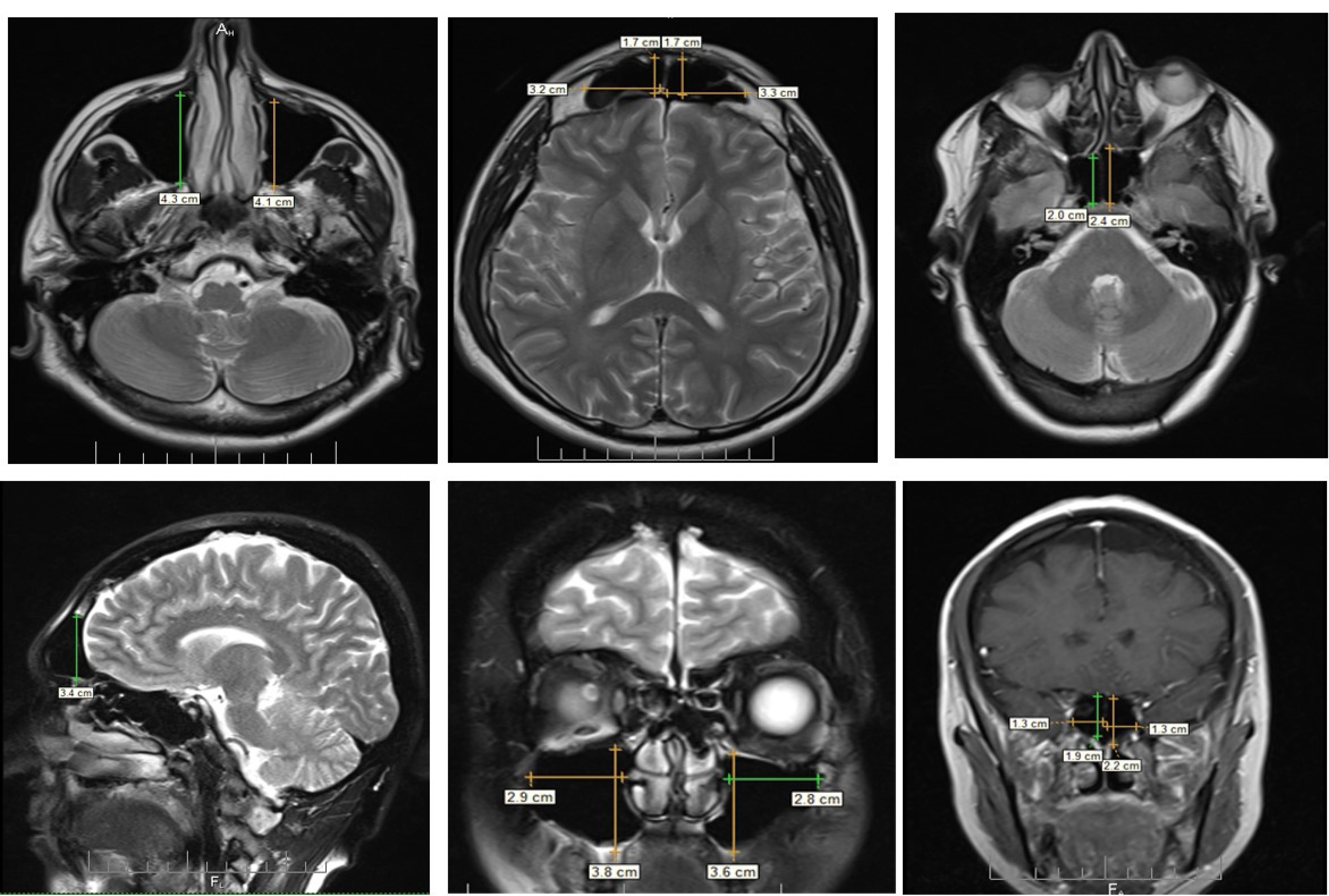
Figure 1.
(A) Depth of Left and Right Maxillary Sinus, (B) Width and Depth of Left and Right Frontal Sinuses, (C) Depth of Left and Right Sphenoid Sinus, (D) Left Frontal Sinus Height, (E) Width and Height of Left and Right Maxillary Sinuses, and (F) Width and Height of Left and Right Sphenoid Sinuses
.
(A) Depth of Left and Right Maxillary Sinus, (B) Width and Depth of Left and Right Frontal Sinuses, (C) Depth of Left and Right Sphenoid Sinus, (D) Left Frontal Sinus Height, (E) Width and Height of Left and Right Maxillary Sinuses, and (F) Width and Height of Left and Right Sphenoid Sinuses
All statistical analyses were performed using SPSS 21.0 (SPSS, Chicago, IL, USA) software. Descriptive statistics and statistical tests, such as the t-test, one-way analysis of variance, Tukey’s post hoc test, and Pearson correlation coefficient, were used to analyze the data, and a significance level was considered to be 0.05.
Results
In general, 310 (200 females and 110 males) patients with a mean age of 35.63 ± 12.37 were included in this study. The youngest and oldest patients were 10 and 70 years old, respectively (Table 1).
Table 1.
Distribution of Participants According to Age and Gender
|
Age Group (y)
|
Female
|
Male
|
Total
|
| 10-20 |
26 |
12 |
38 |
| 20-30 |
42 |
23 |
65 |
| 30-40 |
74 |
29 |
103 |
| 40-50 |
34 |
31 |
65 |
| 50-60 |
21 |
12 |
33 |
| 60-70 |
3 |
3 |
6 |
| Total |
200 |
110 |
310 |
A t-test was utilized to evaluate the relationship between the dimensions of paranasal sinuses and gender. There are significant differences in the dimensions of the paranasal sinuses between men and women (Table 2).
Table 2.
Comparison of Paranasal Sinus Dimensions Based on Gender
|
Sinus Dimensions
|
Gender
|
Number
|
Mean±SD
|
Mean Difference±SE
|
P
Value
|
| Right maxillary sinus width |
Male |
110 |
2.30 ± 0.43 |
0.08 ± 0.04 |
0.078 |
| Female |
200 |
2.22 ± 0.38 |
| Right maxillary sinus height |
Male |
110 |
3.71 ± 0.60 |
0.29 ± 0.07 |
< 0.001 |
| Female |
200 |
3.41 ± 0.57 |
| Right maxillary sinus depth |
Male |
110 |
3.58 ± 0.38 |
0.17 ± 0.04 |
< 0.001 |
| Female |
200 |
3.41 ± 0.34 |
| Left maxillary sinus width |
Male |
110 |
2.22 ± 0.39 |
0.10 ± 0.04 |
0.018 |
| Female |
200 |
2.11 ± 0.35 |
| Left maxillary sinus height |
Male |
110 |
3.69 ± 0.59 |
0.28 ± 0.06 |
< 0.001 |
| Female |
200 |
3.40 ± 0.55 |
| Left maxillary sinus depth |
Male |
110 |
3.62 ± 0.38 |
0.19 ± 0.04 |
< 0.001 |
| Female |
200 |
3.42 ± 0.35 |
| Right frontal sinus width |
Male |
110 |
2.88 ± 0.82 |
0.44 ± 0.09 |
< 0.001 |
| Female |
200 |
2.43 ± 0.75 |
| Right frontal sinus height |
Male |
110 |
2.54 ± 0.76 |
0.18 ± 0.08 |
0.032 |
| Female |
200 |
2.35 ± 0.70 |
| Right frontal sinus depth |
Male |
110 |
1.24 ± 0.71 |
0.34 ± 0.06 |
< 0.001 |
| Female |
200 |
0.89 ± 0.34 |
| Left frontal sinus width |
Male |
110 |
2.96 ± 0.98 |
0.37 ± 0.10 |
< 0.001 |
| Female |
200 |
2.59 ± 0.80 |
| Left frontal sinus height |
Male |
110 |
2.54 ± 0.83 |
0.11 ± 0.09 |
0.238 |
| Female |
200 |
2.43 ± 0.77 |
| Left frontal sinus depth |
Male |
110 |
1.21 ± 0.32 |
0.30 ± 0.03 |
< 0.001 |
| Female |
200 |
0.90 ± 0.24 |
| Right sphenoid sinus width |
Male |
110 |
1.64 ± 0.38 |
0.09 ± 0.04 |
0.040 |
| Female |
200 |
1.54 ± 0.39 |
| Right sphenoid sinus height |
Male |
110 |
2.05 ± 0.39 |
0.16 ± 0.04 |
0.001 |
| Female |
200 |
1.89 ± 0.37 |
| Right sphenoid sinus depth |
Male |
110 |
2.24 ± 0.64 |
0.16 ± 0.07 |
0.027 |
| Female |
200 |
2.08 ± 0.60 |
| Left sphenoid sinus width |
Male |
110 |
1.59 ± 0.35 |
0.07 ± 0.04 |
0.073 |
| Left sphenoid sinus height |
Male |
200 |
2.06 ± 0.38 |
0.18 ± 0.04 |
< 0.001 |
| Female |
110 |
1.87 ± 0.37 |
| Left sphenoid sinus depth |
Male |
200 |
2.19 ± 0.65 |
0.12 ± 0.07 |
0.085 |
| Female |
110 |
2.06 ± 0.60 |
Note. SD: Standard deviation; SE: Standard error.
Moreover, a one-way analysis of variance was employed to investigate the relationship between paranasal sinus dimensions within different age groups. Due to the small number of patients in the age groups of 50–60 and 60–70, the two groups were merged and placed in the group over 50 years. Only 5 variables, including the width of the left and right maxillary sinuses, the height of the left maxillary sinus, the depth of the left frontal sinus, and the depth of the right sphenoid sinus, were significantly related to different age groups (Tables 3, 4, and 5).
Table 3.
Different Age Groups for Maxillary Sinus Measurements
|
Sinus Dimensions
|
Age Group (y)
|
Number
|
Mean±SD
|
Min.
|
Max.
|
P
Value
|
| Right sinus width |
10-20 |
38 |
2.37 ± 0.32 |
1.6 |
3.1 |
0.030 |
| 20-30 |
65 |
2.22 ± 0.37 |
1.4 |
3.2 |
| 30-40 |
103 |
2.29 ± 0.45 |
1.2 |
3.9 |
| 40-50 |
65 |
2.21 ± 0.36 |
1.4 |
3.3 |
| Over 50 |
39 |
2.10 ± 0.39 |
1.4 |
2.9 |
| Right sinus height |
10-20 |
38 |
3.57 ± 0.57 |
1.8 |
4.4 |
0.154 |
| 20-30 |
65 |
3.60 ± 0.61 |
1.9 |
5.0 |
| 30-40 |
103 |
3.57 ± 0.58 |
1.6 |
4.9 |
| 40-50 |
65 |
3.39 ± 0.57 |
2.1 |
5.0 |
| Over 50 |
39 |
3.41 ± 0.69 |
2.2 |
4.7 |
| Right sinus depth |
10-20 |
38 |
3.46 ± 0.39 |
2.2 |
4.2 |
0.708 |
| 20-30 |
65 |
3.45 ± 0.34 |
2.3 |
4.3 |
| 30-40 |
103 |
3.50 ± 0.37 |
2.1 |
4.6 |
| 40-50 |
65 |
3.48 ± 0.35 |
2.6 |
4.4 |
| Over 50 |
39 |
3.41 ± 0.40 |
2.7 |
4.3 |
| Left sinus width |
10-20 |
38 |
2.33 ± 0.26 |
1.9 |
2.8 |
0.001 |
| 20-30 |
65 |
2.17 ± 0.31 |
1.5 |
2.9 |
| 30-40 |
103 |
2.17 ± 0.42 |
1.2 |
3.3 |
| 40-50 |
65 |
2.09 ± 0.38 |
1.1 |
3.3 |
| Over 50 |
39 |
2.00 ± 0.32 |
1.3 |
2.9 |
| Left sinus height |
10-20 |
38 |
3.50 ± 0.56 |
2.3 |
4.4 |
0.048 |
| 20-30 |
65 |
3.61 ± 0.54 |
1.8 |
5.0 |
| 30-40 |
103 |
3.57 ± 0.57 |
1.6 |
5.0 |
| 40-50 |
65 |
3.37 ± 0.54 |
2 |
4.5 |
| Over 50 |
39 |
3.35 ± 0.68 |
2.2 |
4.8 |
| Left sinus depth |
10-20 |
38 |
3.38 ± 0.49 |
2.7 |
4.2 |
0.600 |
| 20-30 |
65 |
3.50 ± 0.31 |
2.8 |
4.1 |
| 30-40 |
103 |
3.53 ± 0.41 |
2.1 |
4.9 |
| 40-50 |
65 |
3.47 ± 0.34 |
2.9 |
4.4 |
| Over 50 |
39 |
3.41 ± 0.40 |
2.6 |
4.2 |
Note. SD: Standard deviation; Min.: Minimum; Max.: Maximum.
Table 4.
Different Age Groups for Frontal Sinus Measurements
|
Sinus Dimensions
|
Age Group (y)
|
Number
|
Mean±SD
|
Min.
|
Max.
|
P
Value
|
| Right sinus width |
10-20 |
38 |
2.47 ± 0.72 |
1.1 |
4.5 |
0.778 |
| 20-30 |
65 |
2.61 ± 0.70 |
1.4 |
4.5 |
| 30-40 |
103 |
2.58 ± 0.81 |
1.1 |
5.0 |
| 40-50 |
65 |
2.68 ± 0.96 |
1.2 |
5.3 |
| Over 50 |
39 |
2.54 ± 0.78 |
0.9 |
4.7 |
| Right sinus height |
10-20 |
38 |
2.25 ± 0.63 |
0.8 |
3.7 |
0.180 |
| 20-30 |
65 |
2.51 ± 0.76 |
0.7 |
4.7 |
| 30-40 |
103 |
2.40 ± 0.63 |
0.9 |
4.0 |
| 40-50 |
65 |
2.54 ± 0.85 |
1.4 |
5.0 |
| Over 50 |
39 |
2.29 ± 0.73 |
0.9 |
4.8 |
| Right sinus depth |
10-20 |
38 |
0.94 ± 0.57 |
0.4 |
4.0 |
0.144 |
| 20-30 |
65 |
0.99 ± 0.32 |
0.3 |
1.8 |
| 30-40 |
103 |
0.95 ± 0.31 |
0.4 |
2.2 |
| 40-50 |
65 |
1.07 ± 0.31 |
0.5 |
1.9 |
| Over 50 |
39 |
1.18 ± 0.15 |
0.6 |
8.0 |
| Left sinus width |
10-20 |
38 |
2.52 ± 0.76 |
1.3 |
4.1 |
0.145 |
| 20-30 |
65 |
2.82 ± 0.86 |
1.1 |
4.8 |
| 30-40 |
103 |
2.81 ± 0.88 |
1.1 |
5.1 |
| 40-50 |
65 |
2.74 ± 0.97 |
1.1 |
5.0 |
| Over 50 |
39 |
2.47 ± 0.85 |
1.4 |
4.2 |
| Left sinus height |
10-20 |
38 |
2.35 ± 0.63 |
0.9 |
3.2 |
0.686 |
| 20-30 |
65 |
2.52 ± 0.92 |
0.7 |
5.1 |
| 30-40 |
103 |
2.52 ± 0.77 |
0.8 |
4.6 |
| 40-50 |
65 |
2.46 ± 0.84 |
1.0 |
5.0 |
| Over 50 |
39 |
2.37 ± 0.73 |
1.3 |
4.6 |
| Left sinus depth |
10-20 |
38 |
0.85 ± 0.26 |
0.4 |
1.5 |
0.002 |
| 20-30 |
65 |
1.05 ± 0.30 |
0.5 |
1.8 |
| 30-40 |
103 |
1.01 ± 0.28 |
0.5 |
1.9 |
| 40-50 |
65 |
1.10 ± 0.34 |
0.5 |
2.0 |
| Over 50 |
39 |
0.97 ± 0.31 |
0.4 |
2.0 |
Note. SD: Standard deviation; Min.: Minimum; Max.: Maximum.
Table 5.
Different Age Groups for Sphenoid Sinus Measurements
|
Sinus Dimensions
|
Age Group (years)
|
Number
|
SD±Mean
|
Min.
|
Max.
|
P
Value
|
| Right sinus width |
10-20 |
38 |
1.57 ± 0.32 |
0.9 |
2.4 |
0.314 |
| 20-30 |
65 |
1.59 ± 0.37 |
0.9 |
2.5 |
| 30-40 |
103 |
1.63 ± 0.40 |
0.9 |
3.0 |
| 40-50 |
65 |
1.54 ± 0.40 |
0.6 |
2.4 |
| Over 50 |
39 |
1.47 ± 0.41 |
0.7 |
2.4 |
| Right sinus height |
10-20 |
38 |
2.00 ± 0.36 |
1.0 |
2.8 |
0.324 |
| 20-30 |
65 |
1.96 ± 0.43 |
0.8 |
3.2 |
| 30-40 |
103 |
1.95 ± 0.35 |
0.7 |
2.8 |
| 40-50 |
65 |
1.95 ± 0.38 |
1.1 |
2.8 |
| Over 50 |
39 |
1.82 ± 0.43 |
0.6 |
2.6 |
| Right sinus depth |
10-20 |
38 |
2.21 ± 0.57 |
0.7 |
3.5 |
0.018 |
| 20-30 |
65 |
2.24 ± 0.58 |
1.1 |
3.3 |
| 30-40 |
103 |
2.16 ± 0.63 |
1.0 |
3.2 |
| 40-50 |
65 |
2.14 ± 0.64 |
0.8 |
3.5 |
| Over 50 |
39 |
1.83 ± 0.60 |
0.8 |
3.1 |
| Left sinus width |
10-20 |
38 |
1.52 ± 0.37 |
0.8 |
2.4 |
0.568 |
| 20-30 |
65 |
1.60 ± 0.35 |
0.9 |
2.6 |
| 30-40 |
103 |
1.53 ± 0.37 |
0.7 |
2.8 |
| 40-50 |
65 |
1.54 ± 0.37 |
0.8 |
2.6 |
| Over 50 |
39 |
1.47 ± 0.30 |
0.8 |
2.3 |
| Left sinus height |
10-20 |
38 |
1.98 ± 0.44 |
1.2 |
2.8 |
0.172 |
| 20-30 |
65 |
2.00 ± 0.37 |
1.1 |
3.2 |
| 30-40 |
103 |
1.94 ± 0.34 |
1.0 |
2.8 |
| 40-50 |
65 |
1.92 ± 0.36 |
1.1 |
2.8 |
| Over 50 |
39 |
1.81 ± 0.47 |
0.5 |
2.6 |
| Left sinus depth |
10-20 |
38 |
2.13 ± 0.59 |
0.7 |
3.5 |
0.094 |
| 20-30 |
65 |
2.28 ± 0.54 |
1.2 |
3.4 |
| 30-40 |
103 |
2.06 ± 0.65 |
1.0 |
3.7 |
| 40-50 |
65 |
2.04 ± 0.65 |
0.9 |
3.4 |
| Over 50 |
39 |
1.99 ± 0.60 |
0.8 |
3.0 |
Note. SD: Standard deviation; Min.: Minimum; Max.: Maximum.
Tukey’s post hoc test was used to compare the pairs between the age groups. There was a significant difference in the width of the right maxillary sinus in the age group of 10–20 years with the age group over 50 years. Similarly, this significant difference was observed in the width of the left maxillary sinus in the age group of 10–20 years with age groups of 40–50 years and over 50 years. In addition, there were significant differences in the depth of the left frontal sinus in the age group of 10–20 years with age groups of 20–30 years and 40–50 years, as well as in the depth of the right sphenoid sinus in the age group over 50 years with age groups of 20–30 years and 30–40 years. Contrarily, no significant difference was found in the height of the maxillary sinus between age groups.
A Pearson correlation coefficient test was utilized to examine the relationship between dimensions of different sinuses, and a weak correlation was observed between each sinus dimensions and other sinuses, while there was a stronger correlation between one side of sinus dimensions and the opposite side.
Based on the data in Table 6, the highest correlation was recorded between the left and right maxillary sinus dimensions, while the lowest correlation was observed between the left and right sphenoid sinus dimensions. All correlations between the left and right maxillary and frontal sinuses were positive. A negative correlation was found only in the sphenoid sinus.
Table 6.
Dimensions of Different Sinuses on the Left and Right According to Pearson Correlation Coefficient
|
Sinus Type
|
Variable
|
Left Sinus Width
|
Left Sinus Height
|
Left Sinus Depth
|
| Maxillary |
Right sinus width |
0.400* |
0.396* |
0.332* |
| Right sinus height |
0.451* |
0.588* |
0.418* |
| Right sinus depth |
0.193* |
0.189* |
0.429* |
| Frontal |
Right sinus width |
0.400* |
0.396* |
0.332* |
| Right sinus height |
0.451* |
0.588* |
0.418* |
| Right sinus depth |
0.193* |
0.189* |
0.429* |
| Sphenoid |
Right sinus width |
-0.179* |
0.057 |
-0.016 |
| Right sinus height |
0.080 |
0.564* |
0.273* |
| Right sinus depth |
0.058 |
0.328* |
0.379* |
Note. Correlation numbers with the sign * are statistically significant.
According to the findings, the highest and lowest symmetries were observed between the left and right maxillary sinuses and between the left and right sphenoid sinuses, respectively. On the other hand, the highest correlation between the dimensions of the left and right sinuses was found in the height, depth, and width, respectively. There was a negative correlation between the width of the right sphenoid sinus and the width and depth of the left sphenoid sinus.
Regarding the correlation of each of the desired dimensions in the two genders, there was a stronger relationship between the height of the left frontal sinus and the height of the right frontal sinus in males compared to females. Furthermore, a stronger negative correlation was found between the depth of the right sphenoid sinus and the depth of the left sphenoid sinus in females than in males.
Discussion
Comprehensive knowledge and appropriate visualization of the paranasal sinus anatomy are crucial for success in the treatment of sinus disorders, and head and neck surgeries, especially otolaryngology surgery, and cranial base surgery to prevent their complications (4,6). Most previously published data in this field have examined one or two pairs of paranasal sinuses in a small sample size (less than 100 samples) using CT or CBCT images. Although these images provide a realistic vision of sinus structure, they have disadvantages such as the high cost and use of ionizing radiation (5,7).
Overall, in this study, the relations between the dimensions (width, height, and depth) of the paranasal sinuses (maxillary, frontal, and sphenoid) were examined according to gender and age using the non-ionizing nature of MRI with a larger study population (310 MRI images).
The results demonstrated that, except for the width of the right maxillary sinus, height of the left frontal sinus, and width and depth of the left sphenoid sinus, there was a significant relationship between the other dimensions of paranasal sinuses and gender. Additionally, the difference in sinus dimensions between males and females was more considerable in the frontal and maxillary sinuses, and the sphenoid sinus had a less significant relationship with gender. The width of the frontal sinus and the height of the maxillary sinus highly differed between males and females compared to the other dimensions.
The results of this study are in line with those of Rani et al, measuring the dimensions of the maxillary sinus on 60 MRI radiographs with SIEMENS software. Based on their findings, the volume of maxillary sinuses was significantly higher in male cases than in female cases (8).
In another study, Abdalla examined different CT images of 330 patients for a three-dimensional evaluation of the maxillary sinus and reported that mean values for measuring the human maxillary sinus in males are significantly higher in length, width, and height than in females (9).
The results of a study by Robles et al conform to the findings of the two above studies. They obtained CT images from 30 patients for three-dimensional evaluation of the maxillary, sphenoid, ethmoid, and frontal sinuses and concluded that there was no statistically significant difference between the volume of maxillary, sphenoid, and ethmoid sinuses in males and females, but this difference is observed in the frontal sinus (10).
In this study, a significant relationship was observed between age and the width of the left and right maxillary sinuses, the height of the left maxillary sinus, the depth of the left frontal sinus, and the depth of the right sphenoid sinus, so that these 5 variables decreased with age. Many studies have investigated the relationship between age and sinus volume.
The findings of the current study corroborate those of Cohen et al, demonstrating that older patients had a significantly lower sinus volume on 201CT images and a decreased volume of maxillary and sphenoid sinuses was expected with increasing age (2). In addition, Aktuna Belgin et al examined CBCT images of maxillary sinuses in 200 patients in 5 age groups and concluded that the volume of maxillary sinuses decreases with age (4).
Conversely, Marino et al found no correlation between age and pneumatization based on 323 CT scans (11). Likewise, in a study performed by Gulec et al on CBCT images of 133 individuals between 8 and 51 years old, there was no significant correlation between maxillary sinus volume and age (12). The contradictions in the results of different studies may be related to the different age distributions of the samples and the dentition status of the individuals.
The results confirmed a low correlation between the dimensions of each sinus with the other ones, while there was a stronger correlation between the dimensions of one side and the opposite side.
The main limitation of this manuscript was our inability to calculate the volume of each sinus. Considering that the MRI files cannot be transformed into a DICOM format, we could not send them to volume measurement software and had to measure each dimension of the sinuses separately.
Conclusion
Based on the results of this study, there was a significant relationship between the dimensions of paranasal sinuses and gender, so that the average size of paranasal sinuses was larger in males than in females. In addition, there was a significant relationship between age and only 5 dimensions of paranasal sinuses. Moreover, the highest correlation was observed between the dimensions of the left and right maxillary sinuses, while the lowest correlation was found between the dimensions of the left and right sphenoid sinuses. Among the dimensions of the sinuses, the height of the left and right sinuses had the highest correlation with each other.
Authors’ Contribution
Conceptualization: Faezeh Yousefi.
Data Curation: Sima Rahimi.
Formal Analysis: Maryam Farhadian.
Funding Acquisition: Faezeh Yousefi.
Investigation: Faezeh Yousefi, Sima Rahimi.
Methodology: Faezeh Yousefi,Maryam Farhadian.
Project administration: Faezeh Yousefi.
Resources: Faezeh Yousefi.
Software: Faezeh Yousefi.
Supervision: Faezeh Yousefi.
Validation: Faezeh Yousefi, Maryam Farhadian.
Visualization: Faezeh Yousefi, Sima Rahimi.
Writing–original draft: Faezeh Yousefi, Sima Rahimi.
Writing–review & editing: Faezeh Yousefi, Sima Rahimi, Maryam Farhadian.
Competing Interests
The authors declare that they have no conflict of interests.
Ethical Approval
This study was approved by Hamadan University of Medical Scieneces (code: IR.UMSHA.REC.1398.592).
Funding
This study was part of an MD thesis in Maxillofacial Radiology (thesis number: 9807305686), which was supported by the Vice-chancellor of Research and Technology, Hamadan University of Medical Sciences, Hamadan, Iran.
References
- Lorkiewicz-Muszyńska D, Kociemba W, Rewekant A, Sroka A, Jończyk-Potoczna K, Patelska-Banaszewska M. Development of the maxillary sinus from birth to age 18 Postnatal growth pattern. Int J Pediatr Otorhinolaryngol 2015; 79(9):1393-400. doi: 10.1016/j.ijporl.2015.05.032 [Crossref] [ Google Scholar]
- Cohen O, Warman M, Fried M, Shoffel-Havakuk H, Adi M, Halperin D. Volumetric analysis of the maxillary, sphenoid and frontal sinuses: a comparative computerized tomography-based study. Auris Nasus Larynx 2018; 45(1):96-102. doi: 10.1016/j.anl.2017.03.003 [Crossref] [ Google Scholar]
- Dkhar W, Pradhan A, Shajan M. Measurement of different dimension of maxillary and frontal sinus through computed tomography. Online J Health Allied Sci 2017; 16(1):5. [ Google Scholar]
- Aktuna Belgin C, Colak M, Adiguzel O, Akkus Z, Orhan K. Three-dimensional evaluation of maxillary sinus volume in different age and sex groups using CBCT. Eur Arch Otorhinolaryngol 2019; 276(5):1493-9. doi: 10.1007/s00405-019-05383-y [Crossref] [ Google Scholar]
- White SC, Pharoah MJ. White and Pharoah’s Oral Radiology E-Book: Principles and Interpretation. Elsevier Health Sciences; 2018.
- Demiralp KO, Kursun Cakmak S, Aksoy S, Bayrak S, Orhan K, Demir P. Assessment of paranasal sinus parameters according to ancient skulls’ gender and age by using cone-beam computed tomography. Folia Morphol (Warsz) 2019; 78(2):344-50. doi: 10.5603/FM.a2018.0089 [Crossref] [ Google Scholar]
- Okşayan R, Sökücü O, Yeşildal S. Evaluation of maxillary sinus volume and dimensions in different vertical face growth patterns: a study of cone-beam computed tomography. Acta Odontol Scand 2017; 75(5):345-9. doi: 10.1080/00016357.2017.1310294 [Crossref] [ Google Scholar]
- Rani SU, Rao GV, Kumar DR, Sravya T, Sivaranjani Y, Kumar MP. Age and gender assessment through three-dimensional morphometric analysis of maxillary sinus using magnetic resonance imaging. J Forensic Dent Sci 2017; 9(1):46. doi: 10.4103/0975-1475.206481 [Crossref] [ Google Scholar]
- Abdalla MA. Maxillary sinus dimensions of different human age groups by CT scan imaging. Mod Med 2021; 28(2):235-41. [ Google Scholar]
- Robles M, Rando C, Morgan RM. The utility of three-dimensional models of paranasal sinuses to establish age, sex, and ancestry across three modern populations: a preliminary study. Aust J Forensic Sci 2022; 54(3):326-45. doi: 10.1080/00450618.2020.1805014 [Crossref] [ Google Scholar]
- Marino MJ, Riley CA, Wu EL, Weinstein JE, Emerson N, McCoul ED. Variability of paranasal sinus pneumatization in the absence of sinus disease. Ochsner J 2020; 20(2):170-5. doi: 10.31486/toj.19.0053 [Crossref] [ Google Scholar]
- Gulec M, Tassoker M, Magat G, Lale B, Ozcan S, Orhan K. Three-dimensional volumetric analysis of the maxillary sinus: a cone-beam computed tomography study. Folia Morphol (Warsz) 2020; 79(3):557-62. doi: 10.5603/FM.a2019.0106 [Crossref] [ Google Scholar]