Avicenna J Dent Res. 15(1):32-35.
doi: 10.34172/ajdr.2023.553
Case Report
Concurrence of Maxillary Fused Primary Central Incisor With Permanent Supernumerary Tooth: A Case Report
Fahimeh Daneshyar 1  , Shaghayegh Golshani 2, * , Zahra Bagheri 3 , Soudeh Tayebi 1 , Zahra Khosravi 1
Author information:
1DDS, Assistant Professor of Pediatric Dentistry, Department of Pediatric Dentistry, Faculty of Dentistry, Hamadan University of Medical Sciences, Hamadan, Iran
2DDS, Postgraduate Student of Pediatric Dentistry, Department of Pediatric Dentistry, Faculty of Dentistry, Hamadan university of Medical Science, Hamedan, Iran
3DDS, Assistant Professor of Prosthodontics, Department of Prosthodontics, Faculty of Dentistry, Hamadan University of Medical Sciences, Hamadan, Iran
Abstract
A non-common case of unilateral fusion between the left upper central tooth and the supernumerary deciduous tooth, which also has an extra maxillary impacted tooth, was reported in the present study. The patient was a 9-year-old Iranian boy. The left lateral maxillary tooth was found during the oral examination. In the radiographic presentations, the fused teeth showed separate roots, pulpal chambers, and separate root canals. Delayed eruption of the first and second maxillary permanent incisors was experienced due to the presence of an extra impacted tooth. In the management of this condition, both the deciduous fused teeth and extra impacted teeth were removed, and an appointment was scheduled for three months to check for spontaneous tooth eruption.
Keywords: Fused teeth, Primary tooth, Supernumerary tooth
Copyright and License Information
© 2023 The Author(s); Published by Hamadan University of Medical Sciences.
This is an open-access article distributed under the terms of the Creative Commons Attribution License (
http://creativecommons.org/licenses/by/4.0), which permits unrestricted use, distribution, and reproduction in any medium provided the original work is properly cited.
Please cite this article as follows: Daneshyar F, Golshani S, Bagheri Z, Tayebi S, Khosravi Z. Concurrence of maxillary fused primary central incisor with permanent supernumerary tooth: A case report. Avicenna J Dent Res. 2023; 15(1):32-35. doi:10.34172/ajdr.2023.553
Introduction
Tooth developmental abnormalities can involve abnormalities in the amount, size, shape, structure, and teeth position (1). Dental anomalies in primary teeth can affect permanent tooth eruption and occlusion stability. Dental abnormalities such as primary double teeth may be frequently observed in daily clinical practice (2). Description of the primary double teeth was in various forms such as fusion, gemination, and double and twin teeth. Fusion and gemination can be defined according to the eruption of the tooth. Unlike gemination where a distinctive tooth bud is divided, fusion can be created through the union of two separate tooth buds (3).
The fusion of teeth involves the union of two different enamels or enamel and dentine of two or more separately maturing tooth buds, (4) and germination is defined as the division of a single tooth bud. Fusion is often confused with gemination, especially if there is an extra tooth (5).
Clinically, fused primary teeth can have a crown of double size or a bifid crown, or normal tooth size. In the radiograph, based on the developmental stage of the tooth, a fused tooth root can be present as two separate roots or a single root. Clinically, a geminated tooth appears as two crowns and may be completely or incompletely separated, having a single root and canal on a radiograph (6).
Whenever fusion takes place among two natural teeth, it might lead to malocclusion such as diastema. In addition, if fusion happens in the natural tooth and the extra tooth, it may cause dental irregularities (7). Reduction in arch length, midline deviation, and esthetic problems may also occur due to abnormal morphology in fused primary teeth (8). The most common complications of fused primary teeth include fusion-related dental aplasia and caries in damaged teeth (9).
Case Report
A 9-year-old boy was referred to the children’s ward of Hamadan Dentistry Faculty. Clinical examinations (Figure 1) showed unilateral anomalous maxillary primary incisors with normal soft tissue. The occlusion of the left molars was cl III on the left side and cl II on the right side. There were no dental crossbites and no relevant medical history. Based on radiographic examinations, there was a normal bud for the first permanent incisors beside supernumerary impacted teeth.
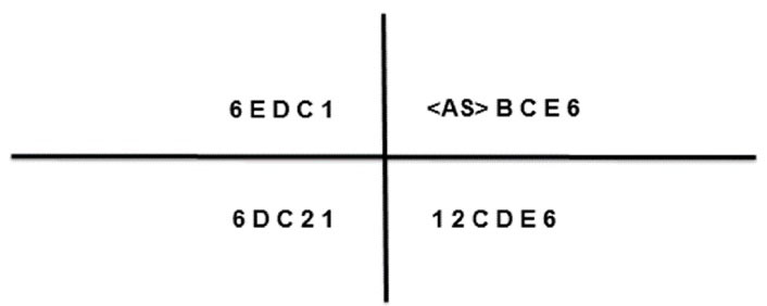
Figure 1.
Present Teeth in his Mouth.
.
Present Teeth in his Mouth.
Parents’ informed consent was taken for treatment and publication of the treatment result as an article.
The left upper deciduous central incisor was fused to supernumerary teeth with grooves in the buccal and palatal sides and the teeth had caries. Moreover, the left upper deciduous second incisor can be observed in Figure 2.
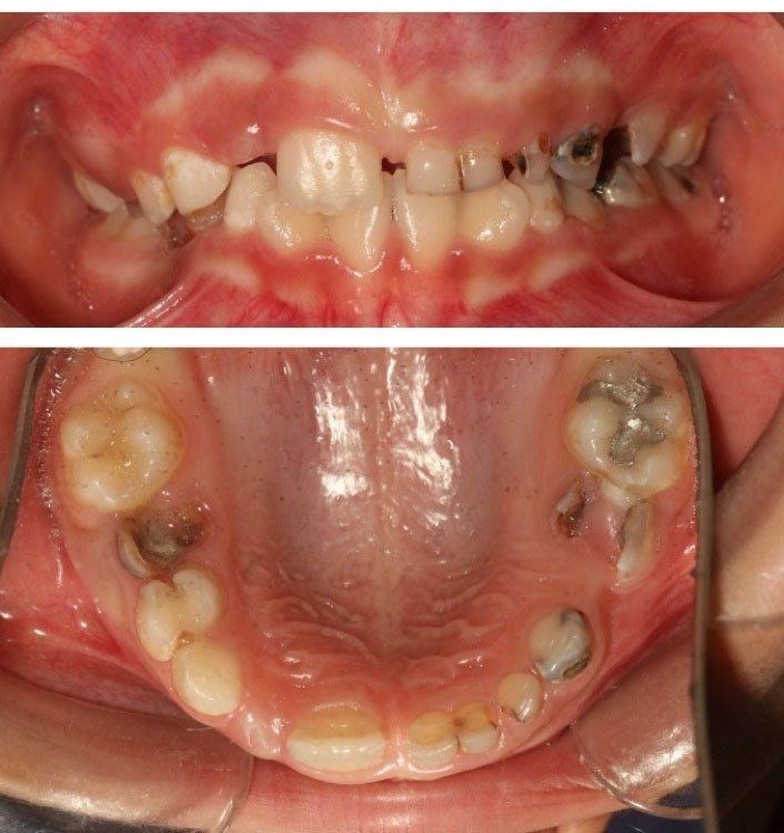
Figure 2.
Anterior and Occlusal Views of Intra-oral Situation.
.
Anterior and Occlusal Views of Intra-oral Situation.
A panoramic view of the X-ray (Figure 3) represented that the crowns and roots of the left maxillary primary incisor and supplemental teeth were fused, and two different pulp cavities and root canals were detected as well. One impacted extra tooth was observed between the unerupted upper secondary lateral incisor, and the roots of the second deciduous tooth were found with more details in one cut of cone-beam computed tomography (Figure 4) of the patient. The crown of the impacted extra tooth was positioned towards the lingual side, but it was located on the buccal side of the bone.
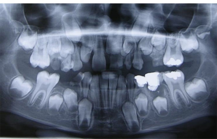
Figure 3.
A Panoramic View.
.
A Panoramic View.
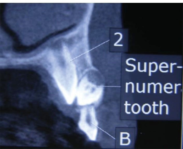
Figure 4.
A Cone-Beam Computed Tomography Radiograph.
.
A Cone-Beam Computed Tomography Radiograph.
Differential Diagnosis: Gemination and Twinning
Treatment: The unilateral fused deciduous tooth and impacted extra teeth were surgically extracted at the age of nine. The patient was recalled three months later for checking spontaneous tooth eruption (Figure 5).
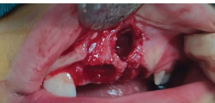
Figure 5.
Frontal View After Finishing Surgical Treatment.
.
Frontal View After Finishing Surgical Treatment.
The removed fused deciduous incisor consisted of different crowns attached to the entire root area. No root canal resorption was observed, and the mesial part was recognized as the primary central incisor considering its crown shape. The two parts of the fused tooth were divided by the vertical grooves of the labial and lingual aspects, and caries were evident in the vertical grooves. The formation of extra impacted teeth was limited to the crown tissue without root formation (Figure 6).
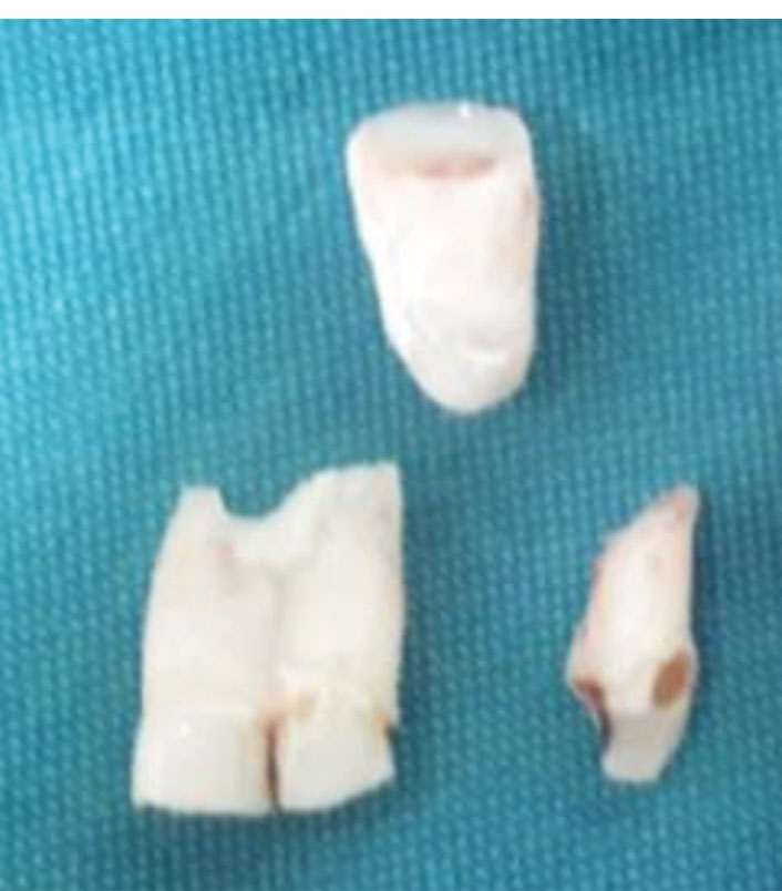
Figure 6.
Labial View of the Extracted Teeth.
.
Labial View of the Extracted Teeth.
Discussion
The present study presented a case of a rare unilateral fusion between central and extra primary teeth with a left maxillary extra impacted tooth.
We call our case fusion because germination is defined as a situation where tooth bud tries to separate; however, fusion is known as the union of two enamels or enamel and dentin of 2 or more maturingtooth buds separately (10). Fusion is frequently confused with germination, especially if there is an extra tooth (5). Mader’s law might be a practical way to distinguish fusion and germination (11). The term primary double teeth, which regularly refers to fusion and germination, is one of the most common developmental abnormalities in deciduous teeth (12). Fusion is more common in deciduous teeth than permanent teeth (13). Double teeth are frequently observed in the anterior part and usually involve the lower second incisors and canines and are found unilaterally more frequently. No considerable statistical difference was detected among the genders, and its prevalence was reported to be 0.74%. The incidence of fusion is lower in Caucasians than in Asians (14).
If fusion occurs between two natural teeth, it may cause malocclusion such as diastema. It can also lead to irregular morphology between a natural tooth and an extra tooth (7).
The maturation of secondary teeth after the fusion of a primary tooth and an extra tooth can take place in 3 ways, including an additional successor, a tooth with an abnormal shape, or without an anomaly. In reported cases of unilateral fusion in the central maxilla and extra teeth, few had extra permanent teeth while there were no abnormalities in the other cases (15).
The cause of tooth fusion is still unknown, but it is speculated that it occurs during tooth formation, trauma, or inflammatory processes due to the metabolic problems of genetic factors. Some researchers believe that physical pressure and force created during eruption cause contact between the two tooth buds. Viral infection and the use of thalidomide during pregnancy are also considered possible causes. From an embryological point of view, there is a direct relationship between fusion in deciduous teeth and agenesis in permanent successors (16).
In some cases, clinical examination and radiography and a simple determination of the total number of the arch teeth may provide sufficient information to differentiate between fusion, gemination, and concurrence. Following fusion in deciduous teeth, we encounter clinical problems in permanent teeth. Consequently, the root resorption of fused deciduous teeth is delayed and leads to delay or irregular eruption of permanent successors (10).
The eruption of the first permanent central maxillary tooth was delayed in the present case, and despite the patient’s age, there was no root resorption in fused deciduous teeth. It is thought that the eruption of the permanent central incisor is delayed due to the position of the extra tooth. Fused deciduous teeth and extra incisors were surgically extracted, and it was recommended to refer again three months later to check for the spontaneous eruption of the secondary first incisors.
Conclusions
When faced with fused deciduous teeth in the clinic, it is recommended that fissure sealant be used in the pits, fissures, and grooves to stop further tooth decay. Radiographic pictures should be taken to observe the eruption of the succeeding teeth. A thorough examination and prompt surgical removal of the affected teeth at a suitable moment remain essential in preventing delayed exfoliation of deciduous teeth and delayed eruption of the successor.
Authors’ Contribution
Conceptualization: Zahra Khosravi, Soudeh Tayebi.
Methodology: Fahimeh Daneshyar.
Validation: Shaghayegh Golshani.
Formal Analysis: Shaghayegh Golshani.
Investigation: Shaghayegh Golshani.
Resources: Shaghayegh Golshani.
Data Curation: Zahra Bagheri.
Writing—Original Draft Preparation: Shaghayegh Golshani.
Writing—Review And Editing: Shaghayegh Golshani.
Visualization: Shaghayegh Golshani.
Supervision: Fahimeh Daneshyar.
Project Administration: Fahimeh Daneshyar.
Competing Interests
The authors declare that they have no conflict of interests.
Ethical Approval
Informed consent was obtained from the parents of the patient for publication of this report.
References
- Folayan MO, Alade M, Adeniyi A, El Tantawi M, Finlayson TL. Association between developmental dental anomalies, early childhood caries and oral hygiene status of 3-5-year-old children in Ile-Ife, Nigeria. BMC Oral Health 2019; 20(1):1. doi: 10.1186/s12903-019-0991-2 [Crossref] [ Google Scholar]
- Bernardi S, Bianchi S, Bernardi G, Tchorz JP, Attin T, Hellwig E. Clinical management of fusion in primary mandibular incisors: a systematic literature review. Acta Odontol Scand 2020; 78(6):417-24. doi: 10.1080/00016357.2020.1734233 [Crossref] [ Google Scholar]
- Ben Salem M, Chouchene F, Masmoudi F, Baaziz A, Maatouk F, Ghedira H. Fusion or gemination? Diagnosis and management in primary teeth: a report of two cases. Case Rep Dent 2021; 2021:6661776. doi: 10.1155/2021/6661776 [Crossref] [ Google Scholar]
- Cunha RS, Junaid A, Mello I. Unilateral fusion of a supernumerary tooth to a maxillary permanent lateral incisor: a report of a rare case. J Endod 2015; 41(3):420-3. doi: 10.1016/j.joen.2014.10.021 [Crossref] [ Google Scholar]
- Tewari N, Pandey RK. Bilateral fusion in primary mandibular teeth: a report of two cases. J Indian Soc Pedod Prev Dent 2011; 29(1):50-2. doi: 10.4103/0970-4388.79936 [Crossref] [ Google Scholar]
- Santos LM, Forte FD, Rocha MJ. Pulp therapy in a maxillary fused primary central incisor--report of a case. Int J Paediatr Dent 2003; 13(4):274-8. doi: 10.1046/j.1365-263x.2003.00464.x [Crossref] [ Google Scholar]
- Milano M, Seybold SV, McCandless G, Cammarata R. Bilateral fusion of the mandibular primary incisors: report of case. ASDC J Dent Child 1999; 66(4):280-2. [ Google Scholar]
- Lochib S, Indushekar KR, Saraf BG, Sheoran N, Sardana D. Occlusal characteristics and prevalence of associated dental anomalies in the primary dentition. J Epidemiol Glob Health 2015; 5(2):151-7. doi: 10.1016/j.jegh.2014.07.001 [Crossref] [ Google Scholar]
- Açıkel H, İbiş S, Şen Tunç E. Primary fused teeth and findings in permanent dentition. Med Princ Pract 2018; 27(2):129-32. doi: 10.1159/000487322 [Crossref] [ Google Scholar]
- Castro IO, Estrela C, Souza VR, Lopes LG, de Souza JB. Unilateral fusion of maxillary lateral incisor: diagnosis using cone beam computed tomography. Case Rep Dent 2014; 2014:934218. doi: 10.1155/2014/934218 [Crossref] [ Google Scholar]
- Mader CL. Fusion of teeth. J Am Dent Assoc 1979; 98(1):62-4. doi: 10.14219/jada.archive.1979.0037 [Crossref] [ Google Scholar]
- Aguiló L, Gandia JL, Cibrian R, Catala M. Primary double teeth A retrospective clinical study of their morphological characteristics and associated anomalies. Int J Paediatr Dent 1999; 9(3):175-83. doi: 10.1046/j.1365-263x.1999.00131.x [Crossref] [ Google Scholar]
- Cheng RB, Chen X, Liu SJ, Pan L, Wu XG. [An epidemiological survey on fusion of deciduous teeth of 4286 kindergarten children in Shenyang city]. Shanghai Kou Qiang Yi Xue 2003;12(6):424-6. [Chinese].
- Aydinbelge M, Sekerci AE, Caliskan S, Gumus H, Sisman Y, Cantekin K. Clinical and radiographic evaluation of double teeth in primary dentition and associated anomalies in the permanent successors. Niger J Clin Pract 2017; 20(7):847-51. doi: 10.4103/1119-3077.183246 [Crossref] [ Google Scholar]
- Tomizawa M, Shimizu A, Hayashi S, Noda T. Bilateral maxillary fused primary incisors accompanied by succedaneous supernumerary teeth: report of a case. Int J Paediatr Dent 2002; 12(3):223-7. doi: 10.1046/j.1365-263x.2002.00351.x [Crossref] [ Google Scholar]
- Buenviaje TM, Rapp R. Dental anomalies in children: a clinical and radiographic survey. ASDC J Dent Child 1984; 51(1):42-6. [ Google Scholar]