Avicenna J Dent Res. 14(4):185-189.
doi: 10.34172/ajdr.2022.548
Case Report
Crown Lengthening Procedure in Epidermolysis Bullosa: A Case Report
Kamran Lahoorpoor 1  , Nazli Rabienejad 1 , Sayed Ali Moosavi 1, *
Author information:
1Department of Periodontology, School of Dentistry, Hamadan University of Medical Sciences, Hamadan, Iran
Abstract
Epidermolysis bullosa (EB) is a spectrum of conditions characterized by mechanical fragility and blistering of the skin. Individuals suffering from EB display a wide range of symptoms based on affected proteins in different organs and tissues in the body, including the craniofacial complex and the oral cavity. In this case-report, a 22-year-old girl with Dominant Dystrophic Epidermolysis Bullosa is presented. She suffered no other medical complication and psychological examination was normal. Atraumatic extraction of hopeless teeth was performed and patient was referred for endodontic treatment of maintainable teeth. Surgical crown lengthening in anterior segments was performed in two sessions with specific precautions to avoid soft tissue trauma and irritation both during surgery and after it. Post-surgery healing was uneventful and acceptable. In conclusion, with a few precautions, surgical crown lengthening can be performed in these patients with minimal soft tissue trauma and optimal healing post-surgery.
Keywords: Epidermolysis Bullosa, Crown Lengthening, Surgery
Copyright and License Information
© 2022 The Author(s); Published by Hamadan University of Medical Sciences.
This is an open-access article distributed under the terms of the Creative Commons Attribution License (
http://creativecommons.org/licenses/by/4.0), which permits unrestricted use, distribution, and reproduction in any medium provided the original work is properly cited.
Please cite this article as follows: Lahoorpoor K, Rabienejad N, Moosavi SA. Crown lengthening procedure in epidermolysis bullosa: a case report. Avicenna J Dent Res. 2022; 14(4):185-189. doi:10.34172/ajdr.2022.548
Introduction
Epidermolysis bullosa (EB) consists of a spectrum of conditions that are characterized by mechanical fragility and blistering of the skin (1). EB, which was first reported by Hebra (2), displays a tremendous genetic and clinical phenotype variety in its family of disorders. The main types of EB are intraepidermal (simplex EB), Junctional, dermolytic (dystrophic EB), and mixed (Kindler syndrome) EB (1).
EB symptoms are often manifested at birth or during the first year of life (3). Tremendous diversity is observed in involved organs and tissues and phenotypic severity in individuals suffering from EB disorders (4-6). Likewise, different EB types result in various craniofacial and oral manifestations with varying characteristics and severity (7,8). There can be significant morbidity and complications involving both soft and hard tissues of the craniofacial complex depending on the type of EB (1).
A specific molecular basis is currently known for 13 EB subtypes (1). The clinical phenotype of the disorder in affected individuals and the involved tissues and organs depends largely on the specific absent or abnormal proteins as a result of causative genetic mutations (9). For example, although type VII collagen is critical for maintaining the integrity of the skin and the oral mucosa, it is not essential in the development of the forming tooth bud. Therefore, individuals with mutations affecting type VII collagen have normally developed dentition but may show severe oral soft tissue involvement. On the other hand, laminin 322 plays an essential role during tooth development, and hence, individuals with mutations affecting laminin 322 may demonstrate defects in the development of the enamel of their teeth (9).
Case Report
A 22-year-old girl was referred to the periodontology ward with the chief complaint of pain in her anterior teeth. The patient had dominant dystrophic EB, which is one of the mildest forms of dystrophic EB. Her parents were identified to be first-degree cousins. On routine medical examination, the patient appeared normal. She had a few blisters and multiple disfiguring scars on her body (Figure 1).
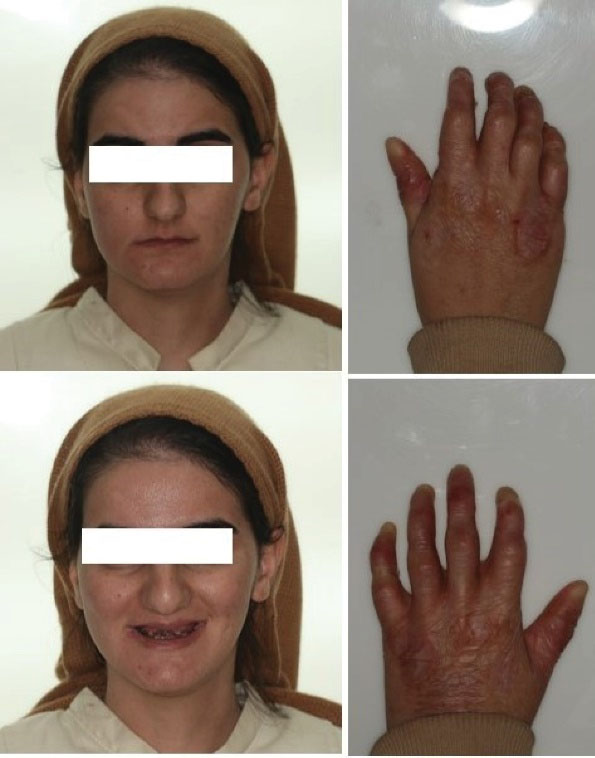
Figure 1.
General appearance of the patient
.
General appearance of the patient
Psychologically, the patient appeared to have a normal cognitive function and was attending college. She was taking supplements and vitamins but not any medications. The radiological panoramic view represented numerous remaining roots in the maxilla and mandible (Figure 2).
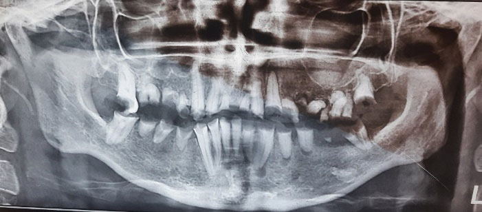
Figure 2.
Initial panoramic radiography
.
Initial panoramic radiography
The patient was very knowledgeable about her condition. She reported frequent intraoral bullae formation as a result of inflicted minor traumas. Minor dental procedures such as oral prophylaxis often caused bullae formation with subsequent discomfort. Cutaneous involvement was limited, and bullae would form under heavier traction forces. Consultation with the patient’s dermatologist conveyed no contraindications to periodontal surgery, with a recommendation to minimize the manipulation of soft tissues.
Oral evaluations revealed multiple ulcers on the buccal mucosa, tongue, and palate. The palate was dystrophic and had severe scarring due to chronic trauma and bullae formation (Figure 3).
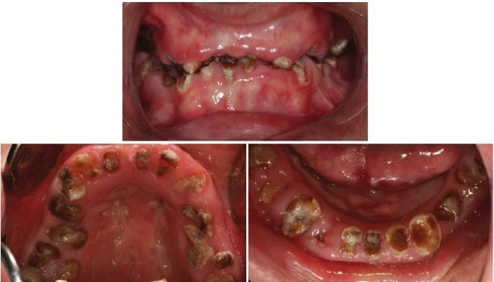
Figure 3.
Initial intra-oral view
.
Initial intra-oral view
The treatment plan consisted of the extraction of hopeless teeth, root canal therapy, crown lengthening, and fixed partial prosthesis. The treatment plan was presented and accepted by the patient.
Local anesthesia was obtained using 2% lidocaine with 1:80,000 epinephrine (Persocaine-E, Darou Pakhsh Pharmaceutical Mfg Company, Tehran, Iran). All hopeless roots were atraumatically extracted in two sessions (Figure 4).
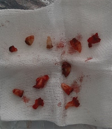
Figure 4.
Extracted hopeless teeth
.
Extracted hopeless teeth
The patient was referred to the endodontics ward to undergo root canal treatment of the remaining periodontally maintainable teeth. Four weeks after the completion of root canal therapy sessions (Figure 5), the patient underwent surgical crown lengthening.
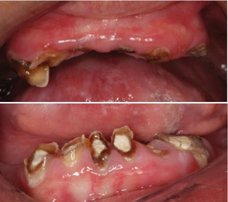
Figure 5.
Intra-oral pre-op view
.
Intra-oral pre-op view
For the crown lengthening of the anterior segment of the maxilla, anesthesia was achieved using buccal and palatal supra-periosteal infiltration of lidocaine 2% with epinephrine 1:80000 (Persocaine-E). First, sub-marginal scalloped internal bevel incisions were made 1 or 2 mm apically from the remaining caries-free tooth structure; sufficient tooth structure would be available for a ferrule effect and consider leaving at least 2 mm of keratinized gingiva. Then, sulcular incisions were performed to detach the attachment apparatus from the root. Next, interdental incisions were made to detach the interdental tissue and the gingival collar from the bone. Full-thickness flap reflection was performed to expose the alveolar bone beyond the mucogingival junction. Bone contouring was mainly performed using a long-shank round diamond bur on a high-speed dental handpiece by utilizing water spray as a coolant. The two hopeless central incisors were atraumatically extracted, and final adjustments to the bony contour were made using hand instruments such as chisels and bone files. After ensuring a minimum of 4 millimeters of the available supracrestal tooth structure, all root surfaces were checked for the remaining bone spicules and widow peaks. Following achieving the optimal bone contour, the soft tissue was adjusted by trimming and thinning so that it optimally adapts to the underlying bone and allows for primary closure, except for the extraction sites. For wound closure, 4-0 absorbable polyglycolate-coated suture (Supabon, SUPA Medical Devices Company, Tehran, Iran) was selected, and closure was obtained using interrupted simple-loop sutures (Figure 6).
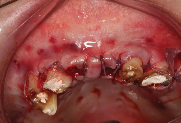
Figure 6.
Maxillary immediate post-op results
.
Maxillary immediate post-op results
Crown lengthening of the mandibular anterior segment was performed after the initial healing of the maxillary mucosa. Anesthesia was achieved using lidocaine 2% with epinephrine 1:80000 (Persocaine-E) by means of bilateral incisive/mental nerve block and lingual infiltration. Moreover, incisions and flap reflections were performed as described previously. Bone recontouring was also conducted based on previous explanations. No teeth needed to be extracted in this region. Copious irrigation was performed after verifying sufficient available supracrestal tooth structure, and wound closure was achieved, as previously described, using 4-0 absorbable polyglycolate-coated sutures.
To minimize soft tissue irritation and manipulation during and after surgeries, a few steps were taken as follows:
-
Incisions were performed sharply and continuously, accompanied by firm pressure to complete the incision in a single stroke.
-
Tongue, cheek, and lip retraction were conducted using large-surface smooth blunt vaseline-coated instruments to avoid pin-point pressure on these tissues.
-
Flap reflection was solely performed using large blunt periosteal elevators leaning directly on the bone.
-
If there was a need for a flap to be held or secured in place (e.g., when a needle had to be passed through), it was attempted to be conducted using instruments applying force from inside the flap (i.e., connective tissue + periosteum surface), and the use of instruments which would cause pressure on the epithelial side (e.g., tissue forceps) was kept to a minimum.
-
Absorbable sutures were selected to avoid an extra session of suture removal and the resulting soft-tissue irritation.
-
No periodontal dressing was placed to minimize soft tissue irritation and trauma and avoid the possible separation of epithelium/bullae formation.
Post-operation instructions included general oral surgery post-op instructions with an emphasis on minimizing soft-tissue trauma and irritation. Medications prescribed post-operatively included amoxicillin 500 mg three times per day for one week, ibuprofen 400 mg three times per day or as needed, and normal saline rinsing 3-4 times a day. Chlorhexidine mouthwash was prescribed as it would reduce the oral cavity microflora which may lead to less inflammation. The patient was revisited 3 days post-operation after each surgery to ensure proper wound healing. Figure 7 displays soft tissue healing was uneventful and without complications. The soft tissue condition at 1-month post-operation immediately after the cementation of two cast posts and before the prophylaxis session.
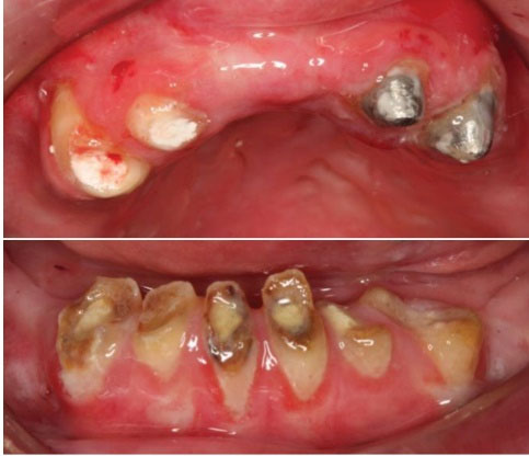
Figure 7.
One month post-op results
.
One month post-op results
Discussion
Several researchers have reported various dental procedures performed on EB patients. However, to the best of our knowledge, periodontal plastic surgery and related complications in patients with EB are rarely mentioned in the literature. In dystrophic EB, blisters are found beneath the basement membrane within the dermis, exposing the underlying connective tissue (10-13).
The presented case demonstrated widespread dental caries that are directly related to the inability to maintain proper oral hygiene (14,15). Wright et al found that susceptibility to caries was significantly higher in EB patients (12). This might be due to the severity of the soft tissue ulceration and consumption of a soft diet rich in sucrose -necessary to provide sufficient energy intake- and the difficulty in mechanical removal of dental plaques due to their incapability in holding a toothbrush (3). The periodontal status of these patients could be well managed by accurate treatment and a good maintenance program (16). Comprehensive oral care instructions should be given to these patients as soon as possible, and they should be put in a preventive program. To enhance the oral clearance of food debris, an increased fluid intake during meals could be helpful (17). Water jets and water flossers, soft toothbrushes, and mouthwashes containing fluoride are recommended (3), all of which can help fight cavities. These patients tolerate the topical application of neutral sodium fluoride and fluoride varnish well (17). Chlorhexidine mouthwash may also help fight cavities (3,17). Sucralfate (the drug of choice for the treatment of active duodenal ulcers) has been proposed as an effective prophylactic and therapeutic agent for reducing pain and blistering in these patients (18).
Rapidly rupturing vesicles frequently develop in EB patients, leaving erosive surfaces on the mucous membranes of the lips and mouth. The other common oral manifestations of EB are vestibular obliteration, denudation of the tongue, microstomia, and ankyloglossia (3,17,19,20).
Ankyloglossia, severe scarring of the mouth and around the mouth, limited opening of the mouth, and vestibular obstruction of the mouth can impede or complicate dental care. Mouth ulcers are inevitable during dental treatment; however, they can be prevented by lubricating the mucosa with hydrocortisone cream, triamcinolone, or petrolatum before starting the procedure (21). Furthermore, retractors can be coated with petrolatum to minimize friction and soft tissue trauma.
Local anesthesia during outpatient dental treatment can also cause blisters. Therefore, it should be avoided whenever possible (20). The injections of local anesthetic solutions should also be slow and deep into the tissue to prevent mechanical damage to the tissue (3). Superficial injections can cause tissue ballooning and possible subsequent bullae formation.
The principles of surgical crown lengthening in EB patients do not differ from systemically healthy patients, but precautions and measures should be taken to minimize soft tissue irritation. These measures include but are not limited to sharp, continuous, and single-stroke incisions, using blunt instruments for flap retraction, flap retraction by applying force to the inner surface of the flap, avoiding tissue forceps whenever possible, using absorbable sutures, and avoiding periodontal dressings.
Conclusions
EB depicts a group of rare genetic disorders that involves the skin and mucous membranes by vesicle and bullae formation. The oral and dental manifestations of EB disrupt the patient’s oral health and hygiene and exhibit a challenge for dental professionals. The administration of general anesthesia for patients with EB requires distinctive attention and inclusive care. Surgical crown lengthening is possible in these patients with some precautions to minimize soft tissue irritation.
Acknowledgements
The authors would like to thank the Dental Research Center and the Vice-chancellor of Research, Hamadan University of Medical Sciences for their support.
Conflict of Interest Disclosures
The authors of this manuscript declare that they have no financial or other competing interests concerning this article.
Ethical Statement
Informed consent was obtained from the patient for the publication of this report.
References
- Fine JD, Eady RA, Bauer EA, Bauer JW, Bruckner-Tuderman L, Heagerty A. The classification of inherited epidermolysis bullosa (EB): report of the Third International Consensus Meeting on Diagnosis and Classification of EB. J Am Acad Dermatol 2008; 58(6):931-50. doi: 10.1016/j.jaad.2008.02.004 [Crossref] [ Google Scholar]
- Crawford EG Jr, Burkes EJ Jr, Briggaman RA. Hereditary epidermolysis bullosa: oral manifestations and dental therapy. Oral Surg Oral Med Oral Pathol 1976; 42(4):490-500. doi: 10.1016/0030-4220(76)90296-6 [Crossref] [ Google Scholar]
- Torres CP, Gomes-Silva JM, Mellara TS, Carvalho LP, Borsatto MC. Dental care management in a child with recessive dystrophic epidermolysis bullosa. Braz Dent J 2011; 22(6):511-6. doi: 10.1590/s0103-64402011000600012 [Crossref] [ Google Scholar]
- Gedde-Dahl T Jr. Sixteen types of epidermolysis bullosa On the clinical discrimination, therapy and prenatal diagnosis. Acta Derm Venereol Suppl (Stockh) 1981; 95:74-87. [ Google Scholar]
- Wolff K, Johnson R. Fitzpatrick’s Color Atlas and Synopsis of Clinical Dermatology. McGraw-Hill Professional; 2009.
- Lai-Cheong JE, Tanaka A, Hawche G, Emanuel P, Maari C, Taskesen M. Kindler syndrome: a focal adhesion genodermatosis. Br J Dermatol 2009; 160(2):233-42. doi: 10.1111/j.1365-2133.2008.08976.x [Crossref] [ Google Scholar]
- Wright JT, Fine JD, Johnson LB. Oral soft tissues in hereditary epidermolysis bullosa. Oral Surg Oral Med Oral Pathol 1991; 71(4):440-6. doi: 10.1016/0030-4220(91)90426-d [Crossref] [ Google Scholar]
- Wiebe CB, Petricca G, Häkkinen L, Jiang G, Wu C, Larjava HS. Kindler syndrome and periodontal disease: review of the literature and a 12-year follow-up case. J Periodontol 2008; 79(5):961-6. doi: 10.1902/jop.2008.070167 [Crossref] [ Google Scholar]
- Meneguzzi G, Marinkovich MP, Aberdam D, Pisani A, Burgeson R, Ortonne JP. Kalinin is abnormally expressed in epithelial basement membranes of Herlitz’s junctional epidermolysis bullosa patients. Exp Dermatol 1992; 1(5):221-9. doi: 10.1111/j.1600-0625.1992.tb00080.x [Crossref] [ Google Scholar]
- Ciccarelli AO, Rothaus KO, Carter DM, Lin AN. Plastic and reconstructive surgery in epidermolysis bullosa: clinical experience with 110 procedures in 25 patients. Ann Plast Surg 1995; 35(3):254-61. doi: 10.1097/00000637-199509000-00006 [Crossref] [ Google Scholar]
- Liu HH, Chen CJ, Miles DA. Epidermolysis bullosa simplex: review and report of case. ASDC J Dent Child 1998; 65(5):349-53. [ Google Scholar]
- Wright JT, Fine JD, Johnson L. Hereditary epidermolysis bullosa: oral manifestations and dental management. Pediatr Dent 1993; 15(4):242-8. [ Google Scholar]
- Sedano HO, Gorlin RJ. Epidermolysis bullosa. Oral Surg Oral Med Oral Pathol 1989; 67(5):555-63. doi: 10.1016/0030-4220(89)90272-7 [Crossref] [ Google Scholar]
- Hitchin AD. The defects of cementum in epidermolysis bullosa dystrophica. Br Dent J 1973; 135(10):437-42. doi: 10.1038/sj.bdj.4803098 [Crossref] [ Google Scholar]
- Album MM, Gaisin A, Lee KW, Buck BE, Sharrar WG, Gill FM. Epidermolysis bullosa dystrophica polydysplastica A case of anesthetic management in oral surgery. Oral Surg Oral Med Oral Pathol 1977; 43(6):859-72. doi: 10.1016/0030-4220(77)90078-0 [Crossref] [ Google Scholar]
- Torkzaban P, Moradihaghgoo J, Shams B, Gholami L, Sabzeghabaie M, Faramarzi F. Periodontal management of a patient with kindler syndrome. Avicenna J Dent Res 2012; 4(2):67-71. [ Google Scholar]
- Prabhu VR, Rekka P, Swathi S. Dental and anesthetic management of a child with epidermolysis bullosa. J Indian Soc Pedod Prev Dent 2011; 29(2):155-60. doi: 10.4103/0970-4388.84690 [Crossref] [ Google Scholar]
- Marini I, Vecchiet F. Sucralfate: a help during oral management in patients with epidermolysis bullosa. J Periodontol 2001; 72(5):691-5. doi: 10.1902/jop.2001.72.5.691 [Crossref] [ Google Scholar]
- Serrano-Martínez MC, Bagán JV, Silvestre FJ, Viguer MT. Oral lesions in recessive dystrophic epidermolysis bullosa. Oral Dis 2003; 9(5):264-8. doi: 10.1034/j.1601-0825.2003.03971.x [Crossref] [ Google Scholar]
- Kostara A, Roberts GJ, Gelbier M. Dental maturity in children with dystrophic epidermolysis bullosa. Pediatr Dent 2000; 22(5):385-8. [ Google Scholar]
- Esfahanizade K, Mahdavi AR, Ansari G, Fallahinejad Ghajari M, Esfahanizadeh A. Epidermolysis bullosa, dental and anesthetic management: a case report. J Dent (Shiraz) 2014; 15(3):147-52. [ Google Scholar]