Avicenna J Dent Res. 14(2):58-62.
doi: 10.34172/ajdr.2022.11
Original Article
Enamel Color Changes After Bleaching Treatment Using Common Bleaching Agents With Er, Cr:YSGG Laser
Zahra Khamverdi 1  , Loghman Rezaei-Soufi 1, Maryam Farhadian 2, Masoud Sharifian 3, Alireza Mazaheri 3, *
Author information:
1Professor, Department of Restorative Dentistry, Dental School, Hamadan University of Medical Sciences, Hamadan, Iran
2Assistant Professor, Department of Biostatistics, School of Public Health, Hamadan University of Medical Sciences, Hamadan, Iran
3DDS, Dentist, Hamadan, Iran
Abstract
Background: Reports indicate that lasers accelerate tooth bleaching by activating bleaching agents. Due to the lack of sufficient information about the application of the Er, Cr:YSGG laser, the present study was conducted to compare the degree of enamel color changes after teeth bleaching treatment using chemical whitening agents alone and with Er, Cr:YSGG laser.
Methods: In this laboratory study, several human molars were cut into 4 parts after cutting and removing the pulp. Based on laser application, the samples were randomly divided into two groups (N=20), including the chemical bleaching group (G1) and the chemical bleaching group with activated laser (G2). In G1, the bleaching process was performed only with 35% hydrogen peroxide for two weeks (3 times, 15 minutes each week). In G2, the gel bleaching was activated by the Er, Cr:YSGG laser with a wavelength of 2780 nm. It was placed 2.5 cm from the sample surface with bleaching agents and applied twice for 15 seconds. Color changes were recorded using a spectrophotometer before bleaching, immediately after, 1 month, and 3 months after bleaching. Data were analyzed by SPSS software (version 18), Repeated measures ANOVA, and Tukey’s tests (α=0.05).
Results: Mean and standard deviation of changes in Δa showed a significant difference between the gel group and the gel group with laser over time (P<0.05), but this difference was not observed in ΔL and ΔB (P>0.05). However, the intragroup comparison demonstrated significant changes in Δa and Δb in both groups over time, but not in ΔL (P>0.05). The ΔE changes in both the laser and bleaching gel groups were above the threshold of 3.3. The results indicated no significant difference between G1 and G2 in terms of the ΔE (P>0.05). Finally, the results revealed that 1 and 3 months after teeth whitening, ΔE changes in both groups were greater than 3.3.
Conclusions: Overall, the application of the Er, Cr:YSGG laser had no positive effect on bleaching efficacy when using a 35% hydrogen peroxide gel. Based on the findings, color changes were stable in the studied groups for up to 3 months after teeth bleaching.
Keywords: Tooth bleaching, Laser therapy, Enamel, Hydrogen peroxide
Copyright and License Information
© 2022 The Author(s); Published by Hamadan University of Medical Sciences.
This is an open-access article distributed under the terms of the Creative Commons Attribution License (
http://creativecommons.org/licenses/by/4.0), which permits unrestricted use, distribution, and reproduction in any medium provided the original work is properly cited.
Please cite this article as follows: Khamverdi Z, Rezaei-Soufi L, Farhadian M, Sharifian M, Mazaheri A. Enamel color changes after bleaching treatment using common bleaching agents with er, cr:ysgg laser. Avicenna J Dent Res. 2022; 14(2):58-62. doi:10.34172/ajdr.2022.11
Introduction
Generally, the aesthetics and health of teeth are important for individuals. The white teeth can provide a good appearance (1), and any color change may affect the aesthetic of teeth in the smile because tooth color is an essential factor in having an attractive smile. Tooth discoloration can occur on the surface of the enamel (external discoloration) or inside the tooth structure (internal discoloration). Enamel discoloration is often caused by the deposition of food pigments on the bacterial plaque or the surface of the enamel (2). In addition, internal pigments may be caused by pre- or post-emergence factors (1). The color of tooth structures results from the interaction between different light effects in hard tissues; these structures include scattering, reflection, transmission, and absorption (3). Therefore, the final color is the result of the optical properties of the enamel and dentin, including translucency and color characteristics (3,4).
Teeth bleaching is performed through several methods such as whitening toothpaste, over-the-counter gels and strips, whitening mouthwashes, and teeth bleaching in the office and at home (5).
Another method that can be considered for teeth whitening is the use of lasers by both photothermal and photochemical methods. Today, the photothermal bleaching method is better known and is the most frequently used method for tooth bleaching (6). The Er, Cr:YSGG (2780 nm) laser is one of the most widely applied lasers in dentistry. The wavelengths of this laser have the highest water absorption in hydroxyapatite, thus it is the treatment of choice for hard tooth tissues (7). Dionysopoulos et al showed that Er, Cr:YSGG laser bleaching treatment is more effective than conventional methods in terms of color change and treatment duration (8).
Kiomars et al investigated the effect of the diode laser on the discoloration of tooth enamel by applying different percentages of hydrogen peroxide and different wavelengths of diode lasers. They found no significant difference in the degree of enamel discoloration in the studied groups, and various wavelengths of diode laser light provided the same improvement in enamel discoloration (9).
Cesar reported that using laser light and tooth whitening products changes tooth color and increases light reflection. As a result, laser light can aid the teeth whitening process by increasing the light reflection on the enamel surface (10).
To the best of our knowledge, few studies have focused on the laser bleaching process by laser, and the obtained results are occasionally contradictory (11-14). Therefore, this study aimed to determine the effect of the Er, Cr:YSGG laser on tooth enamel discoloration after bleaching treatment and compare it with the degree of color changes after bleaching treatment without laser at different times.
Materials and Methods
In this laboratory study, some extracted intact posterior teeth were collected in a 10% formalin solution over 3 months from some offices and dental centers due to periodontal problems or orthodontic treatments. From these teeth, 20 healthy and non-cracked teeth without caries or restorative lesions were selected and cleaned using a periodontal curette and placed in normal saline at room temperature.
Twenty-four hours before initiation, the teeth were kept in distilled water. For this purpose, the teeth were cut from CEJ using a high-speed disc, and their pulp was then removed, and each sample was cut parallel to the tooth’s longitudinal axis in the mesiodistal direction by a disc and divided into two halves. Next, the samples were examined under a light microscope at 10 × magnification to ensure the lack of any cracks or fractures.
A mold was used to place the samples in a similar position during the measurement and standardize the measurements during the study. Then, all samples were randomly assigned to two equal groups based on the applied laser (N = 20). The samples in G1 were bleached using a whitening gel of 35% hydrogen peroxide (Whiteness HP Maxx, FGM, Brazil). The bleaching gel was applied three times for 15 minutes with an interval of 1 minute for 1 week (15). After each bleaching process, the samples were washed with water for 1 minute and gently dried with air spray. Next, the 35% hydrogen peroxide bleaching gel with Er: Cr:YSGG (Bio Lase, USA) laser (with a wavelength of 2780 nm, a frequency of 10 Hz, and power of 10 W in S Mode from a distance of 2.5 cm from the sample surface without air with Gold handpiece and MZ8 type) was employed twice for 15 seconds in G2 (16). A spectrophotometer (Easy Shade Advance, Bad Säckingen, Germany Vita) was used to evaluate the tooth color before (T1), 1 month (T2), and 3 months (T3) after bleaching.
L*, a*, and b* were evaluated for all teeth, indicating the value, green-red, and yellow-blue parameters, respectively. The difference color change of each sample was calculated by the following formula:
∆E = [(∆L*)2 + (∆a*)2 + (∆b*)2]1/2
where ∆L*, ∆a*, and ∆b* are changes in L*, a*, and b* at intervals before bleaching and subsequent measurements, respectively.
Then, the data were entered into SPSS software (version 18) and analyzed by Kolmogorov-Smirnov, repeated measures ANOVA, and Tukey’s HSD post hoc tests. The significance level was considered less than 0.05.
Results
Figure 1 displays color variations in the studied groups. The Δa mean revealed a significant difference between G1 and 2 over time (P < 0.05). Based on the results of the intragroup comparison, significant changes were found in Δa in both groups over time. Conversely, the mean (standard deviation, SD) of changes in ΔL demonstrated no statistically significant difference between the gel group and the gel group with laser over time (P > 0.05). Additionally, the intragroup comparison represented no significant changes regarding ΔL in both groups at times 1, 2, and 3. Similarly, there was no significant difference between the gel group and the gel group accompanied by laser over time based on the mean (SD) of changes in Δb. The P values obtained in the intragroup comparison indicated significant changes for Δb in both groups over time (Table 1).
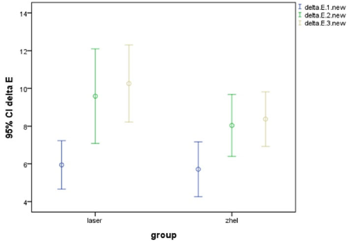
Figure 1.
The Trend of ΔE Index Changes in the Gel Group and Gel Group With Laser Over Time.
.
The Trend of ΔE Index Changes in the Gel Group and Gel Group With Laser Over Time.
Table 1.
Repeated-measureANOVA Results for Comparing Δa, ΔL, and Δb in the Gel Group and Gel Group With Laser at the Studied Durations
|
Variable
|
Time
|
Gel With Laser
|
Whitening Gel
|
Intergroup Significance
|
|
Mean±SD
|
Mean±SD
|
| Δa |
1 |
-0.19 ± 1.02 |
0.94 ± 2.12 |
0.012 |
| 2 |
-2.29 ± 1.30 |
-1.00 ± 1.19 |
| 3 |
- 1.73 ± 1.36 |
-1.19 ± 1.76 |
| Intragroup significance |
< 0.001 |
< 0.001 |
|
| ΔL |
1 |
1.00 ± 5.35 |
2.39 ± 3.97 |
0.127 |
| 2 |
2.07 ± 3.83 |
1.85 ± 4.45 |
| 3 |
- 1.27 ± 6.45 |
2.61 ± 3.72 |
| Intragroup significance |
0.11 |
0.811 |
|
| Δb |
1 |
-1.79 ± 3.24 |
0.82 ± 3.98 |
0.057 |
| 2 |
-7.45 ± 5.35 |
-4.59 ± 5.67 |
| 3 |
-7.57 ± 4.37 |
-6.34 ± 3.90 |
| Intragroup significance |
< 0.001 |
< 0.001 |
|
Note. SD: Standard deviation.
Table 2 presents the mean (SD) of changes in ΔE. The results showed there was no statistically significant difference between G1 and G2 over time (P > 0.05). According to P values obtained for ΔE in the intragroup comparison, generally, significant changes were observed in ΔE over time in the gel group and the gel group with the laser (P˂0.05).
The variation trend in the ΔE index in the gel group and the gel group with the laser over time is shown in Figure 1. The results of Tukey’s post hoc test are provided in Table 3.
Table 2.
Comparison Results of the ΔE Parameter in the Gel Group and Gel Group With Laser Over Time
|
Variable
|
Time
|
Group 2
|
Group 1
|
Intergroup Significance
|
|
Mean±SD
|
Mean±SD
|
| ΔE |
1 |
5.94 ± 2.74 |
5.71 ± 3.09 |
0.169 |
| 2 |
9.58 ± 5.35 |
8.03 ± 3.51 |
| 3 |
10.25 ± 4.37 |
8.37 ± 3.08 |
| Intragroup significance |
< 0.001 |
0.010 |
|
Note. SD: Standard deviation.
Table 3.
Paired Comparison of Color Changes in the Studied Times
|
Variable
|
Time
|
Gel With Laser
|
Whitening Gel
|
|
Mean Difference±SE
|
P
|
Mean Difference±SE
|
P
|
| ΔE |
1 versus 2 |
3.64 ± 1.04 |
0.002 |
2.32 ± 0.99 |
0.031 |
| 1 versus 3 |
4.31 ± 1.04 |
0.001 |
2.65 ± 0.801 |
0.004 |
| 2 versus 3 |
0.669 ± 1.25 |
0.601 |
0.335 ± 0.879 |
0.708 |
| Δa |
1 versus 2 |
-2.10 ± 0.29 |
0.001 |
-1.94 ± 0.51 |
0.001 |
| 1 versus 3 |
-1.54 ± 0.28 |
0.001 |
-2.13 ± 0.38 |
< 0.001 |
| 2 versus 3 |
0.56 ± 0.18 |
0.006 |
- 0.19 ± 0.40 |
0.644 |
| Δb |
1 versus 2 |
-5.66 ± 0.97 |
0.001 |
-5.41 ± 1.92 |
0.011 |
| 1 versus 3 |
-5.77 ± 0.68 |
0.001 |
-7.17 ± 1.21 |
< 0.001 |
| 2 versus 3 |
-0.12 ± 0.82 |
0.888 |
-1.75 ± 1.17 |
0.15 |
Note. SE: Standard error.
Discussion
The present study evaluated the effect of using the Er, Cr:YSGG laser (2780 nm) with 35% hydrogen peroxide on tooth discoloration in comparison with the bleaching method using 35% hydrogen peroxide alone.
Based on the results, the Er, Cr:YSGG laser (2780 nm) showed high efficiency. Dionysopoulos et al studied the safety of using the Er, Cr:YSGG laser (2780 nm). They investigated the surface of tooth enamel and its mineral composition with a scanning electron microscope (SEM) and found that using laser simultaneously with hydrogen peroxidase does not have destructive effects on tooth enamel. They concluded that it is safe to use in vitro; nonetheless, more studies are needed to evaluate the effects of this laser in vivo (8). The results of the mentioned study and the present study indicate that using the Er, Cr:YSGG laser has high efficiency in changing the tooth enamel color and is safer compared to other lasers. Moreover, it is easy to use and does not reduce the minerals in tooth enamel. Using the Er, Cr:YSGG laser has less penetration power and is less likely to cause tissue damage due to its higher wavelength in comparison to most lasers used in dentistry (7). Dionysopoulos et al reported that using the Er, Cr:YSGG laser has a significant effect on teeth bleaching compared to routine chemical methods after 24 hours (16).
In another study, Papadopoulos et al used the Er, Cr:YSGG laser with a power of 1.25 and 2.5 W to bleach teeth and reported no statistically significant difference in tooth discoloration after one week (17). The power of the used Er, Cr:YSGG laser in this study was 1.25 W.
ΔE is the most important factor in the efficiency of teeth bleaching. The acceptable threshold for the clinical efficacy of the bleaching agents for the ΔE is 3.3 (18). The results of the present study showed changes above the threshold for G1 and G2. In the study by Kiomars et al, a diode laser with 810 and 980 nm wavelengths was employed for tooth bleaching. At both wavelengths, the changes in the ΔE were greater than the threshold of 3.3. Based on their results, no significant difference was observed in the color change in the two wavelengths (9). Likewise, Ergin et al applied the Er: YAG laser to whiten teeth and revealed that the Er: YAG laser, similar to the diode and LED lasers, significantly affects the rate of enamel discoloration, which is comparable to bleaching compounds (19), which is in line with the findings of this study.
In the current study, no significant difference was observed in ΔL between the two groups. Variable L indicates the amount of the value, and the tooth appears brighter with the higher number of L (20). However, the results of this study revealed that using a laser with 35% hydrogen peroxide gel or 35% hydrogen peroxide gel alone did not affect the brightness of the teeth, and thus ΔL did not change significantly in both groups. In addition, the degree of ΔL changes at different times was not significant in the studied groups. This result implies that using a laser with 35% hydrogen peroxide gel or 35% hydrogen peroxide gel alone could not affect the brightness of the teeth (enamel) over time. Consistent with the results of this study, Wetter et al reported no change in ΔL when using diode and LED lasers, although it was associated with a decrease in a + b values in the laser group (20). Similar results were observed in the study by Zhang et al (21).
Another finding of this study showed that Δa changes in the laser group with 35% hydrogen peroxide gel were greater than in the 35% hydrogen peroxide gel group alone. Further, this parameter was associated with a decrease in both groups until the third month. These changes confirmed that the amount of green-blue color in the enamel decreased over time in both groups. Furthermore, Δb changes reduced over time in both groups up to the third month, suggesting that the amount of yellow-blue color in tooth enamel decreased over time in both groups.
One of the advantages of the present study over these studies was studying color changes at different times and up to 3 months after laser application. In the study by Sağlam et al, color changes were examined immediately after bleaching (22). Kiomars et al observed only a slight increase in ΔE after using a diode laser. They reported reductions in the values of Δa and Δb (9), while we noticed a decrease mostly in Δb.
Polydorou et al also investigated the effect of a diode laser on tooth discoloration after 3 months and found that discoloration remains constant after 3 months, and the effects of laser use on tooth discoloration during this period are irreversible (23), which conforms to the results of the present study.
Considering that only color changes and laser efficiency in teeth bleaching were studied in this research, it is recommended that another study investigates translucency in laser-assisted bleaching. In this study, mineral composition in the bleached enamel was not evaluated using a laser, which can be investigated in future studies.
Conclusions
Under the limitations of this study, the results showed that the application of the Er, Cr:YSGG laser had no positive effect on bleaching efficacy when using 35% hydrogen peroxide gel. Finally, the color was stable in the studied groups for up to 3 months after teeth bleaching.
Acknowledgments
This article is extracted from a dissertation in the General Dentistry course approved by Hamadan University of Medical Sciences (No. 980210679).
Authors’ Contribution
ZK has contributed to the performance of the tests and suggestion of the main idea, LRS has applicated the laser, MF has carried out statistical analysis, MS has performed tests, AM has collected the data and written the manuscript.
Conflict of Interests
The authors declare that there is no conflict of interests
Ethical Statement
The study was approved by the Ethics Committee of Hamadan
University of Medical Sciences IR.UMSHA.REC.1397.714
References
- Samorodnitzky-Naveh GR, Geiger SB, Levin L. Patients’ satisfaction with dental esthetics. J Am Dent Assoc 2007; 138(6):805-8. doi: 10.14219/jada.archive.2007.0269 [Crossref] [ Google Scholar]
- Addy M, Moran J, Newcombe R, Warren P. The comparative tea staining potential of phenolic, chlorhexidine and anti-adhesive mouthrinses. J Clin Periodontol 1995; 22(12):923-8. doi: 10.1111/j.1600-051x.1995.tb01796.x [Crossref] [ Google Scholar]
- Villarroel M, Fahl N, De Sousa AM, De Oliveira OB Jr. Direct esthetic restorations based on translucency and opacity of composite resins. J Esthet Restor Dent 2011; 23(2):73-87. doi: 10.1111/j.1708-8240.2010.00392.x [Crossref] [ Google Scholar]
- Hasegawa A, Ikeda I, Kawaguchi S. Color and translucency of in vivo natural central incisors. J Prosthet Dent 2000; 83(4):418-23. doi: 10.1016/s0022-3913(00)70036-9 [Crossref] [ Google Scholar]
- Carey CM. Tooth whitening: what we now know. J Evid Based Dent Pract 2014; 14 Suppl:70-6. doi: 10.1016/j.jebdp.2014.02.006 [Crossref] [ Google Scholar]
- Abdelfattah MM. Different types of laser use in teeth bleaching. J Med Med Sci 2014; 5(10):230-7. doi: 10.14303/jmms.2014.207 [Crossref] [ Google Scholar]
- Harashima T, Kinoshita J, Kimura Y, Brugnera A, Zanin F, Pecora JD. Morphological comparative study on ablation of dental hard tissues at cavity preparation by Er: YAG and Er, Cr: YSGG lasers. Photomed Laser Surg 2005; 23(1):52-5. doi: 10.1089/pho.2005.23.52 [Crossref] [ Google Scholar]
- Dionysopoulos D, Strakas D, Koliniotou-Koumpia E, Koumpia E. Effect of Er, Cr: YSGG laser irradiation on bovine enamel surface during in-office tooth bleaching ex vivo. Odontology 2017; 105(3):320-8. doi: 10.1007/s10266-016-0273-2 [Crossref] [ Google Scholar]
- Kiomars N, Azarpour P, Mirzaei M, Hashemi Kamangar SS, Kharazifard MJ, Chiniforush N. Evaluation of the Diode laser (810nm, 980 nm) on color change of teeth after external bleaching. Laser Ther 2016; 25(4):267-72. doi: 10.5978/islsm.16-OR-21 [Crossref] [ Google Scholar]
- Cesar IC, Soares LE, Alves LP, Martin AA, Munin E, Liporoni PC. Fourier transform-Raman and reflectance studies on dental enamel bleached with hydrogen peroxide activated using a light-emitting diode-laser system. Photomed Laser Surg 2009; 27(6):913-9. doi: 10.1089/pho.2008.2409 [Crossref] [ Google Scholar]
- Sulieman MA. An overview of tooth-bleaching techniques: chemistry, safety and efficacy. Periodontol 2000 2008; 48:148-69. doi: 10.1111/j.1600-0757.2008.00258.x [Crossref] [ Google Scholar]
- Buchalla W, Attin T. External bleaching therapy with activation by heat, light or laser--a systematic review. Dent Mater 2007; 23(5):586-96. doi: 10.1016/j.dental.2006.03.018 [Crossref] [ Google Scholar]
- Goldberg M, Grootveld M, Lynch E. Undesirable and adverse effects of tooth-whitening products: a review. Clin Oral Investig 2010; 14(1):1-10. doi: 10.1007/s00784-009-0302-4 [Crossref] [ Google Scholar]
- Joiner A. Review of the effects of peroxide on enamel and dentine properties. J Dent 2007; 35(12):889-96. doi: 10.1016/j.jdent.2007.09.008 [Crossref] [ Google Scholar]
- Kossatz S, Dalanhol AP, Cunha T, Loguercio A, Reis A. Effect of light activation on tooth sensitivity after in-office bleaching. Oper Dent 2011; 36(3):251-7. doi: 10.2341/10-289-c [Crossref] [ Google Scholar]
- Dionysopoulos D, Strakas D, Tolidis K, Tsitrou E, Koumpia E, Koliniotou-Koumpia E. Spectrophotometric analysis of the effectiveness of a novel in-office laser-assisted tooth bleaching method using Er, Cr: YSGG laser. Lasers Med Sci 2017; 32(8):1811-8. doi: 10.1007/s10103-017-2274-y [Crossref] [ Google Scholar]
- Papadopoulos A, Dionysopoulos D, Strakas D, Koumpia E, Tolidis K. Spectrophotometric evaluation of the effectiveness of Er, Cr: YSGG laser-assisted intracoronal tooth bleaching treatment using different power settings. Photodiagnosis Photodyn Ther 2021; 34:102272. doi: 10.1016/j.pdpdt.2021.102272 [Crossref] [ Google Scholar]
- Joiner A. The bleaching of teeth: a review of the literature. J Dent 2006; 34(7):412-9. doi: 10.1016/j.jdent.2006.02.002 [Crossref] [ Google Scholar]
- Ergin E, Ruya Yazici A, Kalender B, Usumez A, Ertan A, Gorucu J. In vitro comparison of an Er: YAG laser-activated bleaching system with different light-activated bleaching systems for color change, surface roughness, and enamel bond strength. Lasers Med Sci 2018; 33(9):1913-8. doi: 10.1007/s10103-018-2555-0 [Crossref] [ Google Scholar]
- Wetter NU, Barroso MC, Pelino JE. Dental bleaching efficacy with diode laser and LED irradiation: an in vitro study. Lasers Surg Med 2004; 35(4):254-8. doi: 10.1002/lsm.20103 [Crossref] [ Google Scholar]
- Zhang C, Wang X, Kinoshita J, Zhao B, Toko T, Kimura Y. Effects of KTP laser irradiation, diode laser, and LED on tooth bleaching: a comparative study. Photomed Laser Surg 2007; 25(2):91-5. doi: 10.1089/pho.2006.2025 [Crossref] [ Google Scholar]
- Sağlam BC, Koçak MM, Koçak S, Türker SA, Arslan D. Comparison of Nd: YAG and diode laser irradiation during intracoronal bleaching with sodium perborate: color and Raman spectroscopy analysis. Photomed Laser Surg 2015; 33(2):77-81. doi: 10.1089/pho.2014.3845 [Crossref] [ Google Scholar]
- Polydorou O, Wirsching M, Wokewitz M, Hahn P. Three-month evaluation of vital tooth bleaching using light units-a randomized clinical study. Oper Dent 2013; 38(1):21-32. doi: 10.2341/12-041-c [Crossref] [ Google Scholar]