Avicenna J Dent Res. 14(1):20-24.
doi: 10.34172/ajdr.2022.04
Original Article
In Vitro Effect of Strontium-Doped 45S5 Bioglass and Nd:YAG Laser on Microhardness of Demineralized Enamel
Bahareh Asgartooran 1, *, Erfan Akbari 2, Loghman Rezaei-Soufi 1, Roya Najafi-Vosough 3
Author information:
1Department of Restorative Dentistry, Dental School, Hamedan University of Medical Sciences, Hamedan, Iran
2Dentist
3Department of Biostatistics and Epidemiology, School of Public Health, Hamedan University of Medical Sciences, Hamedan, Iran
*
Corresponding author: Bahareh Asgartooran, Assistant Professor, Dental School of Hamedan University of Medical Sciences, Besat Boulevard, Hamadan, Iran, 65176-59114, Tel:+98-8138381086, Email:
basgartooran@yahoo.com
Abstract
Background: This study aimed to assess the effect of strontium-doped 45S5 bioglass (BG) and Nd:YAG laser on the microhardness of demineralized enamel.
Materials and Methods: In this in vitro, experimental study, 65 sound enamel samples were prepared of human premolars and polished, and then immersed in a demineralizing solution for 10 weeks. The samples were randomly divided into 5 groups of control, BG, laser, laser plus BG, and BG plus laser. Two samples of each group underwent assessment of surface morphology under a scanning electron microscope. Finally, the microhardness of samples was measured using a Vickers hardness tester, and data were analyzed by ANOVA.
Results: The mean microhardness of the BG group was significantly higher than that of other groups (P<0.05). The lowest microhardness was noted in the control group. The difference in microhardness was significant between laser and laser plus BG groups (P<0.05). Further, the BG plus laser group had a significant difference in microhardness with BG and control groups (P<0.05). The difference between laser plus BG and control groups was also significant in this respect (P<0.05). Eventually, maximum morphological changes were observed in the BG group.
Conclusions: Overall, BG seems to be effective for the treatment of incipient enamel caries. It effectively increases enamel microhardness and decreases mineral loss while preserving the integrity of the enamel surface.
Keywords: Bioactive glass, Laser, Enamel, Microhardness, Caries
Copyright and License Information
© 2022 The Author(s); Published by Hamadan University of Medical Sciences.
This is an open-access article distributed under the terms of the Creative Commons Attribution License (
http://creativecommons.org/licenses/by/4.0), which permits unrestricted use, distribution, and reproduction in any medium provided the original work is properly cited.
Please cite this article as follows: Asgartooran B, Akbari E, Rezaei-Soufi L, Najafi-Vosough R. In vitro effect of strontium-doped 45s5 bioglass and nd:yag laser on microhardness of demineralized enamel. Avicenna J Dent Res. 2022; 14(1):20-24. doi:10.34172/ajdr.2022.04
Background
Dental health plays an important role in oral health and can significantly affect the general health and wellbeing of individuals. Carious teeth are often managed by dental restorations. However, preventive approaches such as fissure-sealant and fluoride therapy are gaining increasing popularity (1). Dental enamel is composed of compact hydroxyapatite crystals that form enamel rods. These rods have a key-shaped cross-section and are synthesized by ameloblasts. Each rod starts from the dentinoenamel junction and reaches the crown surface (2,3). White spot lesions are primary demineralized enamel lesions with an off-white color, which can progress and degrade the enamel structure in the case of poor oral hygiene. According to Gorelick et al (4), carious lesions are present in 24% of individuals. Detection of patients at high risk of enamel demineralization is important prior to the onset of treatment. Patients with a plaque index > 3 or DMFT > 8 and those with more than 4 incipient carious lesions require preventive procedures and professional fluoride therapy or chlorhexidine administration (5).
Despite the mechanical methods of oral hygiene maintenance, chemical methods such as the use of different forms of fluoride (e.g., varnishes), fluoride-releasing adhesives, and casein phosphopeptide amorphous calcium phosphate and newer methods such as laser irradiation have been suggested for the treatment of incipient enamel lesions.
The application of different forms of fluoride is a conventional chemical method of caries prevention. This method has been confirmed to reinforce remineralization. Fluoride ions can replace the hydroxyl groups in hydroxyapatite and form fluorapatite and reinforce the tooth structure accordingly (6).
Enamel demineralization may be reversed by the application of mineralizing agents. Bioactive glasses are among the commonly used remineralizing agents. Evidence shows that these glasses can inhibit and reverse the progression of incipient enamel lesions. They further can form a protective layer of hydroxyl carbonate apatite on the surface of demineralized enamel and enhance enamel remineralization. The 45S5 bioglass (BG), produced by Hench, has high biocompatibility and can form hydroxyl carbonate apatite (a biologic mineral) in body fluids. It is extensively applied for bone tissue regeneration (7,8).
Fredholm et al (9) evaluated the effect of the substitution of calcium with strontium (due to having a larger ionic radius than calcium) and found that it resulted in further expansion of the glass network and increased the formation of apatite. Furthermore, they reported that the incorporation of strontium into the BG structure enhanced enamel remineralization.
Laser irradiation is a new method of enamel remineralization with successful results. This method increases the temperature of the substrate surface and the underlying layers and causes chemical and structural changes in the enamel, including a reduction in carbonate and fusion and the re-crystallization of hydroxyapatite crystals. Thus, the tooth structure becomes more resistant to acid attacks (10).
The present study sought to assess the effect of strontium-doped 45S5 BG and Nd:YAG laser irradiation on the microhardness of demineralized enamel and its surface morphology in vitro.
Materials and Methods
This in vitro, experimental study evaluated sound human premolars extracted for orthodontic reasons. The teeth were free from caries, enamel defects, hypoplastic lesions, cracks, and fractures. The sample size was calculated to be 13 in each group according to previous studies (11,12), assuming a type one error of 5% and 85% study power (a total of 65 samples).
The teeth were then immersed in 0.1% thymol solution at room temperature for 2 months for disinfection. Tissue residues were removed from the tooth surface using a #15 scalpel. The plaque was removed using a Prophy brush and low-speed handpiece. Next, the teeth were immersed in a 0.2% chlorhexidine solution for 60 seconds and kept in distilled water at room temperature during the study period.
To prevent microbial contamination, distilled water was refreshed weekly. The teeth were then mounted in empty amalgam caps using dental stone to enhance their sectioning. Subsequently, the teeth were mesiodistally sectioned at the cementoenamel junction under water coolant using a diamond saw.
Afterward, the teeth were mesiodistally sectioned into buccal and lingual halves and embedded in orthodontic resin (13). The labial and lingual surfaces were polished using 600, 1000, and 2000-grit silicon carbide abrasive papers and a Prophy brush with diamond paste (1-5 µm). The samples were rinsed with distilled water and evaluated under a microscope to ensure the absence of cracks or defects (7). Cracked or defective samples were excluded from the analysis. They were then immersed in demineralizing solution including 2 mM CaCl2, 2 mM NaHPO4, and 50 mM acetic acid (pH = 4.8) for 10 weeks (13). The solution was refreshed every 5 days (14). Eventually, the samples were stored in distilled water and divided into 5 groups of 13 as follows:
-
Group 1 served as the control group, received no treatment, and was immersed in distilled water for 20 minutes.
-
Group 2 samples were immersed in a solution of 1 g BG (Med Zist Company, Tehran, Iran) in 10 cc distilled water for 20 minutes.
-
The samples of group 3 were subjected to Nd:YAG laser irradiation with 1064 nm wavelength, 1 W power, 300 µs pulse width, 15 Hz frequency, 60 seconds duration with 300 µ handpiece tip, and 294 J/cm2 energy density at a 5 cm distance with swiping motion for 5 minutes.
-
Group 4 samples were immersed in the BG solution for 20 minutes and then subjected to Nd:YAG laser at 5 cm distance for 1 minute.
-
The samples of Group 5 were subjected to Nd:YAG laser irradiation for 5 minutes at a 5 cm distance, followed by immersion in the BG solution for 20 minutes.
Next, the samples underwent a Vickers hardness test using a Vickers hardness tester (Laizhou Huayi Testing Instrument Co, Ltd. Shandong China), and a 200 g load was applied for 15 seconds by a diamond indenter. The cross-sectional area of the created indentations at three points was calculated, and the mean of the three values was considered as the hardness number of the samples expressed in kilogram-force per square millimeters (kgf/mm2).
Vickers hardness number =
Of each group, two samples were subjected to scanning electron microscopy (SEM)-energy dispersive X-ray spectroscopy (7,13). The samples were cleaned with distilled water, air-dried, and gold sputter-coated.
The morphological changes of the enamel surface due to demineralization and remineralization were evaluated using SEM (XL30 ESEM, Phillips, Amsterdam, Netherlands) according to previous studies (7,13).
Data Analysis
Data were analyzed using descriptive statistics. The normal distribution of data was evaluated by the Kolmogorov-Smirnov test. Further, the ANOVA test was employed to compare the mean microhardness of the five groups. Tukey’s post hoc test was applied for pairwise comparisons. All statistical analyses were performed by SPSS (SPSS Inc., IL, USA, version 23) at a 0.05 level of significance.
Results
Table 1 presents the mean microhardness of the groups. The mean microhardness had the highest and lowest levels in the BG and control groups, respectively. The results of the Kolmogorov-Smirnov test showed the normal distribution of data in all groups (P > 0.05). Levene’s test was applied to assess the homogeneity of variances, indicating that the assumption of the homogeneity of variances was not met (P = 0.014). However, this assumption was disregarded since the number of samples in the five groups was the same, and ANOVA was employed to compare the microhardness of the five groups, and a significant difference was observed in this regard (P = 0.000). Thus, Tukey’s post-hoc test was used for pairwise comparisons (Table 2).
Table 1.
Mean Microhardness of the Groups
|
Group
|
Mean
|
Standard Deviation
|
Minimum
|
Maximum
|
| Laser |
130.05 |
5.16 |
123.67 |
138.33 |
| Bioglass plus laser |
128.35 |
5.08 |
119.00 |
135.33 |
| Bioglass |
137.69 |
5.02 |
128.33 |
145.33 |
| Laser plus bioglass |
125.38 |
2.81 |
121.33 |
130.00 |
| Control |
101.23 |
2.06 |
97.33 |
105.33 |
Note. Each group included 13 samples.
Table 2.
Pairwise Comparisons by Tukey’s Test
|
Group I
|
Group J
|
Mean Difference (I-J)
|
Std. Error
|
P
Value
|
95% Confidence Interval
|
|
Lower Bound
|
Upper Bound
|
| Laser |
BG plus laser |
1.69231 |
1.66290 |
.846 |
-2.9845 |
6.3692 |
| BG |
-7.64103* |
1.66290 |
.000 |
-12.3179 |
-2.9642 |
| Laser plus BG |
4.66667 |
1.66290 |
.051 |
-.0102 |
9.3435 |
| Control |
28.82051* |
1.66290 |
.000 |
24.1437 |
33.4974 |
| BG plus laser |
BG |
-9.33333* |
1.66290 |
.000 |
-14.0102 |
-4.6565 |
| Laser plus BG |
2.97436 |
1.66290 |
.390 |
-1.7025 |
7.6512 |
| Control |
27.12821* |
1.66290 |
.000 |
22.4514 |
31.8050 |
| BG |
Laser plus BG |
12.30769* |
1.66290 |
.000 |
7.6308 |
16.9845 |
| Control |
36.46154* |
1.66290 |
.000 |
31.7847 |
41.1384 |
| Laser plus BG |
Control |
24.15385* |
1.66290 |
.000 |
19.4770 |
28.8307 |
Note. BG: Bioglass. *The mean difference is significant at the 0.05 level.
Figures 1 to 5 illustrate the SEM micrographs of the surface of samples in the five groups. As shown, the enamel surface in the no-treatment control group had much more microcracks and porosities compared to other groups (Figure 1).
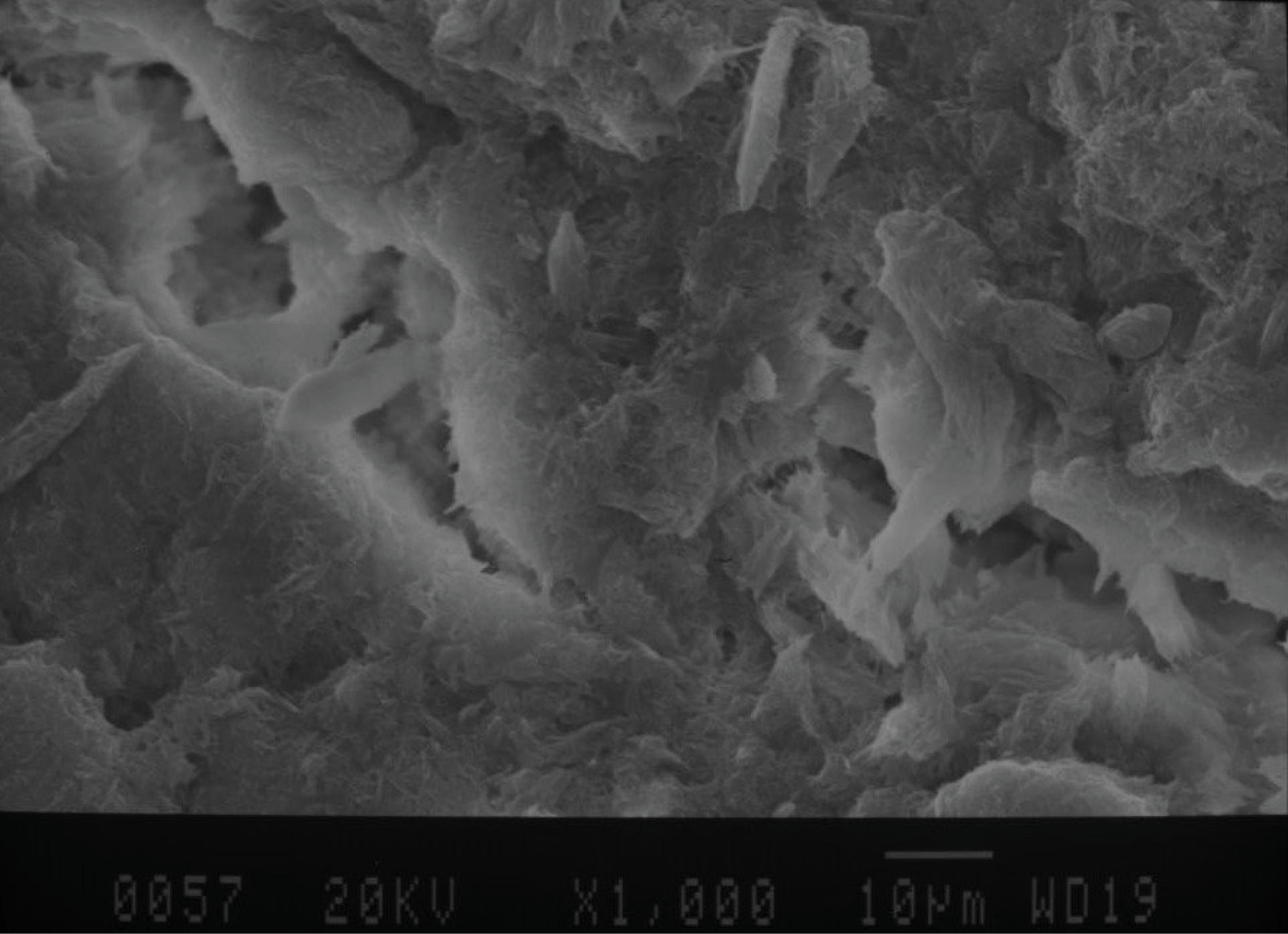
Figure 1.
SEM Micrograph of the Surface of Samples in the Control Group. Note. SEM: Scanning electron microscope.
.
SEM Micrograph of the Surface of Samples in the Control Group. Note. SEM: Scanning electron microscope.
In the laser plus BG group, the amount of porosities and cracks was lower compared to the control group, but the difference was not considerable (Figures 2 and 3). In the laser group, the enamel surface was more uniform in comparison with the control group and had significantly fewer cracks and porosities (Figure 4). The enamel surface was more uniform in the BG group and had fewer porosities compared to the control group (Figure 5).
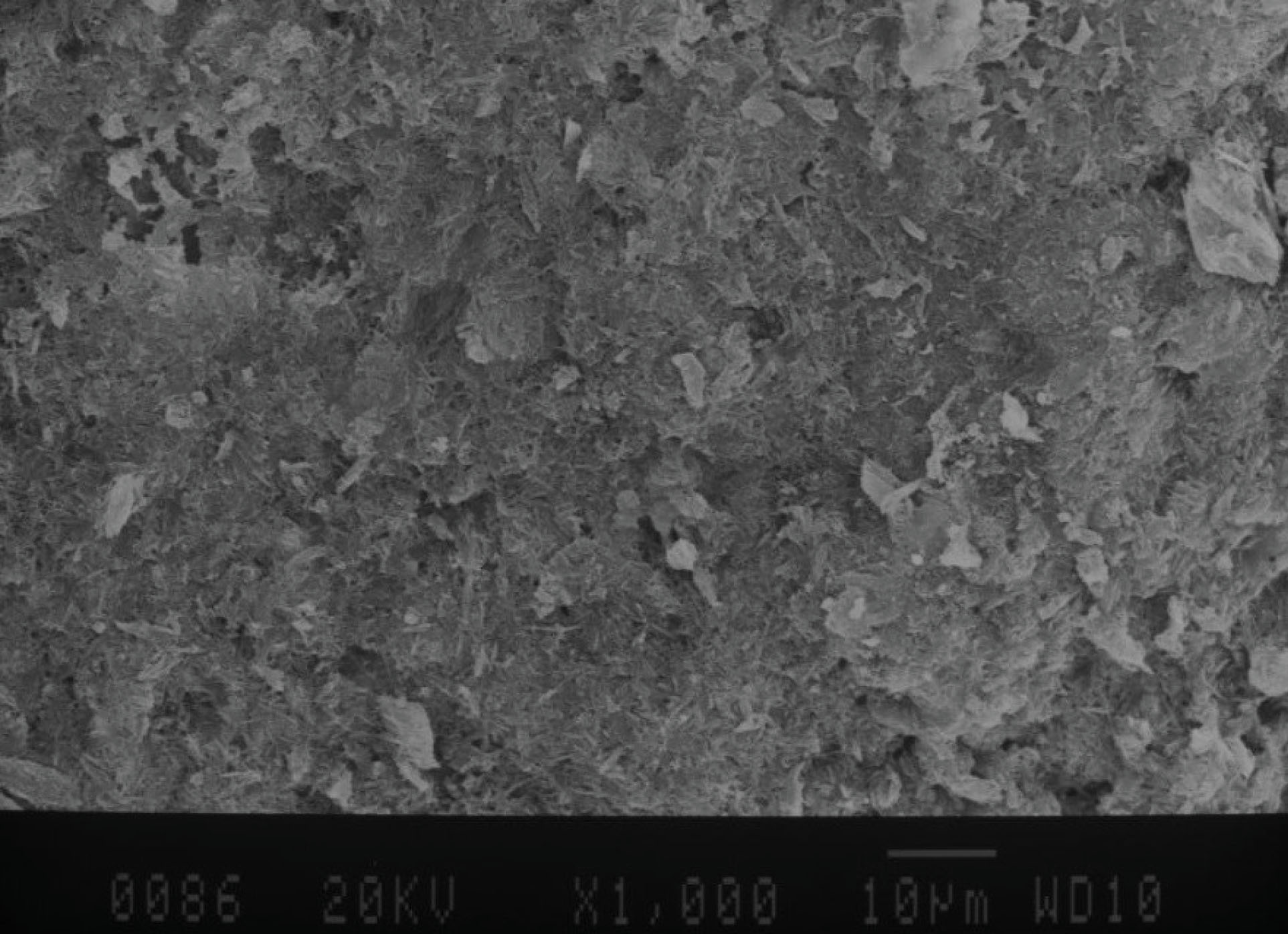
Figure 2.
SEM Micrograph of the Surface of Samples in Bioglass After the Laser Group
Note. SEM: Scanning electron microscope.
.
SEM Micrograph of the Surface of Samples in Bioglass After the Laser Group
Note. SEM: Scanning electron microscope.
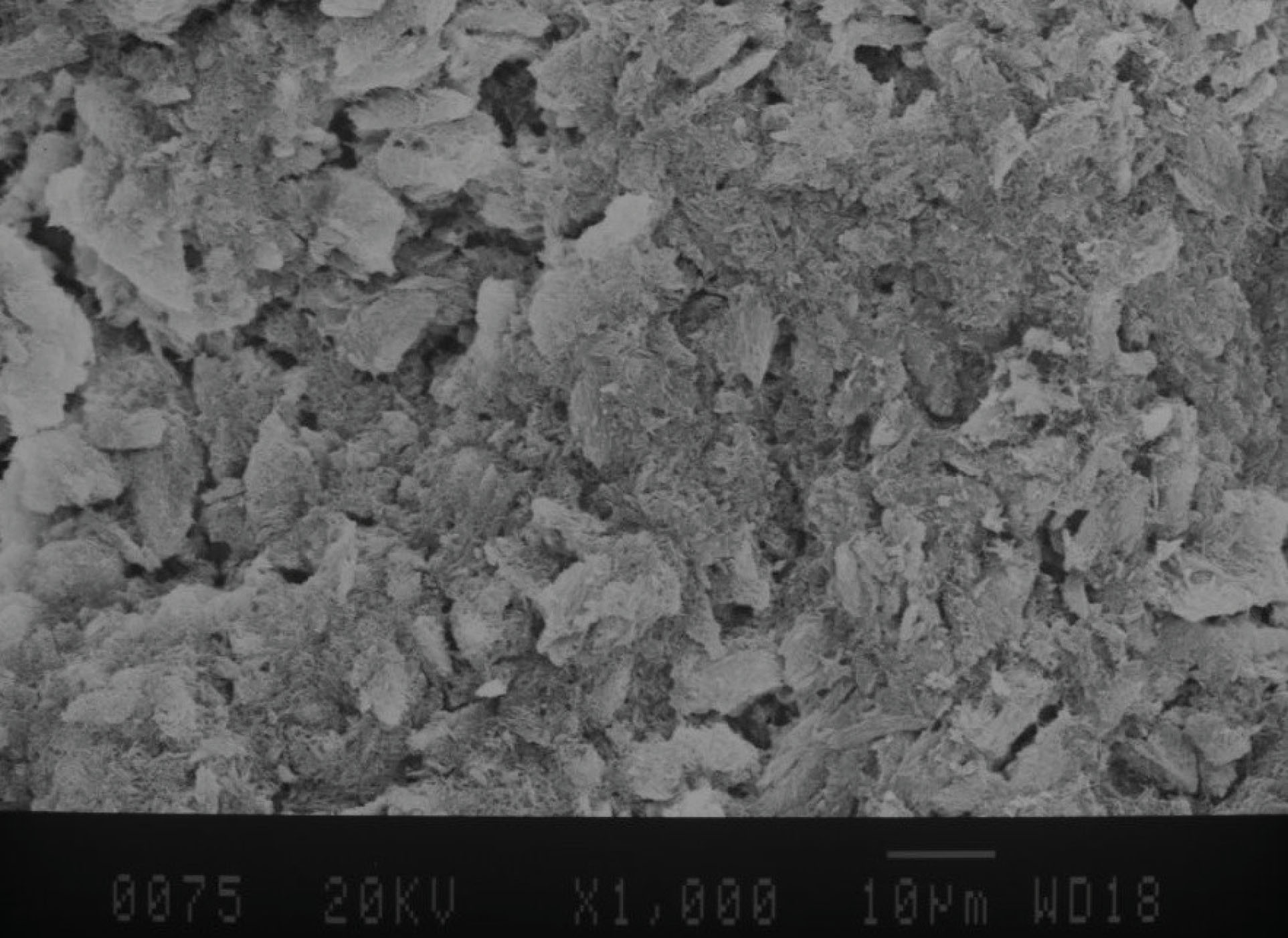
Figure 3.
SEM Micrograph of the Surface of Samples in Bioglass Before the Laser Group
Note. SEM: Scanning electron microscope.
.
SEM Micrograph of the Surface of Samples in Bioglass Before the Laser Group
Note. SEM: Scanning electron microscope.
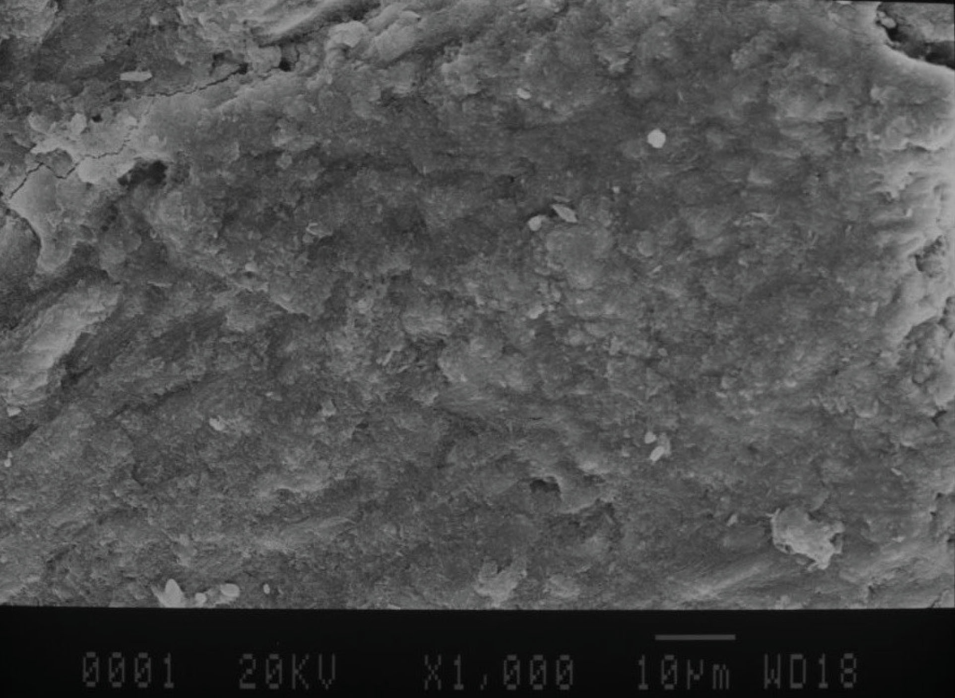
Figure 4.
SEM Micrograph of the Surface of Samples in the Laser Group. Note. SEM: Scanning electron microscope.
.
SEM Micrograph of the Surface of Samples in the Laser Group. Note. SEM: Scanning electron microscope.
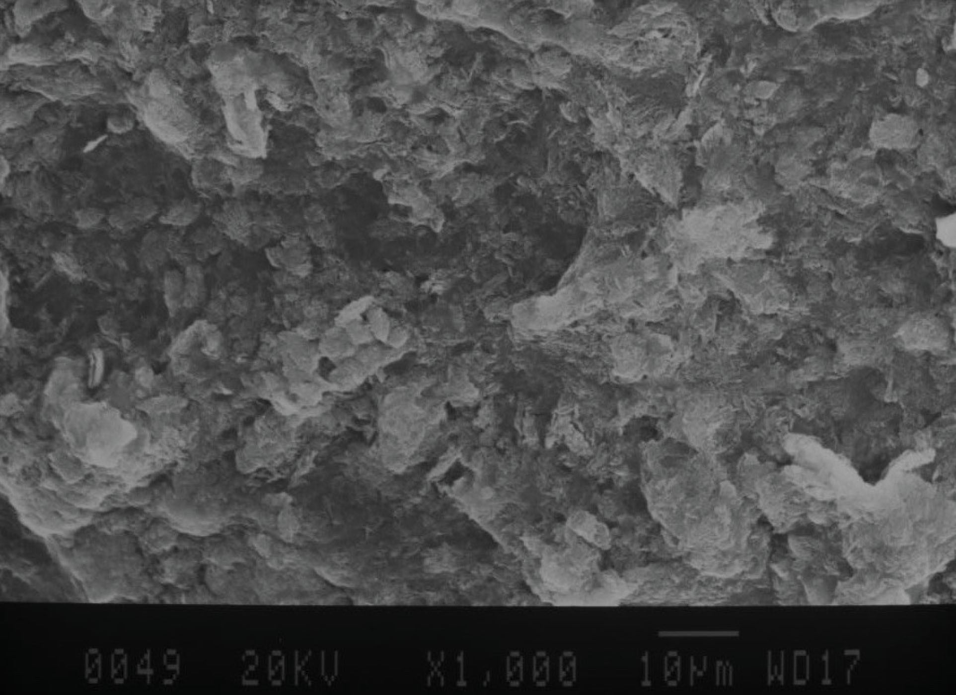
Figure 5.
SEM Micrograph of the Surface of Samples in the Bioglass Group. Note. SEM: Scanning electron microscope.
.
SEM Micrograph of the Surface of Samples in the Bioglass Group. Note. SEM: Scanning electron microscope.
Discussion
This study evaluated the remineralizing effect of 45S5 BG doped with strontium and Nd:YAG laser irradiation on the demineralized enamel surface by measuring the Vickers hardness number and SEM analysis. Sound extracted premolars were used in this study to better simulate the clinical setting (15). The demineralizing solution used in this study well-simulated acid attacks by bacteria in the oral cavity.
Several methods are used for the assessment of enamel microhardness, remineralization, and demineralization, including abrasion, scratch technique, indentation, and atomic force microscopy (16). Vickers microhardness test, which indirectly measures the changes in the strength of the enamel structure due to demineralization and remineralization, was employed in this study (16).
Bioactive glass is a remineralizing agent that can chemically bond to the tooth structure and is composed of calcium, sodium, phosphorous, and silica oxides in ratios suitable for biological activities. Under in vitro conditions, these glasses form a hydroxyapatite layer on their surface (17,18). This feature is associated with its chemical composition that intimates the mineral matter characteristics of the bone and dentin structure (19).
In the present study, 45S5 BG doped with strontium in the form of a suspension in distilled water was applied once on the surface of demineralized enamel for 1 minute. The results represented that the mean microhardness was the highest in the BG group with significant differences from other groups.
The findings of one study analyzing the spot lesion pattern revealed that diverse types of bioactive compounds used in this study can regenerate demineralized enamel primary lesions. Bioactive glasses are regarded as a milestone in tooth regeneration since their current standard treatment and demineralization lesion prevention are still considered slow procedures. Our results would be confirmed by the above-mentioned studies since all BAG-containing slurry treated groups in this study demonstrated a higher intensity average in laser spot photos that were similar to enamel photos (20).
Literature is rich regarding the effect of BG on enamel remineralization (7), and it seems that it can effectively enhance remineralization, especially after dental bleaching if used in adequate frequency and duration. However, further studies are required in this respect.
In one study, the strontium-incorporated 45s5 BG was applied, and the findings indicated that replacement of strontium with calcium increases glass dissolution due to increased network expansion caused by mineral network enlargement compared with calcium. Moreover, such replacement increases secretion and apatite formation in complete glass, leading to an increase in osteoblast attachment. The results of this study showed that the BG-containing group can restore the demineralized enamel surface. As shown by SEM photos, BG sediments are found in the tooth enamel surface, confirming that these sediments could be an ion source for remineralization in this case, even with an existing protective layer on the surface of the tooth enamel (21).
Several laser types such as argon, diode, Nd:YAG, and CO2 lasers are used to prevent enamel demineralization. Evidence regarding the efficacy of Nd:YAG laser in this regard is inconclusive, and further investigations are required in this regard. In our study, the mean microhardness of the laser group was significantly higher compared to the control group, but this value was still significantly lower than that of the BG group. The mean microhardness of the laser plus BG and BG plus laser groups was slightly (but not significantly) lower than that of other groups, except for the control group (they represented significantly higher microhardness in comparison to the control group). The microhardness of the BG plus laser group was lower than that of the BG and laser groups and higher than that of the control and laser plus BG groups. This difference was only significant with BG and control groups. The control group demonstrated minimum microhardness. Laser absorption possessing a thermal effect in the tooth structure affects the structural and chemical characteristics of the tooth hard tissue. Generated heat during laser radiation leads to evaporation, mineral elements oxidation, phosphate acid conversion to pyrophosphate, and a decrease in the carbonate content. These changes result in the formation of compounds with lower solubility and higher stability that may form a more resistant base against demineralization (22).
This study had an in vitro design, which limits the generalization of results to the clinical setting. Clinical trials are required to cast a clinical judgment in this respect.
Conclusions
Within the limitations of this in vitro study, the application of 45S5 BG doped with strontium was superior to Nd:YAG laser and combined use of BG and Nd:YAG laser for the remineralization of incipient enamel lesions and yielded the highest microhardness in our study. Thus, its application as a varnish may be considered for enhancing the remineralization of incipient enamel caries.
Authors’ Contribution
All authors have contributed to the concept and design of the study. EA and LR contributed to the data collectio. The statistical analysis and assessment of data were carried out by EA, BA and RN. BA,EA and LR drafted the manuscript. All the authors completely revised,read and approved the final manuscript.
Conflict of Interest Disclosures
The authors declare that they have no conflict of interests.
Ethical Statement
The study was approved by the Ethics Committee of Hamadan University of Medical Sciences (IR.UMSHA.REC.1396.560).
References
- Rugg-Gunn A. Dental caries: strategies to control this preventable disease. Acta Med Acad 2013; 42(2):117-30. doi: 10.5644/ama2006-124.80 [Crossref] [ Google Scholar]
- Perdigao J, Swift EJ Jr, Walter R. Fundamental concepts of enamel and dentin adhesion. In: Heymann HO, Swift EJ Jr, Ritter AV, eds. Sturdevant’s Art and Science of Operative Dentistry. Mosby; 2013. p. 114-40.
- Costa FS, Silveira ER, Pinto GS, Nascimento GG, Thomson WM, Demarco FF. Developmental defects of enamel and dental caries in the primary dentition: a systematic review and meta-analysis. J Dent 2017; 60:1-7. doi: 10.1016/j.jdent.2017.03.006 [Crossref] [ Google Scholar]
- Gorelick L, Geiger AM, Gwinnett AJ. Incidence of white spot formation after bonding and banding. Am J Orthod 1982; 81(2):93-8. doi: 10.1016/0002-9416(82)90032-x [Crossref] [ Google Scholar]
- Zimmer BW, Rottwinkel Y. Assessing patient-specific decalcification risk in fixed orthodontic treatment and its impact on prophylactic procedures. Am J Orthod Dentofacial Orthop 2004; 126(3):318-24. doi: 10.1016/j.ajodo.2003.09.031 [Crossref] [ Google Scholar]
- Artun J, Thylstrup A. A 3-year clinical and SEM study of surface changes of carious enamel lesions after inactivation. Am J Orthod Dentofacial Orthop 1989; 95(4):327-33. doi: 10.1016/0889-5406(89)90166-2 [Crossref] [ Google Scholar]
- Deng M, Wen HL, Dong XL, Li F, Xu X, Li H. Effects of 45S5 bioglass on surface properties of dental enamel subjected to 35% hydrogen peroxide. Int J Oral Sci 2013; 5(2):103-10. doi: 10.1038/ijos.2013.31 [Crossref] [ Google Scholar]
- Andersson OH, Kangasniemi I. Calcium phosphate formation at the surface of bioactive glass in vitro. J Biomed Mater Res 1991; 25(8):1019-30. doi: 10.1002/jbm.820250808 [Crossref] [ Google Scholar]
- Fredholm YC, Karpukhina N, Brauer DS, Jones JR, Law RV, Hill RG. Influence of strontium for calcium substitution in bioactive glasses on degradation, ion release and apatite formation. J R Soc Interface 2012; 9(70):880-9. doi: 10.1098/rsif.2011.0387 [Crossref] [ Google Scholar]
- Kuroda S, Fowler BO. Compositional, structural, and phase changes in in vitro laser-irradiated human tooth enamel. Calcif Tissue Int 1984; 36(4):361-9. doi: 10.1007/bf02405347 [Crossref] [ Google Scholar]
- Desu M. Sample Size Methodology. Elsevier; 2012.
- Kirk RE. Experimental Design. Wiley Online Library; 1982.
- Heravi F, Ahrari F, Mahdavi M, Basafa S. Comparative evaluation of the effect of Er:YAG laser and low level laser irradiation combined with CPP-ACPF cream on treatment of enamel caries. J Clin Exp Dent 2014; 6(2):e121-6. doi: 10.4317/jced.51309 [Crossref] [ Google Scholar]
- Hassanein OE, El-Brolossy TA. An investigation about the remineralization potential of bio-active glass on artificially carious enamel and dentin using Raman spectroscopy. Egypt J Sol 2006; 29(1):69-80. [ Google Scholar]
- de Souza-e-Silva CM, Parisotto TM, Steiner-Oliveira C, Kamiya RU, Rodrigues LK, Nobre-dos-Santos M. Carbon dioxide laser and bonding materials reduce enamel demineralization around orthodontic brackets. Lasers Med Sci 2013; 28(1):111-8. doi: 10.1007/s10103-012-1076-5 [Crossref] [ Google Scholar]
- Gutiérrez-Salazar M, Reyes-Gasga J. Microhardness and chemical composition of human tooth. Mater Res 2003; 6(3):367-73. doi: 10.1590/s1516-14392003000300011 [Crossref] [ Google Scholar]
- Gjorgievska E, Nicholson JW. Prevention of enamel demineralization after tooth bleaching by bioactive glass incorporated into toothpaste. Aust Dent J 2011; 56(2):193-200. doi: 10.1111/j.1834-7819.2011.01323.x [Crossref] [ Google Scholar]
- Hemagaran G, Neelakantan P. Remineralization of the tooth structure-the future of dentistry. Int J PharmTech Res 2014; 6(2):487-93. [ Google Scholar]
- Sawant K, Pawar AM. Bioactive glass in dentistry: a systematic review. Saudi J Oral Sci 2020; 7(1):3-10. doi: 10.4103/sjos.SJOralSci_56_19 [Crossref] [ Google Scholar]
- Angelini Sfalcin R, da Silva JVP, Oliva Pessoa V, Santos J, Garcia Olivan SR, Porta Santos Fernandes K. Remineralization of early enamel caries lesions induced by bioactive particles: an in vitro speckle analysis. Photodiagnosis Photodyn Ther 2019; 28:201-9. doi: 10.1016/j.pdpdt.2019.07.022 [Crossref] [ Google Scholar]
- Saffarpour M, Asgartooran B, Tahriri MR, Mohammadi Savadroudbari M, Khabazkhoob M. Effect of modified 45S5 bioglass on physical and chemical properties of bleached enamel. Braz J Oral Sci 2019; 18:e191424. doi: 10.20396/bjos.v18i0.8655314 [Crossref] [ Google Scholar]
- Moghaddas MJ, Moosavi H, Yaghoubirad S, Chiniforush N. The effect of the bioactive glass and the Er:YAG laser on the remineralization of the affected dentin: a comparative in vitro study. J Lasers Med Sci 2020; 11(2):160-6. doi: 10.34172/jlms.2020.27 [Crossref] [ Google Scholar]