Avicenna J Dent Res. 12(3):71-75.
doi: 10.34172/ajdr.2020.15
Original Article
Evaluating the Effect of Working in the Dental Radiology Center on Micronucleus Frequency in Buccal Mucosa
Fatemeh Shahsavari 1  , Mahsa Arianfar 2, Ladan Hafezi 3, *
Author information:
1Department of Oral Pathology, Faculty of Dentistry, Tehran Medical Branch, Islamic Azad University, Tehran, Iran.
2Dentist, Private Practice, Tehran, Iran.
3Department of Oral Radiology, Faculty of Dentistry, Tehran Medical Branch, Islamic Azad University, Tehran, Iran.
Abstract
Background: The present study aimed to investigate the effect of working in the dental radiology department on the frequency of micronucleus.
Methods: In this cross-sectional study, Papanicolaou staining was used to count the micronucleus in 63 individuals working in the dental radiology department. The presence of micronucleus cells and their mean frequency in each cell were investigated. Mann- Whitney U test, t test, and chi-square test were used to determine the effect of age, gender, and employment duration on micronucleus frequency per cell.
Results: The mean frequency of micronucleus per cell in the control and case groups was not statistically significant (P = 0.4). In addition, no statistically significant difference was observed in males and females regarding the mean frequency of micronucleus per cell (P = 0.6). Employment background and age had no significant impact on the percentage of micronucleus-containing cells and the mean frequency of micronucleus per cell.
Conclusions: Working in a dental radiology center had no impact on the percentage of micronucleus-containing cells and the mean frequency of micronucleus per cell.
Keywords: Micronucleus, Cytotoxicity, Buccal mucosal cells, Occupation, Dental radiology center
Copyright and License Information
© 2020 The Author(s); Published by Hamadan University of Medical Sciences.
This is an open-access article distributed under the terms of the Creative Commons Attribution License (
http://creativecommons.org/licenses/by/4.0), which permits unrestricted use, distribution, and reproduction in any medium provided the original work is properly cited.
Citation: Shahsavari F, Arianfar M, Hafezi L. Evaluating the effect of working in the dental radiology center on micronucleus frequency in Buccal Mucosa. Avicenna J Dent Res. 2020;12(3):71-75. doi: 10.34172/ajdr.2020.15.
Background
Highlights
In the recent decade, x-rays have been increasingly used to prepare radiographic images for diagnostic dentistry. The effects of ionizing radiation on the DNA structure and crosslink DNA proteins that cause cell death cannot be overlooked (1,2). Recently, medical exposure to such radiations is estimated to be increasing in advanced countries (3); and various methods have been used to examine the side effects of radiation on cell health and cancer screening and diagnosis (4).
Exposure to harmful substances results in the formation of small nuclear objects called micronucleus (MN) in the interphase during the cycle of nuclear division. It contains fragments of chromosomes or complete chromosomes. The MN measurement is one of the most sensitive markers of DNA damage so that MN tests have been used as one of the most reliable markers to assess occupational and environmental damages in several human epidemiological studies (5-9).
MN test is a mutagenic test to detect factors leading to the formation of small pieces of DNA membrane. This MN can be derived from eccentric components (chromosomal pieces lacking a single centromere) or chromosomes that are unable to migrate to other chromosomes during cell division anaphase. When the eccentric components lack a centromere, they do not move toward the nucleus of the female cells when the cell is being divided, and remain in the cytoplasm as MN (10).
MN test has already been used to evaluate the genotoxicity of chemicals and cigarettes (9). The MN test, which is being run in the interphase, is more appropriate than other cytogenetic markers examining sister chromatid exchange or chromosomal abnormalities since it is not limited to metaphase cells and allows the rapid screening of many cells (6).
The MN test uses some criteria developed by Schmidt. According Schmidt, MN is similar to cell nucleus, but smaller in size. They are circle or oval in shape with a clear margin and the same color as the cell nucleus; however, their dimensions are about 3.1 times as large as the cell nucleus (11).
In maxillofacial research, there have been several studies evaluating the effects and side-effects of radiography; however, limited studies have evaluated the dosimeter effects of employment in the radiology ward on the frequency of MN. Despite the increased frequency of MN after radiation, these studies reported no significant findings (12).
Considering the effect of x-rays on cellular changes and the radiology staff’s long-term exposure to such rays, as well as controversial findings regarding MN changes in radiologists (13,14), compared to the control group, the researchers of the present study attempted to evaluate the frequency of MN and its relevant factors in the buccal mucus among the staff of the Oral Radiology Department at Islamic Azad University, Tehran Medical Branch and Shahid Beheshti University, Iran in 2018.
Materials and Methods
This cross-sectional study was carried out using an analytic approach and target base sampling method. The subjects having a systemic illness, taking drugs, having a recent viral infection, and a history of radiation therapy and chemotherapy were removed from the study through face-to-face interviews. A total of 63 individuals were included and divided into two groups: 31 subjects in the case group (all the staff of dental radiology centers in the Faculty of Dentistry at Islamic Azad University and Shahid Beheshti University, with a minimum experience of working for one year in the radiology department) and 32 subjects in the control group (employees in Periodontology and Oral Diseases department). All participants submitted a written consent to participate in the study and were homogenous in terms of age and smoking. The required data was collected through interviewing the participants, completing the demographic forms, taking specimens from the participants’ buccal mucosa by using a wet tongue depressor, and observing the specimens by an optical microscope (Nikon-YS100, Japan with ×400). To present the MN through Papanicolaou staining (15), xylenol, absolute alcohol, alcohol 97%, 80%, and 70%, hematoxylin color, acid alcohol, lithium carbonate solution, colored Orange Og6 solution, EA50 color, Canad bondadhesive, and an optical microscope were used.
Prior to collecting buccal mucosa cells, the participants were asked to wash their mouths with water. The buccal mucosa cells were scratched by a damp spatula, and the cells were spread over small and clean glass slides. The smears prepared on the slides were then fixed using a pathofix spray. Next, the slides were allowed to be dried at room temperature. Papanicolaou staining was used to measure MN. There are other methods to stain cells and nuclei, and the most specific DNA staining methods are Feulgen and Acridine orange. Some studies have also used DAPI and propidium iodide. About 30% of studies have used non-specific colors such as Giemsa and Hoechst (16-20).
Papanicolaou staining included the following phases: 1) Fixation, 2) Nucleus staining, 3) Cytoplasmic staining, and 4) Dehydration.
Cell examination was performed by optical microscope with a magnification of ×400 to count the MN as described below (21):
-
It is a smooth and circular space that represents a membrane.
-
It is smaller than one third of the nucleus diameter, but large enough to characterize the shape and color.
-
It has staining intensity similar to that of the nucleus.
-
It has consolidation and tissues like the nucleus.
-
It had a focal length similar to the nucleus.
-
It has no overlapping or connection with the nucleus.
For MN counting, cells that were characterized by specified margins and nuclei were used, and the MN count was not performed in areas where the cells overlapped. Dead or degenerated cells and nuclear bubbles were also excluded from counting (22).
Accordingly, 500 cells per sample were counted and the presence or absence of MN cells in the specimens was determined. The results were reported in percentage (15). It should be noted that the samples were counted by two oral and maxillofacial pathologists, and the mean counts were used in case of inconsistency.
Finally, to evaluate the effect of the variables such as age, gender, smoking, and the duration of employment in the radiology department on the frequency of MN-containing cells, the frequency of MNs in each group was calculated using t test, Mann-Whitney U, and chi-square tests.
Results
Table 1 shows the distribution of the participants in terms of their specifications and occupation in the radiology center. The results of the t test and chi-square tests revealed that the two groups were homogenous in terms of age (P = 0.4) and smoking (P = 0.9).
Table 1.
Distribution of Participants by Their Specifications
|
Employment in Radiology Ward
|
Specifications
|
|
Age
|
Smoking
|
|
Yes
|
No
|
Employed
(n=32) |
34.46±9.22 |
3 |
29 |
Non-employed
(n=31) |
38.03±11.46 |
2 |
29 |
| Test result |
P = 0.4 |
P = 0.88 |
The distribution of the case and control groups by the frequency of MN-containing cells and the mean frequency of MN per cell are presented in Table 2.
Table 2.
Distribution of Participants by Percentage of MN-Containing Cells and Mean Frequency of MN Per Cell
Employment in
Radiology Ward
|
MN-Containing Cells
|
|
Frequency of MN-Containing Cells (%) |
Mean Frequency of
MN
|
| Control group |
17.81±10.36 |
2.07±0.53 |
| Case group |
18.83 ±12.88 |
2.25±0.73 |
| Mann-Whitney U test |
P = 0.58 |
P = 0.37 |
The percentage of MN-containing cells was 17.5% in the control group and 18.83% in the case group (Figure 1), and the statistical test showed no statistically significant difference (P = 0.6).
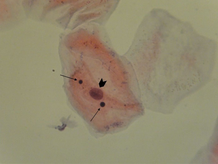
Figure 1.
Photomicrograph Showing Exfoliated Buccal Epithelial Cell in Case Group. Nucleus (arrowhead), micronucleus (arrows) ( Papanicolaou staining x400).
.
Photomicrograph Showing Exfoliated Buccal Epithelial Cell in Case Group. Nucleus (arrowhead), micronucleus (arrows) ( Papanicolaou staining x400).
The mean frequency of MN per cell was 2.7 in the control group and 2.25 in the case group, and the statistical tests showed that the difference was not statistically significant (P = 0.4).
Table 3 shows the distribution of the participants in the case group in terms of the percentage of MN-containing cells and the mean frequency of MN per cell according to the variables age, gender, and duration of employment in the radiology ward. According to this table, it can be inferred that:
-
The case group consisted of 4 males and 27 females. The percentage of MN-containing cells in females was 2.5 times larger than that of males. According to the test results, the difference was not statistically significant (P = 0.6); however, there was no difference between females and males (P = 0.6) regarding the mean frequency of MN per cell.
-
The mean duration of employment in the radiology department was 9 years, and this variable had no correlation with the frequency of MN cells and the mean frequency of MN per cell (P = 0.4).
-
The mean age in the case group was 38 years, and the difference in the percentage of MN-containing cells and the mean frequency of MN per cell for the age groups below the mean and those above the mean was not statistically significant (P = 0.9).
Table 3.
Distribution of the Case Group by the Percentage of MN-Containing Cells and the Mean Frequency of Micronucleus Per Cell Considering Age, Gender, and Duration of Employment
|
Relevant Variables
|
|
Indicators
|
|
Percentage of MN-Containing Cells
|
Mean ofMN
|
| Gender |
Male |
8.25±6.5 |
0.86±2.06 |
| Female |
20.4±12.19 |
2.28±0.72 |
| Mann-Whitney U test |
P = 0.8 |
P = 0.6 |
| Age |
< Mean |
16.47±9.32 |
2.12±0.66 |
| > Mean |
21.71±16.11 |
2.41±0/8 |
| Mann-Whitney U test |
P = 0.9 |
P = 0.4 |
| Duration of employment |
< Mean |
17.31±8.93 |
2.05±0.65 |
| > Mean |
20.46±16.26 |
2.47±0.77 |
| Mann-Whitney U test |
P = 0.4 |
P = 0.9 |
Discussion
The results of this study suggested that working in the radiology department did not directly influence the frequency of MN-containing cells. Our results are similar to the findings of Vral et al (23) and Joseph et al (24), and in contrast with the results of other studies (12,21,25,26), as the latter group reported an increase in MN-containing cells for those individuals employed in the radiology department. The inconsistency might be due to their larger sample size.
Almost all faculty members and colleagues cooperated in this study; however, the statistical population of the study only included the staff of dental radiology departments in the Faculty of Dentistry at Islamic Azad University and Shahid Beheshti University of Medical Sciences. As far as the researchers investigated, this study is the only research that has investigated this group of individuals so far. In all other studies, the general radiology group was concerned and the specimens were peripheral blood and lymphocyte cells. Giemsa was used for staining in all other studies (16), while we used specific Papanicolaou staining for buccal mucosa cells (Buccal mucus sampling).
The frequency and mean frequency of MN per cell and the standard deviation in the present study were larger than similar studies conducted in other countries, which may be due to differences in quality of life in different countries and other variables including environmental and pollution factors. In the present study, the researchers attempted to eliminate intervening factors affecting the development of MN and nuclear cell changes (e.g., cigarettes, alcohol, chemotherapy, etc); however, the effect of environmental variables, such as particles suspended in air and ionizing waves in the environment and other geographically-induced environmental exposures cannot be ignored. Interestingly, two other similar studies in Iran by Zakeri & Hirobe and Farhadi et al showed a higher incidence of MN compared to non-Iranian studies, indicating the presence of other harmful factors in Iran (26,27).
The results of our study also suggest that gender affects the frequency of MN-containing cells and their frequency is higher in females. This finding is in line with some other studies (12,21,23,25) and contrary to the results of studies by Joseph et al and Zakeri & Hirobe (24,26). This difference might be due to the larger frequency of females in the oral radiology departments, which requires further studies with larger sample sizes. Since some other researchers have reported increased number of MN in females, other underlying factors in females should be assessed. For example, females may work longer or they might be exposed to other harmful factors which have not been considered in this study (e.g., the use of detergents and disinfectants that are commonly used by women in Iran).
According to the findings of this study, age did not directly affect the frequency of MN-containing cells. This is consistent with the findings of Zakeri & Hirobe (26) and contrary to the findings of some other studies (12,21,24,25).
Furthermore, the findings of our study illustrated that employment background in the radiology ward had no direct impact on the frequency of MN-containing cells. This finding is consistent with some studies (21,25,26).
We found that the mean frequency of MN and the frequency of MN-containing cells was 2.25 in the case group and 2.7 in the control group; however, this difference was not statistically significant (P <0.4). In other words, the frequency of MNs per cell did not change significantly. Most studies on MN in radiologists have merely examined the frequency of this nuclear change and the mean frequency of MN per cell in MN-containing cells has not been evaluated in previous studies (23,25). This is considered as the novelty of this study. Hence, our findings could provide the grounds for future research in this filed.
Finally, in this study, no significant relationship was observed between age, gender, and employment background in the radiology department with the mean frequency of MN.
Conclusions
Occupation in oral radiology department had no impact on the percentage of MN-containing cells and the mean frequency of MN per cell.
Conflict of Interest Disclosures
The authors declare that they have no conflict of interests.
Ethical Statement
The ethics committee of dental faculty at Islamic Azad University, Tehran Medical Branch approved the protocol of the study (IR.IAU.DENTAL.RCE.1397.009).
References
- Ribeiro DA, Angelieri F. Cytogenetic biomonitoring of oral mucosa cells from adults exposed to dental X-rays. Radiat Med 2008; 26(6):325-30. doi: 10.1007/s11604-008-0232-0 [Crossref] [ Google Scholar]
- Nakano T, Xu X, Salem AMH, Shoulkamy MI, Ide H. Radiation-induced DNA-protein cross-links: mechanisms and biological significance. Free Radic Biol Med 2017; 107:136-45. doi: 10.1016/j.freeradbiomed.2016.11.041 [Crossref] [ Google Scholar]
- White SC, Pharoah MJ. Oral Radiology: Principles and Interpretation. 7th ed. St. Louis: Elsevier; 2017.
- Winawer S, Fletcher R, Rex D, Bond J, Burt R, Ferrucci J. Colorectal cancer screening and surveillance: clinical guidelines and rationale-update based on new evidence. Gastroenterology 2003; 124(2):544-60. doi: 10.1053/gast.2003.50044 [Crossref] [ Google Scholar]
- Claudio SR, Simas JMM, Souza ACF, M DOCBDA, Yamauchi LY, Ribeiro DA. Genomic instability and cytotoxicity in buccal mucosal cells of workers in banana farming evaluated by micronucleus test. Anticancer Res 2019; 39(3):1283-6. doi: 10.21873/anticanres.13239 [Crossref] [ Google Scholar]
- Ishikawa H, Tian Y, Yamauchi T. Induction of micronuclei formation in preimplantation mouse embryos after maternal treatment with 2-bromopropane. Reprod Toxicol 2001; 15(1):81-5. doi: 10.1016/s0890-6238(00)00112-x [Crossref] [ Google Scholar]
- Alexandrescu I, Havârneanu D, Popa D. New approaches in biomonitoring human populations exposed to genotoxic agents: epithelial cell micronucleus assay. J Prev Med 2006; 14(3-4):57-65. [ Google Scholar]
- Ribeiro D. Evidence of genotoxicity and cytotoxicity of X-rays in the oral mucosa epithelium of adults subjected to cone beam computed tomography. Dentomaxillofac Radiol 2019; 48(6):20180299. doi: 10.1259/dmfr.20180299 [Crossref] [ Google Scholar]
- Abbasi F, Farhadi S, Esmaili M. Efficacy of pilocarpine and bromhexine in improving radiotherapy-induced xerostomia. J Dent Res Dent Clin Dent Prospects 2013; 7(2):86-90. doi: 10.5681/joddd.2013.015 [Crossref] [ Google Scholar]
- Fenech M. The in vitro micronucleus technique. Mutat Res 2000; 455(1-2):81-95. doi: 10.1016/s0027-5107(00)00065-8 [Crossref] [ Google Scholar]
- Kamboj M, Mahajan S. Micronucleus--an upcoming marker of genotoxic damage. Clin Oral Investig 2007; 11(2):121-6. doi: 10.1007/s00784-006-0075-y [Crossref] [ Google Scholar]
- Maffei F, Angelini S, Forti GC, Lodi V, Violante FS, Mattioli S. Micronuclei frequencies in hospital workers occupationally exposed to low levels of ionizing radiation: influence of smoking status and other factors. Mutagenesis 2002; 17(5):405-9. doi: 10.1093/mutage/17.5.405 [Crossref] [ Google Scholar]
- Thomas P, Fenech M. Buccal micronucleus cytome assay. Methods Mol Biol 2011; 682:235-48. doi: 10.1007/978-1-60327-409-8_17 [Crossref] [ Google Scholar]
- Ribeiro DA, de Oliveira G, de Castro G, Angelieri F. Cytogenetic biomonitoring in patients exposed to dental X-rays: comparison between adults and children. Dentomaxillofac Radiol 2008; 37(7):404-7. doi: 10.1259/dmfr/58548698 [Crossref] [ Google Scholar]
- Farhadi S, Mohamadi M, Mohamadi M. Repair index in examination of nuclear changes in the buccal mucosa of smokers: a useful method for screening of oral cancer. Asian Pac J Cancer Prev 2017; 18(11):3087-90. doi: 10.22034/apjcp.2017.18.11.3087 [Crossref] [ Google Scholar]
- Nersesyan A, Kundi M, Atefie K, Schulte-Hermann R, Knasmüller S. Effect of staining procedures on the results of micronucleus assays with exfoliated oral mucosa cells. Cancer Epidemiol Biomarkers Prev 2006; 15(10):1835-40. doi: 10.1158/1055-9965.epi-06-0248 [Crossref] [ Google Scholar]
- Milić M, Gerić M, Nodilo M, Ranogajec-Komor M, Milković Đ, Gajski G. Application of the buccal micronucleus cytome assay on child population exposed to sinus X-ray. Eur J Radiol 2020; 129:109143. doi: 10.1016/j.ejrad.2020.109143 [Crossref] [ Google Scholar]
- Darzynkiewicz Z, Huang X. Analysis of cellular DNA content by flow cytometry. Curr Protoc Immunol 2004;Chapter 5:Unit 5.7. 10.1002/0471142735.im0507s60.
- Lecoeur H. Nuclear apoptosis detection by flow cytometry: influence of endogenous endonucleases. Exp Cell Res 2002; 277(1):1-14. doi: 10.1006/excr.2002.5537 [Crossref] [ Google Scholar]
- Takkouche B, Regueira-Méndez C, Montes-Martínez A. Risk of cancer among hairdressers and related workers: a meta-analysis. Int J Epidemiol 2009; 38(6):1512-31. doi: 10.1093/ije/dyp283 [Crossref] [ Google Scholar]
- Ropolo M, Balia C, Roggieri P, Lodi V, Nucci MC, Violante FS. The micronucleus assay as a biological dosimeter in hospital workers exposed to low doses of ionizing radiation. Mutat Res 2012; 747(1):7-13. doi: 10.1016/j.mrgentox.2012.02.014 [Crossref] [ Google Scholar]
- Chauhan V, Wilkins RC. A comprehensive review of the literature on the biological effects from dental X-ray exposures. Int J Radiat Biol 2019; 95(2):107-19. doi: 10.1080/09553002.2019.1547436 [Crossref] [ Google Scholar]
- Vral A, Decorte V, Depuydt J, Wambersie A, Thierens H. A semi-automated FISH-based micronucleus-centromere assay for biomonitoring of hospital workers exposed to low doses of ionizing radiation. Mol Med Rep 2016; 14(1):103-10. doi: 10.3892/mmr.2016.5265 [Crossref] [ Google Scholar]
- Joseph LJ, Patwardhan UN, Samuel AM. Frequency of micronuclei in peripheral blood lymphocytes from subjects occupationally exposed to low levels of ionizing radiation. Mutat Res 2004; 564(1):83-8. doi: 10.1016/j.mrgentox.2004.07.012 [Crossref] [ Google Scholar]
- Sari-Minodier I, Orsière T, Auquier P, Martin F, Botta A. Cytogenetic monitoring by use of the micronucleus assay among hospital workers exposed to low doses of ionizing radiation. Mutat Res 2007; 629(2):111-21. doi: 10.1016/j.mrgentox.2007.01.009 [Crossref] [ Google Scholar]
- Zakeri F, Hirobe T. A cytogenetic approach to the effects of low levels of ionizing radiations on occupationally exposed individuals. Eur J Radiol 2010; 73(1):191-5. doi: 10.1016/j.ejrad.2008.10.015 [Crossref] [ Google Scholar]
- Farhadi S, Jolehar M, Safapour F. Micronucleus assay of buccal mucosal cells in hairdressers: the importance of occupational exposure. Asian Pac J Cancer Prev 2018; 19(8):2131-4. doi: 10.22034/apjcp.2018.19.8.2131 [Crossref] [ Google Scholar]