Avicenna J Dent Res. 12(1):19-24.
doi: 10.34172/ajdr.2020.05
Original Article
Age Estimation Based on the Pulp Chamber Volume of Multi-rooted Teeth Using Cone Beam Computed Tomography
Faezeh Yousefi 1  , Sima Lari 2, Abbas Shokri 3, Soroush Hashemi 4, *, Mehdi Hosseini 5
Author information:
1Assistant Professor, Department of Oral and Maxillofacial Radiology, Dental School, Hamadan University of Medical Science, Hamadan, Iran.
2General Dentist, Department of Oral and Maxillofacial Radiology, Dental School, Hamadan University of Medical Science, Hamadan, Iran.
3Associate Professor, Dental Implant Research Center, Department of Oral and Maxillofacial Radiology, Dental School, Hamadan University of Medical Science, Hamadan, Iran.
4General Dentist, Private Clinic, Tehran, Iran. 5 Department of Biostatistics, Hamadan University of Medical Science, Hamadan, Iran.
5Department of Biostatistics, Hamadan University of Medical Science, Hamadan, Iran.
Abstract
Background: Age estimation is a critical issue in forensic medicine for identifying corpses and to determining fake identities. The present study aimed to estimate the age based on the pulp chamber volume of multi-rooted teeth using cone-beam computed tomography (CBCT) images.
Methods: CBCT of 142 patients, consisting of 77 males and 65 females with an age range of 10-70 years, were selected. The images of 84 maxillary first molars and 79 mandibular first molars were evaluated. All the CBCT images were taken using the CRANEX 3D system and saved in OnDemand software. The images were converted into the DICOM format and saved in a semi-automatic segmentation software (ITK-SMAP version 3.6.0-beta). Based on the results of logarithmic regression analysis, age, as a dependent variable, was correlated with the pulp chamber volume, as a predicting factor, which can be used in preparing a statistical model for estimating the human age.
Results: The correlation coefficient between age and all the morphological variables was negative, indicating a decrease in the mean of all these variables with age. The results of ANOVA showed a significant difference in the means of all these variables between the different age groups. In addition, the means of all these variables decreased with age. There was a relatively high correlation between age and the pulp chamber volume of the first molars (R2 =0.513-0.543, depending on the tooth type and gender).
Conclusions: There was a linear correlation between the volume of maxillary and mandibular pulp chambers and the chronological age of the population studied. The regression models achieved in the present study could be used to predict the subjects’ age with 0.54% and 0.51% accuracy based on the maxilla and mandible, respectively. The mean pulpal volume of the maxilla was a little larger than that of the mandible. Furthermore, the mean volume of the pulp chamber decreased with age.
Keywords: Age determination by teeth, Cone beam computed tomography, First molar, Pulp chamber
Copyright and License Information
© 2020 The Author(s); Published by Hamadan University of Medical Sciences.
This is an open-access article distributed under the terms of the Creative Commons Attribution License (
http://creativecommons.org/licenses/by/4.0), which permits unrestricted use, distribution, and reproduction in any medium provided the original work is properly cited.
Citation: Yousefi F, Lari S, Abbas Shokri, Hashemi S, Hosseini M. Age Estimation Based on the Pulp Chamber Volume of Multi-rooted Teeth Using Cone Beam Computed Tomography. Avicenna J Dent Res. 2020;12(1):19-24. doi: 10.34172/ ajdr.2020.05.
Background
Highlights
Age estimation is a critical issue in forensic medicine for identifying corpses and to determining fake identities (1). Different body parts can be used to estimate age; however, since these body parts undergo injuries and changes under different conditions, they cananot always be used as a fixed pattern (2). Since teeth are greatly resistant to mechanical and chemical agents, physical contact, the passage of time (1,3), they remain almost intact for a long time after death (1,2) and undergo minor age-related changes and changes due to nutrition, the environment, and the individual’s living conditions (4,5). Therefore, a large number of age estimation techniques have been developed based on teeth.
The dentin–pulp complex is one of the tooth structures that reflects age-related physiological and pathological changes (2). The synthesis of secondary dentin is an age-related process and begins in the final stages of tooth calcification (6,7), resulting in the narrowing of the pulpal canals of all the teeth. Measuring these morphologic changes involves destructive processes, such as tooth extraction and tooth sectioning, which are not feasible in living individuals. Therefore, conservative techniques are used, which mainly depend on the radiographic examination of teeth (8-15). It is possible to use three-dimensional (3D) images to calculate pulp-to-tooth volume (16). Cone-beam computed tomography (CBCT) images are used to estimate the age for two reasons as follows:
-
The analysis of the pulp and tooth volumes is more reliable than the surface characteristics because the secondary dentin might not form uniformly on all the pulp surfaces; therefore, measurements made on the surfaces might yield an incorrect image of this process.
-
CBCT is an accurate technique for studying the pulp chamber and the root canal system, and it produces 3D data of teeth in living human beings by a single scanning procedure. This technique can be used in a non-destructive manner (17).
All the teeth can be used for the estimation of age. However, since the majority of previous studies have evaluated single-rooted teeth to estimate age and the human dentition consists of multi-rooted teeth too, the present study was designed to estimate age based on the pulp chamber volume of multi-rooted teeth using the CBCT technique because only limited studies are available on age estimation using the CBCT technique. In addition, no studies have been undertaken in our country on this subject based on multi-rooted teeth with a large sample size.
Materials and Methods
In the present cross-sectional study, 142 CBCT images from 77 males and 65 females, with an age range of 10-70 years, were evaluated. The images consisted of 84 maxillary first molars and 79 mandibular first molars.
The inclusion criteria were as follows:
-
Teeth without extensive caries because it might induce pulp chamber regression, resulting in a decrease in its volume.
-
Teeth without extensive erosion because it affects the pulp chamber volume.
-
Teeth without extensive restorations; the secondary effects of restorative materials result in irreversible structural and biochemical changes in the tooth pulp in some cases.
-
Absence of artifacts related to metallic restorations on the tooth in question because artifacts change the resolution of the images, making it difficult to evaluate the samples
-
Absence of pulpal calcification because it also affects the tooth pulp volume.
Teeth that were not fully erupted, and teeth with open apices were excluded from the study due to the absence of pulpal stability.
In each subject, all the first molar teeth were analyzed. The subjects were assigned to the following age groups: 10–19, 20–29, 30–39, 40–49, 50–59, and 60–70 years.
The sample size was estimated to be 150 based on similar studies (17,18). All the CBCT images were taken using the CRANEX 3D system (Soredex, Tuusula, Finland) at kVp=90, mA=8, and exposure time of 6.12 and saved in OnDemand software. All the images were converted into the DICOM format and saved in the semi-automatic segmentation software (ITK-SNAP 3.60-beta), which is a useful tool to manually or automatically separate the anatomic structures on medical 3D images and to accurately determine the volume of certain structures. The software is free and available at www.itksnap.org. The paintbrush mode of the tool was used to half-automatically segment the images.
To prevent the effect of the root pulps of the first molars and facilitate the procedural steps, the pulp chamber floor was placed on the plane of the section, and the root pulp was eliminated so that only the volume of the pulp chamber was calculated (Figure 1). Finally, the volume of the colored segment was calculated through the segmentation>volumes and statistics pathway (Figure 2).
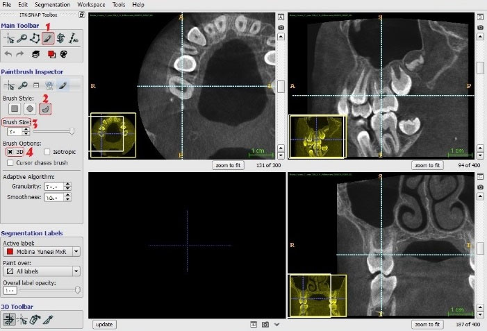
Figure 1.
. (C) The Final 3D Image.
.
. (C) The Final 3D Image.
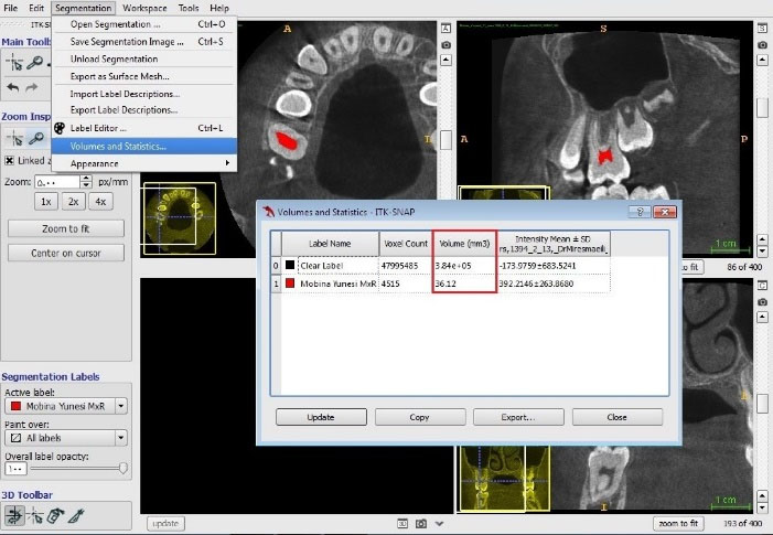
Figure 2.
. Calculation of the Volume of Cross Sections Using the Software.
.
. Calculation of the Volume of Cross Sections Using the Software.
Statistical Analysis
In the logarithmic regression analysis, age, as a dependent variable, and the volume of the pulp chamber, as a predictor variable, were correlated so that a statistical model was constructed for estimating the age of the human subjects.
To create a proper statistical model for estimating age irrespective of gender, in the regression analysis too, age and the pulp chamber volume were correlated, as dependent and predictor variables, respectively, in the maxillary and mandibular first molars.
Descriptive statistics (means, standard deviations, and graphs) and inferential statistics (paired t test, independent t test, one-way ANOVA, Pearson correlation coefficient, and linear regression analysis) were used for the analysis of data. Statistical significance was defined at P<0.05 in all the analyses (α=0.05).
All the measurements were repeated by the same operator. To evaluate intra-examiner agreement, 20 maxillary first molars and 20 mandibular first molars were selected randomly and re-evaluated after three weeks. At the same time, the same samples were re-evaluated by the second operator to evaluate the inter-examiner agreement.
The results of intraclass correlation coefficient (ICC) for the evaluation of intra- and inter-observer agreement showed that the two observers had a high rate of agreement. In addition, both observers showed a high rate of reliability (>88%).
The pulp chamber volume was calculated after the segmentation process explained above. Then, the numeric values of the pulp chamber volume of each case were converted into statistical models to estimate age for each sample, followed by determination of the accuracy of the models created.
Results
In the present study, 142 samples were evaluated to estimate age based on the volume of the tooth pulp chamber. The samples were taken from 77 (54.2%) females and 65 (45.7%) males. Table 1 presents the age distribution of the subjects.
Table 1.
The age distribution of the subjects
|
Age
|
No.
|
Percent
|
| 20> |
131 |
80.3 |
| 20–30 |
13 |
8.0 |
| 31–30 |
9 |
5.5 |
| 41–50 |
6 |
3.7 |
| 50< |
4 |
2.5 |
| Total |
163 |
100.0 |
Table 2 presents the distribution of the first molars, mean volume, and the maximum and minimum volumes of the mandible and maxilla.
Table 2.
The Distribution of the First Molars, Mean Volumes, and Maximum and Minimum Volumes of the Mandible and Maxilla
|
|
Age
|
Maxilla
|
Mandible
|
| N |
163 |
84 |
79 |
| Missing |
0 |
0 |
0 |
| Mean |
20.3103 |
36.8632 |
33.1524 |
| SD |
12.46101 |
12.37932 |
11.07682 |
| Min |
10.00 |
6.45 |
13.20 |
| Max |
68.00 |
79.78 |
70.02 |
According to Table 2, the mean volume of the pulp chamber of the maxillary first molars (36.86) was slightly larger than that of the mandibular first molars (33.15).
Table 3 presents the age distribution, mean volumes, and the maximum and minimum volumes of the mandible and maxilla.
Table 3.
The Distribution of Age, Mean Volumes, and Maximum and Minimum Volumes of the Mandible and Maxilla
|
|
|
N
|
Mean
|
SD
|
Min
|
Max
|
| Maxilla |
20> |
70 |
40.2179 |
10.42335 |
24.11 |
79.78 |
| 20–30 |
2 |
17.6400 |
2.85671 |
15.62 |
19.66 |
| 31–40 |
5 |
19.5900 |
3.78209 |
15.30 |
24.88 |
| 41–50 |
6 |
23.5967 |
5.62085 |
16.92 |
31.24 |
| 50< |
1 |
6.4500 |
. |
6.45 |
6.45 |
| Total |
84 |
36.8632 |
12.37932 |
6.45 |
79.78 |
| Mandible |
20> |
65 |
36.2083 |
9.64769 |
18.57 |
70.02 |
| 20–30 |
7 |
20.6414 |
3.39459 |
16.44 |
25.42 |
| 31–40 |
4 |
16.4075 |
4.09205 |
13.20 |
22.04 |
| 41–50 |
3 |
18.4600 |
3.32271 |
14.64 |
20.68 |
| 50< |
0 |
. |
. |
. |
. |
| Total |
79 |
33.1524 |
11.07682 |
13.20 |
70.02 |
ANOVA was used to compare the means of the study variables in different age groups (Table 4). The results of ANOVA showed significant differences in the mean of all these variables between the different age groups. In addition, the mean of all the different variables decreased with aging.
Table 4.
Comparison of the Means of the Variables in the Maxilla and Mandible between the Different Age Groups (ANOVA)
|
|
|
Sum of squares
|
df
|
Mean Square
|
F
|
P
Value
|
| Maxilla |
Between groups |
4999.609 |
4 |
1249.902 |
12.791 |
0.000 |
| Within groups |
7719.936 |
79 |
97.721 |
|
|
| Total |
12719.545 |
83 |
|
|
|
| Mandible |
Between groups |
3471.845 |
3 |
1157.282 |
14.233 |
0.000 |
| Within groups |
6098.444 |
75 |
81.313 |
|
|
| Total |
9570.289 |
78 |
|
|
|
According to Table 4, the age, as an effective factor, resulted in different volumes of the pulp chamber in the maxillary and mandibular first molars at different ages.
The results of the regression model analysis indicated a relationship between age and different variables evaluated.
In further analyses, the age was considered as a linear function of the variables and modeled in the form of a regression model. In the first stage, all the variables were included in the model for the fitting of the regression model; in the next stage, the forward selection method was used so that only the variables that showed a significant relationship with the response in question (the age) remained in the regression model.
Tables 5 and 6 present the results of the regression model for all the data in the presence of all the variables. Table 5 shows the reliability of the volume of the pulp chamber in the maxillary first molars in predicting age (R2=54%), indicating that the possibility of correct estimation of age based on the volume of the pulp chamber of maxillary first molars was up to 54%. Moreover, the reliability of the volume of the pulp chamber in mandibular first molars for estimating age was found to be R2=51%; i.e., it was possible to correctly estimate age based on the pulp chamber volume of the mandibular first molar up to 51%.
Table 5.
The Results of the Regression Model of all the Data in the Presence of all the Dependent and Predictor Variables
|
|
Model
|
R
|
R Square
|
Adjusted R Square
|
Standard Error of the Estimate
|
| Maxilla |
1 |
0.737a |
0.543 |
0.538 |
7.56894 |
| Mandible |
1 |
0.716a |
0.513 |
0.507 |
5.93843 |
a Predictors: (Constant), Ln(Maxilla).
Table 6.
The results of the Logarithmic Regression Model for the Data in the Presence of All the Dependent and Predictive Variables
|
Model
|
Unstandardized Coefficients
|
Standardized Coefficients
|
t
|
P
Value
|
|
B
|
Std. error
|
Beta
|
Maxilla
1 |
(Constant) |
95.979 |
7.890 |
|
12.165 |
0.000 |
| Ln(Maxilla) |
-21.867 |
2.213 |
-0.737 |
-9.879 |
0.000 |
Mandible
1 |
(Constant) |
77.562 |
6.708 |
|
11.562 |
0.000 |
| Ln(Mandible) |
-17.458 |
1.938 |
-0.716 |
-9.009 |
0.000 |
Note. Dependent variable: Age.
Based on the comparisons, the maxillary first molar was more reliable than the mandibular first molar for estimating age.
According to Table 6, age, as an effective factor, can easily be estimated by the pulp chamber volume of the maxillary first molars.
The regression model analysis in this condition was achieved as follows:
Formula 1: The estimation of age in the maxilla irrespective of gender:
Additionally, according to Table 6, age, as an effective factor, could be estimated based on the pulp chamber volume of the mandibular first molar.
The regression model equation was achieved as follows:
Formula 2: The estimation of age based on the mandible irrespective of gender:
The results showed that the fitted regression models were appropriate models, and the correlation between the chronological age and the predicted age in each condition was high.
Figures 3 and 4 show the relationship between the age and volume of the pulp chamber in maxillary and mandibular first molars.
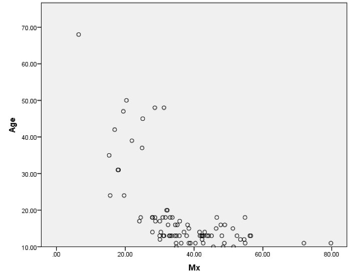
Figure 3(A).
(A). The Relationship Between Volume of the Pulp Chamber in Maxillary First Molars and Age.
.
(A). The Relationship Between Volume of the Pulp Chamber in Maxillary First Molars and Age.
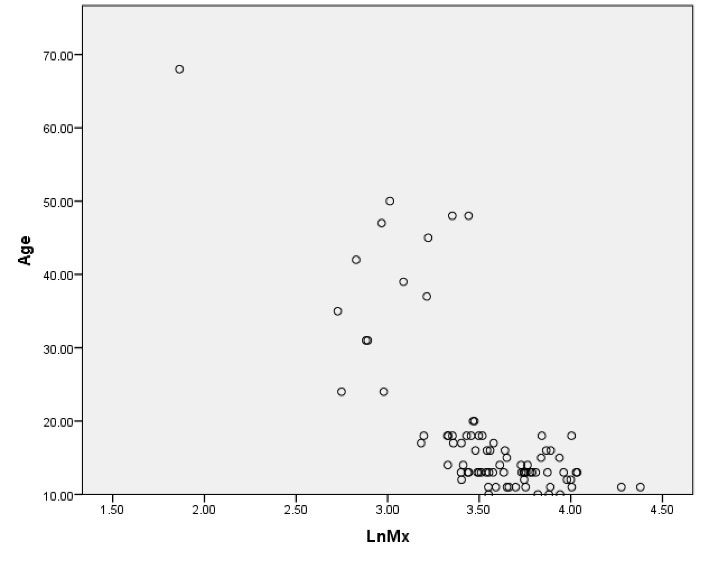
Figure 3(B).
(B). The Logarithmic Relationship Between Volume of the Pulp Chamber in the Maxillary First Molars and Age.
.
(B). The Logarithmic Relationship Between Volume of the Pulp Chamber in the Maxillary First Molars and Age.
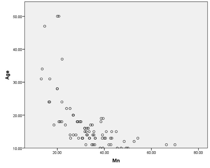
Figure 4(A).
(A). The Relationship Between Volume of the Pulp Chamber in the Mandibular First Molars and Age.
.
(A). The Relationship Between Volume of the Pulp Chamber in the Mandibular First Molars and Age.
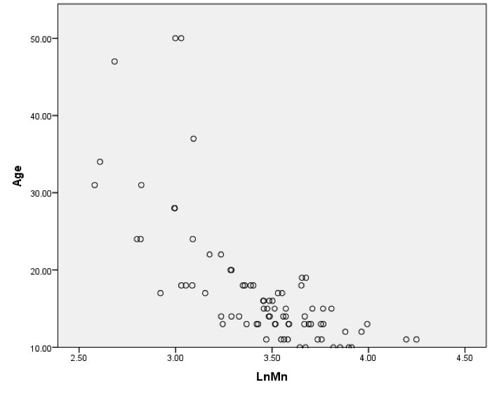
Figure 4(B).
(B). The Logarithmic Relationship Between Volume of the Pulp Chamber in the Mandibular First Molars and Age.
.
(B). The Logarithmic Relationship Between Volume of the Pulp Chamber in the Mandibular First Molars and Age.
Discussion
In the present study, age estimation was evaluated by measuring the volume of the pulp chamber in the maxillary and mandibular first molars. Contrary to other previous studies in which the pulp-to-tooth volume was used as a criterion for estimating human age, in the present study, the volume of the pulp chamber was used.
The pulp chamber volume is an indirect indicator of the precipitation of secondary dentin. In the present study, there was a negative correlation between age and all the morphological variables, indicating a decrease in all these variables with aging. In addition, the results of ANOVA showed a significant difference in the means of these variables between the different study groups. In addition, all the different variables decreased with aging, consistent with the results of a study by Ge et al (1).
Someday et al14 and Agematsu et al (19) applied regression analysis to estimate age based on the correlation between age and a decrease in the volume of the pulp chamber with the use of micro-CT images of mandibular central incisors and second premolars. They reported that the selection of an estimation equation that considers gender might yield a more reliable age estimation scheme compared to the situation in which there is no gender separation. The results of the present study supported those of the above-mentioned studies and provided further evidence for significant gender differences.
Contrary to previous studies, in which linear mathematical models have been created, a logarithmic model was developed in the present study. Linear regression analysis was carried out in the present study with age as a dependent variable and the pulp chamber volume as a predictor variable to compare logarithmic models with linear models.
In the present study, there was a significant difference in the mean volume of the pulp chamber between the maxilla and the mandible, consistent with the results of a study by Ge et al (3).
In our study, age, as an effective factor, resulted in different volumes of the pulp in the first molars of the mandible and maxilla at different ages. In a study by Pinchi et al (2), these changes were observed with aging too.
One of the limitations of the present study was the use of only the first molar teeth. The first molars undergo more restorative procedures due to dental caries compared to other teeth and are generally lost at a higher rate compared to other teeth, which might limit the use of a more developed model for estimating age, especially in the elderly. However, the young subjects might be affected by the above-mentioned factor at a lower rate because they usually have all four first molars on both sides of the dental arch. Only in sporadic cases, all four first molars might be lost at the same time or undergo restorative procedures.
Conclusions
There was a linear relationship between the volumes of the pulp chambers of maxillary and mandible first molars and the chronological age in the subjects evaluated. The volume of the first molars pulp chamber in the maxilla and mandible could be used to estimate the human age with 54% and 51% accuracy, respectively. The mean volume of the pulp chamber in the maxillary first molars was higher than that in the mandible. In addition, in the different age groups, the mean volume of the pulp chamber decreased with aging.
Conflict of Interest Disclosures
The authors declare that they have no conflict of interests.
Acknowledgments
This study is part of the MD thesis in Maxillofacial Radiology (thesis number: 9509165371) which was supported by the Vice-chancellor of Research and Technology of Hamadan university of medical sciences, Hamadan, Iran.
Ethical Statement
Ethics committee of Hamadan university of medical science was approved this article.
Authors’ Contribution
Study conception and design: FY and SL. Acquisition of data and drafting of the manuscript: SH and AS. Analysis and interpretation of data: SH and MH. Critical revision: FY.
Funding
None.
References
- Ge ZP, Yang P, Li G, Zhang JZ, Ma XC. Age estimation based on pulp cavity/chamber volume of 13 types of tooth from cone beam computed tomography images. Int J Legal Med 2016; 130(4):1159-67. doi: 10.1007/s00414-016-1384-6 [Crossref] [ Google Scholar]
- Pinchi V, Pradella F, Buti J, Baldinotti C, Focardi M, Norelli GA. A new age estimation procedure based on the 3D CBCT study of the pulp cavity and hard tissues of the teeth for forensic purposes: a pilot study. J Forensic Leg Med 2015; 36:150-7. doi: 10.1016/j.jflm.2015.09.015 [Crossref] [ Google Scholar]
- Ge ZP, Ma RH, Li G, Zhang JZ, Ma XC. Age estimation based on pulp chamber volume of first molars from cone-beam computed tomography images. Forensic Sci Int 2015; 253:133.e1-7. doi: 10.1016/j.forsciint.2015.05.004 [Crossref] [ Google Scholar]
- Panchbhai AS. Dental radiographic indicators, a key to age estimation. Dentomaxillofac Radiol 2011; 40(4):199-212. doi: 10.1259/dmfr/19478385 [Crossref] [ Google Scholar]
- Ardakani F, Bashardoust N, Sheikhha M. The accuracy of dental panoramic radiography as an indicator of chronological age in Iranian individuals. J Forensic Odontostomatol 2007; 25(2):30-5. [ Google Scholar]
- Corradi F, Pinchi V, Barsanti I, Manca R, Garatti S. Optimal age classification of young individuals based on dental evidence in civil and criminal proceedings. Int J Legal Med 2013; 127(6):1157-64. doi: 10.1007/s00414-013-0919-3 [Crossref] [ Google Scholar]
- Corradi F, Pinchi V, Barsanti I, Garatti S. Probabilistic classification of age by third molar development: the use of soft evidence. J Forensic Sci 2013; 58(1):51-9. doi: 10.1111/j.1556-4029.2012.02216.x [Crossref] [ Google Scholar]
- Bagherpour A, Anbiaee N, Partovi P, Golestani S, Afzalinasab S. Dental age assessment of young Iranian adults using third molars: a multivariate regression study. J Forensic Leg Med 2012; 19(7):407-12. doi: 10.1016/j.jflm.2012.04.009 [Crossref] [ Google Scholar]
- Kvaal SI, Kolltveit KM, Thomsen IO, Solheim T. Age estimation of adults from dental radiographs. Forensic Sci Int 1995; 74(3):175-85. doi: 10.1016/0379-0738(95)01760-g [Crossref] [ Google Scholar]
- Bosmans N, Ann P, Aly M, Willems G. The application of Kvaal’s dental age calculation technique on panoramic dental radiographs. Forensic Sci Int 2005; 153(2-3):208-12. doi: 10.1016/j.forsciint.2004.08.017 [Crossref] [ Google Scholar]
- Yang F, Jacobs R, Willems G. Dental age estimation through volume matching of teeth imaged by cone-beam CT. Forensic Sci Int 2006; 159 Suppl 1:S78-83. doi: 10.1016/j.forsciint.2006.02.031 [Crossref] [ Google Scholar]
- Vandevoort FM, Bergmans L, Van Cleynenbreugel J, Bielen DJ, Lambrechts P, Wevers M. Age calculation using X-ray microfocus computed tomographical scanning of teeth: a pilot study. J Forensic Sci 2004; 49(4):787-90. [ Google Scholar]
- Cameriere R, Ferrante L, Belcastro MG, Bonfiglioli B, Rastelli E, Cingolani M. Age estimation by pulp/tooth ratio in canines by mesial and vestibular peri-apical X-rays. J Forensic Sci 2007; 52(5):1151-5. doi: 10.1111/j.1556-4029.2007.00530.x [Crossref] [ Google Scholar]
- Someda H, Saka H, Matsunaga S, Ide Y, Nakahara K, Hirata S. Age estimation based on three-dimensional measurement of mandibular central incisors in Japanese. Forensic Sci Int 2009; 185(1-3):110-4. doi: 10.1016/j.forsciint.2009.01.001 [Crossref] [ Google Scholar]
- Pinchi V, Norelli GA, Pradella F, Vitale G, Rugo D, Nieri M. Comparison of the applicability of four odontological methods for age estimation of the 14 years legal threshold in a sample of Italian adolescents. J Forensic Odontostomatol 2012; 30(2):17-25. [ Google Scholar]
- Yang F, Jacobs R, Willems G. Dental age estimation through volume matching of teeth imaged by cone-beam CT. Forensic Sci Int 2006; 159 Suppl 1:S78-83. doi: 10.1016/j.forsciint.2006.02.031 [Crossref] [ Google Scholar]
- Jagannathan N, Neelakantan P, Thiruvengadam C, Ramani P, Premkumar P, Natesan A. Age estimation in an Indian population using pulp/tooth volume ratio of mandibular canines obtained from cone beam computed tomography. J Forensic Odontostomatol 2011; 29(1):1-6. [ Google Scholar]
- Corradi F, Pinchi V, Barsanti I, Garatti S. Probabilistic classification of age by third molar development: the use of soft evidence. J Forensic Sci 2013; 58(1):51-9. doi: 10.1111/j.1556-4029.2012.02216.x [Crossref] [ Google Scholar]
- Agematsu H, Someda H, Hashimoto M, Matsunaga S, Abe S, Kim HJ. Three-dimensional observation of decrease in pulp cavity volume using micro-CT: age-related change. Bull Tokyo Dent Coll 2010; 51(1):1-6. doi: 10.2209/tdcpublication.51.1 [Crossref] [ Google Scholar]