Avicenna J Dent Res. 12(2):40-45.
doi: 10.34172/ajdr.2020.09
Original Article
Age Estimation in Iranian Population Using Tooth/Pulp Ratio in Mandibular Premolars by Panoramic Radiography
Arash Dabbaghi 1  , Mohammad Kazemi 1, *
Author information:
1Faculty of Dentistry, Ahvaz Jundishapur University of Medical Sciences, Ahvaz, Iran.
Abstract
Background: Age estimation is a fundamental part of forensic science. Considering the fact that pulp size decreases throughout the life because of secondary dentine apposition, many researchers have used it as a method for age estimation with great results. This study aimed to determine the applicability of tooth to pulp ratio in mandibular second premolars in an Iranian population using panoramic radiographs.
Methods: The sample consisted of 153 digital panoramic images of male and female Iranians aged 13-70 years, and the tooth to pulp ratio of both left and right mandibular second premolars was obtained using AutoCAD software. Analysis of variance (ANOVA), Pearson’s correlation, and regression analysis were used to analyze the data. A total of 20 images were reexamined after 30 days to test intra-observer error.
Results: ANOVA test showed that gender had no effect on age estimation, and there was a significant correlation between age and tooth/pulp ratio (r=0.679 for right premolar, r=0.712 for left premolars, and r=0.736 for both premolars). Linear regression formulae were created based on the results and the mean differences between the estimated and actual age were reported to be 10.35±0.51, 9.17±0.54, and 8.83±0.52 for right, left, and both premolars, respectively. Intra-observer error showed no significant difference between repeated measurements.
Conclusions: Tooth/pulp ratio of mandibular second premolars is a reliable and repeatable method for age estimation in Iranian population and using premolars of both sides yields the best results.
Keywords: Age estimation, Cameriere’s method, Chronological age, Panoramic radiograph, Pulp area, Forensic science
Copyright and License Information
© 2020 The Author(s); Published by Hamadan University of Medical Sciences.
This is an open-access article distributed under the terms of the Creative Commons Attribution License (
http://creativecommons.org/licenses/by/4.0), which permits unrestricted use, distribution, and reproduction in any medium provided the original work is properly cited.
Citation: Dabbaghi A, Kazemi M. Age estimation in Iranian population using tooth/pulp ratio in mandibular premolars by panoramic radiography. Avicenna J Dent Res. 2020;12(2):40-45. doi: 10.34172/ajdr.2020.09.
Background
Highlights
-
Pulp/tooth ratio of premolars on panoramic is useful to predict chronological age.
-
Using both premolars’ ratio was better correlated with chronological age than one of them.
-
Upper canine ratio was better correlated with chronological age of premolars.
Age estimation is a fundamental component of forensic medicine. It is used to predict the age of an unidentified person whether dead or alive helps in a lot of situations such as creating a profile and identifying victims in criminal cases and large-scale fatalities due to disasters or accidents. Moreover, it can be used in social cases such as refugees with no identification to grant civil rights (1-3). Therefore, a growing demand for age estimation has been shown.
Currently, the most commonly used methods are based on the analysis of skeletal remains but bones are subject to taphonomic processes and only have acceptable results in individuals in their early adulthood (4-6). Recently, numerous dental methods have been proposed for age estimation because unlike bones, teeth are much less affected by environmental or systemic factors such as drugs or endocrine problems. Additionally, tooth development patterns are less diverse than other developmental symptoms and are preserved for a long time after death; therefore, teeth are excellent parameters for age estimation (4,7-10). Some methods are based on dental development but they do not yield reliable results after the age of 21 (4,11). Methods that are used in adults are based on degenerative processes of the teeth but a lot of them require extraction of the teeth and studying them which can be both expensive and unethical (12-17). However, some changes like secondary dentin deposition in teeth happen during the entire lifespan of a subject and except for caries and specific abrasion, no other factor affects it. Additionally, it can be analyzed using radiographs which can be easily done in living individuals with a low cost. Therefore, it has become a popular parameter for age estimation in recent years (18-23).
Kvaal et al first introduced a method for age estimation based on secondary deposition by taking radiographs of teeth and measuring parameters like width and length of different parts of tooth and pulp. Based on the analysis of the data, they concluded that indeed secondary has a correlation with chronological age and can be used to estimate age, producing acceptable results (19,24). In 2004, Cameriere et al proposed a new method based on the analysis of pulp to tooth area ratio (AR) in radiographs. This method was originally tested on maxillary canines using peri-apical x-rays and showed acceptable results (20). Since Cameriere’s method was introduced, multiple researchers have tested and validated its repeatability and reliability in different cultures and different teeth such as incisors and mandibular premolars (4,18,25-38). The mandibular premolars alongside maxillary canines have shown to be the most appropriate teeth to be analyzed for age estimation (28,29). Panoramic radiographs have also been tested for age estimation with great success (28,39,40). They are taken routinely for clinical examination of teeth and offer a great field of all quadrants of the mouth and unlike peri-apical x-rays, which can differ based on the technique and technician’s experience, they have high repeatability with little or no error (12).
The aim of this study is to determine the accuracy of Cameriere’s method for age estimation in an Iranian population in Ahvaz using panoramic radiographs and to create a formula for age estimation specific to this region. If the results are satisfactory, it can be recommended for future use in forensic science.
Materials and Methods
The sample consisted of 153 digital panoramic radiographs belonging to patients who had taken them for other purposes. They were selected randomly from the archive of the Department of Oral and Maxillofacial Radiology at Ahvaz JondiShapur University of Medical Sciences, Faculty of Dentistry. Panoramic images belonged to 65 male and 88 female patients aged 13-70 years. The subjects were divided into 6 equally distributed groups based on their age with an interval of 10 years each. The patients’ gender, date of birth, and date of x-ray were recorded according to their files, and the chronological age of the patients was obtained by subtracting date of image from date of birth. All panoramic radiographs were taken using a Cranex D X-ray unit (Sordex, Helsinki, Finland). Radiographs with the following criteria were included in the study: having good image quality with visible contours of tooth and pulp and the presence of both mandibular left and right second premolars with fully formed roots. The exclusion criteria were the presence of one of the following conditions: root fillings, crown restorations, severe caries, rotations, severe attrition, large areas of overlap with neighboring teeth, or other developmental anomalies.
All panoramic radiographs were obtained from the imaging software as high-resolution JPEG images. Images were attached to a worksheet in Autodesk AutoCAD 2019. For each of the mandibular second premolars, at least 20 points were selected and connected to determine the outline of the teeth using the polyline tool and at least 10 points were selected to identify the pulp area (Figure 1). Tooth/pulp AR was calculated by dividing tooth area by pulp area.
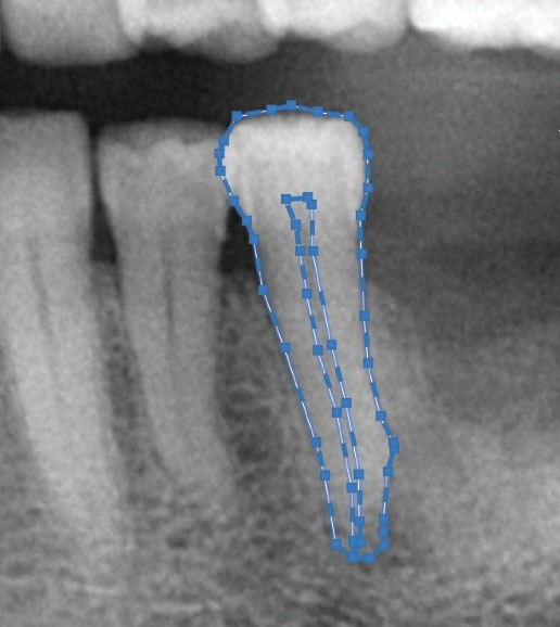
Figure 1.
A Cropped Digital Panoramic Image Showing at Least 20 and 10 Points, Respecitvely, for Tooth and Pulp Area Connected to Show Periphery of the Mentioned Areas Using AutoCad Software.
.
A Cropped Digital Panoramic Image Showing at Least 20 and 10 Points, Respecitvely, for Tooth and Pulp Area Connected to Show Periphery of the Mentioned Areas Using AutoCad Software.
In order to blind the observer, after entering each subject’s chronological age and gender into a Microsoft Office 2013 Excel spreadsheet (Microsoft Corp., Redmond, WA, USA), every image was given a unique identification number and the measurements were performed randomly based on ID numbers. A total of 20 images were randomly selected and measured after 30 days in order to test intra-observer error using a paired sample t-test.
All statistical analyses were performed by Statistical Package for the Social Sciences (SPSS version 23.0) and P < 0.05 was considered statistically significant.
Analysis of variance (ANOVA) test was performed to determine whether any significant difference existed between male and female subjects in terms of AR. Pearson’s correlation coefficient between age and AR for right, left and, and both premolars together was calculated to determine if any relationship existed. Linear regression was developed and used to estimate age based on AR for left, right, and both premolars.
Results
The present study was conducted on 153 digital panoramic radiographs which were divided into 6 age groups of roughly the same size (25±2). Table 1 shows the number of cases and gender distribution in 6 age groups.
Table 1.
Gender and Age Distribution of Subjects
|
Age Group
|
N
|
Males
|
Females
|
| Under 20 |
24 |
11 |
13 |
| 20-29 |
27 |
8 |
19 |
| 30-39 |
27 |
11 |
16 |
| 40-49 |
26 |
19 |
7 |
| 50-59 |
24 |
9 |
15 |
| Over 60 |
25 |
7 |
18 |
Paired sample t test showed no significant difference in intra-observer error between first and second measurements for premolars (P = 0.586 for the right side, P = 0.429 for the left side).
ANOVA test showed no statistically significant difference between AR and gender (P = 0.221, P = 0.896).
Tooth to pulp AR increased with age and ranged from 4.78 to 10.76 for right second premolar and from 4.42 to 10.67 for left second premolar. Figure 2 shows AR for the right and left premolars by age group.
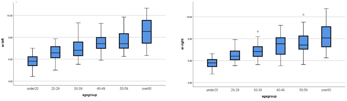
Figure 2.
Boxplots Showing Tooth to Pulp Area Ratio of Right and Left Premolars in Different Age Groups.
.
Boxplots Showing Tooth to Pulp Area Ratio of Right and Left Premolars in Different Age Groups.
Pearson’s correlation coefficient showed a strong and direct relationship between age and tooth to pulp AR (r=0.679 for right premolar, r=0.712 for left premolars, and r=0.736 for both premolars). Table 2 shows Pearson’s correlation coefficient for different teeth and Figure 3 shows the scatter diagram of the correlation between age and AR in the right and left second premolars.
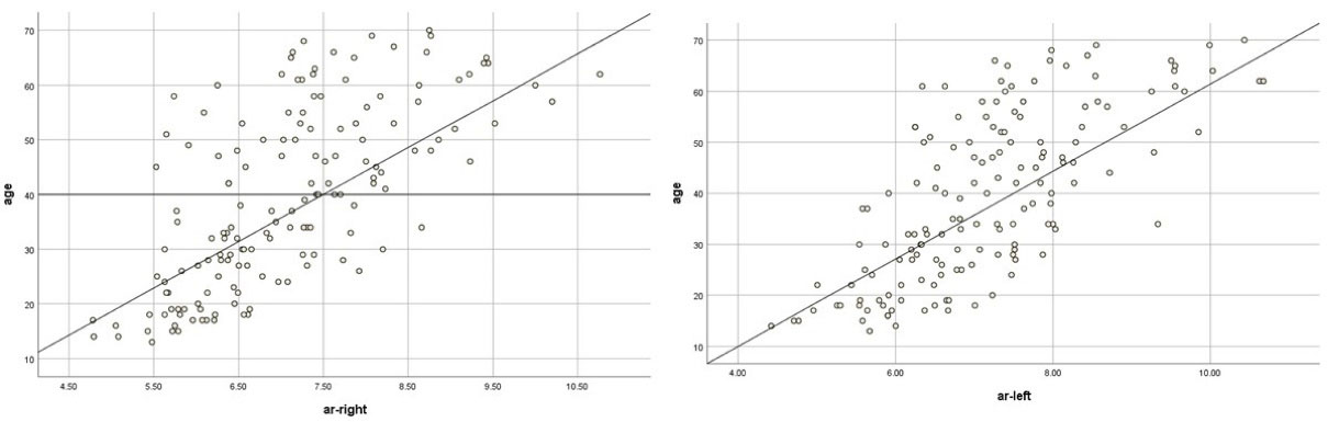
Figure 3.
Scatter Diagram of Correlation Between Age and AR in the Right and Left Second Premolars.
.
Scatter Diagram of Correlation Between Age and AR in the Right and Left Second Premolars.
Table 2.
Pearson’s Correlation Coefficient Between Age and Tooth to Pulp Area Ratio (AR) for Different Teeth
|
Tooth
|
R
|
R Square
|
Standard Error of the Estimate
|
| Right premolar |
0.679 |
0.461 |
11.951 |
| Left premolar |
0.712 |
0.506 |
11.438 |
| Both premolars |
0.736 |
0.542 |
11.055 |
Linear regression formulae were created to estimate the age based on AR of different teeth according to regression model analysis (Tables 3 and 4).
Table 3.
Regression Model Analysis Used to Create Formulae for Age Estimation
|
Model
|
Unstandardized Coefficients
|
t
|
P
Value
|
|
B
|
Standard Error
|
| Right premolar |
(Constant) |
-27.500 |
5.974 |
-4.604 |
0.000 |
| Area ratio-right |
9.480 |
0.834 |
11.364 |
0.000 |
| Left premolar |
(Constant) |
-26.899 |
5.415 |
-4.967 |
0.000 |
| Area ratio-left |
9.271 |
0.745 |
12.443 |
0.000 |
| Both premolars |
(Constant) |
-34.552 |
5.693 |
-6.069 |
0.000 |
| ar-left |
6.075 |
1.180 |
5.146 |
0.000 |
| ar-right |
4.321 |
1.265 |
3.416 |
0.001 |
Table 4.
Formulae Created for Age Estimation
|
Tooth
|
Formula
|
| Right premolar |
(9.48*ARR) - 27.5 |
| Left premolar |
(9.271*ARL) – 26.889 |
| Both premolars |
(4.321*ARR) + (6.075*ARL) -34.552 |
Results of the difference between estimated age using formulae and actual age can be seen in Tables 4 and 5. Best results were found when using both premolars for age estimation (8.83±0.52) compared to the right (10.35±0.51) and left (9.17±0.54).
Table 5.
Mean Difference Between Estimated and Actual Values of Age
|
Age Group
|
Right Premolar
|
Left Premolar
|
Both Premolars
|
| Under 20 |
12.38±0.88 |
9.77±1.14 |
8.87±0.94 |
| 20-29 |
10.92±1.12 |
8.93±1.09 |
8.30±0.93 |
| 30-39 |
7.48±1.13 |
6.60±1.14 |
5.57±1.08 |
| 40-49 |
8.29±0.96 |
5.77±0.80 |
5.93±0.87 |
| 50-59 |
10.37±1.47 |
11.70±1.33 |
11.57±1.72 |
| Over 60 |
12.99±1.61 |
12.74±1.84 |
13.22±1.72 |
| Total |
10.35±0.51 |
9.17±0.54 |
8.83±0.52 |
Discussion
Age estimation is an indispensable factor in forensic medicine for creating the profile of living and dead unidentified persons. Estimating the age of a living person has a lot of challenges because it needs to be ethical, cost-effective, and simple. Secondary dentine apposition in teeth is an excellent biomarker for age estimation because it happens throughout the lifespan of a person and can be analyzed in dental radiographs such as peri-apical and panoramic x-rays thus making it widely acceptable and executable (19,41,42). Cameriere et al have proposed the use of pulp to tooth AR to determine the age and previous studies have shown great acceptability and validity in different ethnic groups (18,20-22,25-28,42).
In the current study, digital panoramic radiographs of Iranians living in Ahvaz were used to determine age based on the tooth to pulp AR. One of the concerns for measurement error in panoramic radiographs is that some observers may have trouble identifying anatomical structures. However, in this study, all images had high image quality with great contrast and tooth and pulp had visible outlines so the issue was resolved. In addition, insignificant statistical value for intra-observer error showed that the method had high repeatability.
Mandibular second premolars were chosen for the assessment of age because previous studies have shown that maxillary canines and mandibular premolars have the highest accuracy for age estimation (18,28,29,36,37,42). Mandibular premolars were chosen instead of canines because no studies have been conducted in Iranian population using premolars. Teeth on both sides of the mandible were analyzed to see if the results improved.
The study showed that gender had no effect on age estimation and tooth to pulp AR, as has been published by other researchers in previous studies (10,18,26,39).
Data showed that tooth to pulp AR increased with age which means that the pulp area decreased with age due to secondary dentine apposition, as has been published in previous studies (27-32,34). Pearson correlation coefficient showed a significant relationship between chronological age and tooth/pulp AR which suggests that the pulp/tooth AR can be a good parameter for age estimation. Tooth to pulp AR was used in the current study unlike other studies which used pulp to tooth AR; therefore, the correlation coefficient was direct as opposed to the inverse correlation reported in other studies. However, the value of regression coefficient was in the same range as reported in other studies, indicating that premolars are significantly correlated with age (28,29). Canines have also been reported to have a strong correlation with age. Dehghani et al reported that the regression value of maxillary canines was r=−0.794 which is close to the regression value reported in our study (ranging from 0.679 to 0.736), which means that premolars have a high correlation with age in Iranian population (25).
Regression coefficient of both premolars (r=0.736) was slightly higher than each premolar alone (r=0.679 for right and r=0.712 for left), showing that using both premolars can produce improved results although not with a high margin.
The mean difference between chronological age and estimated age using AR of both premolars had the highest accuracy (8.83±0.52). According to Cameriere et al (18,20), the mean error of equal or less than 10 years is acceptable for forensic science, and the mean difference in this study is close to the residual standard error reported by Cameriere et al (7.42 years) when using a single type of premolars (28). However, this value decreased to 5.75 when they used two different premolars which could mean that the use of two different teeth would yield better results as opposed to one tooth on both sides. Yet, this conclusion needs to be tested in an Iranian population in the future.
It was observed that the mean difference between chronological age and estimated age obtained via regression formulae yielded the best results in ages between 30 and 50 years (5.57±1.08 in 30-39, 5.93±0.87) which means premolars have the best accuracy for age estimation in adults compared to younger or older individuals.
Conclusions
The results of the study showed that using tooth/pulp ratio of mandibular second premolars can be a simple and cost-efficient method with acceptable results for age estimation in Iranians especially when both right and left premolars are used. The formulae created can be used to determine the age of individuals in a short period of time. The use of panoramic radiographs with higher image quality can improve the results especially if they are used to determine the formulae. Further studies are needed to be done on Iranian population to determine the effects of other teeth or combinations of them.
Conflict of Interest Disclosures
The authors declare that they have no conflict of interests.
Acknowledgments
This article is part of D.D.S undergraduate thesis (project number: U-98030) conducted by Mohammad Kazemi. The authors would like to express their gratitude to Ahvaz Jondishapur University of Medical Sciences, Faculty of Dentistry, Department of Oral and Maxillofacial Radiology for technical assistance and Ahvaz Jondishapur University of Medical Sciences for funding this research.
Ethical Statement
The protocol of the study was reviewed and approved by the Ethics Committee of Research Deputy of Ahvaz JondiShapur University of Medical Sciences, Ahvaz, Iran, and the study was conducted in accordance with the ethical standards of the Declaration of Helsinki.
Authors’ Contribution
MK and AD conceived the idea of the research. MK carried out the experiment and wrote the manuscript with help and supervision of AD.
Funding
This article is part of D.D.S undergraduate thesis (project number: U-98030) conducted by Mohammad Kazemi which used funding granted by Ahvaz Jondishapur university of medical sciences.
References
- Soomer H, Ranta H, Lincoln MJ, Penttilä A, Leibur E. Reliability and validity of eight dental age estimation methods for adults. J Forensic Sci 2003; 48(1):149-52. [ Google Scholar]
- Solheim T, Sundnes PK. Dental age estimation of Norwegian adults--a comparison of different methods. Forensic Sci Int 1980; 16(1):7-17. doi: 10.1016/0379-0738(80)90174-7 [Crossref] [ Google Scholar]
- Baccino E, Schmitt A. Determination of adult age at death in the forensic context. In: Schmitt A, Cunha E, Pinheiro J, eds. Forensic Anthropology and Medicine. Humana Press; 2006. p. 259-80. 10.1007/978-1-59745-099-7_11.
- De Luca S, Alemán I, Bertoldi F, Ferrante L, Mastrangelo P, Cingolani M. Age estimation by tooth/pulp ratio in canines by peri-apical X-rays: reliability in age determination of Spanish and Italian medieval skeletal remains. J Archaeol Sci 2010; 37(12):3048-58. doi: 10.1016/j.jas.2010.06.034 [Crossref] [ Google Scholar]
- Santiago BM, Almeida L, Cavalcanti YW, Magno MB, Maia LC. Accuracy of the third molar maturity index in assessing the legal age of 18 years: a systematic review and meta-analysis. Int J Legal Med 2018; 132(4):1167-84. doi: 10.1007/s00414-017-1766-4 [Crossref] [ Google Scholar]
- Liu Y, Geng K, Chu Y, Xu M, Zha L. Third molar mineralization in relation to chronologic age estimation of the Han in central southern China. Int J Legal Med 2018; 132(5):1427-35. doi: 10.1007/s00414-018-1804-x [Crossref] [ Google Scholar]
- Jain S, Nagi R, Daga M, Shandilya A, Shukla A, Parakh A. Tooth coronal index and pulp/tooth ratio in dental age estimation on digital panoramic radiographs-a comparative study. Forensic Sci Int 2017; 277:115-21. doi: 10.1016/j.forsciint.2017.05.006 [Crossref] [ Google Scholar]
- Nolla CM. The Development Of Permanent Teeth. University of Michigan; 1952.
- Fins P, Pereira ML, Afonso A, Pérez-Mongiovi D, Caldas IM. Chronology of mineralization of the permanent mandibular second molar teeth and forensic age estimation. Forensic Sci Med Pathol 2017; 13(3):272-7. doi: 10.1007/s12024-017-9876-3 [Crossref] [ Google Scholar]
- Shah PH, Venkatesh R. Pulp/tooth ratio of mandibular first and second molars on panoramic radiographs: an aid for forensic age estimation. J Forensic Dent Sci 2016; 8(2):112. doi: 10.4103/0975-1475.186374 [Crossref] [ Google Scholar]
- Shahin KA, Chatra L, Shenai P. Dental and craniofacial imaging in forensics. J Forensic Radiol Imaging 2013; 1(2):56-62. doi: 10.1016/j.jofri.2012.12.001 [Crossref] [ Google Scholar]
- Bosmans N, Ann P, Aly M, Willems G. The application of Kvaal’s dental age calculation technique on panoramic dental radiographs. Forensic Sci Int 2005; 153(2-3):208-12. doi: 10.1016/j.forsciint.2004.08.017 [Crossref] [ Google Scholar]
- Gustafson G. Age determination on teeth. J Am Dent Assoc 1950; 41(1):45-54. doi: 10.14219/jada.archive.1950.0132 [Crossref] [ Google Scholar]
- Bang G, Ramm E. Determination of age in humans from root dentin transparency. Acta Odontol Scand 1970; 28(1):3-35. doi: 10.3109/00016357009033130 [Crossref] [ Google Scholar]
- Kasetty S, Rammanohar M, Raju Ragavendra T. Dental cementum in age estimation: a polarized light and stereomicroscopic study. J Forensic Sci 2010; 55(3):779-83. doi: 10.1111/j.1556-4029.2010.01363.x [Crossref] [ Google Scholar]
- Wochna K, Bonikowski R, Śmigielski J, Berent J. Aspartic acid racemization of root dentin used for dental age estimation in a Polish population sample. Forensic Sci Med Pathol 2018; 14(3):285-94. doi: 10.1007/s12024-018-9984-8 [Crossref] [ Google Scholar]
- Spalding KL, Buchholz BA, Bergman LE, Druid H, Frisén J. Forensics: age written in teeth by nuclear tests. Nature 2005; 437(7057):333-4. doi: 10.1038/437333a [Crossref] [ Google Scholar]
- Cameriere R, Ferrante L, Belcastro MG, Bonfiglioli B, Rastelli E, Cingolani M. Age estimation by pulp/tooth ratio in canines by peri-apical X-rays. J Forensic Sci 2007; 52(1):166-70. doi: 10.1111/j.1556-4029.2006.00336.x [Crossref] [ Google Scholar]
- Kvaal SI, Kolltveit KM, Thomsen IO, Solheim T. Age estimation of adults from dental radiographs. Forensic Sci Int 1995; 74(3):175-85. doi: 10.1016/0379-0738(95)01760-g [Crossref] [ Google Scholar]
- Cameriere R, Ferrante L, Cingolani M. Variations in pulp/tooth area ratio as an indicator of age: a preliminary study. J Forensic Sci 2004; 49(2):317-9. [ Google Scholar]
- Cameriere R, Brogi G, Ferrante L, Mirtella D, Vultaggio C, Cingolani M. Reliability in age determination by pulp/tooth ratio in upper canines in skeletal remains. J Forensic Sci 2006; 51(4):861-4. doi: 10.1111/j.1556-4029.2006.00159.x [Crossref] [ Google Scholar]
- Cameriere R, Ferrante L, Belcastro MG, Bonfiglioli B, Rastelli E, Cingolani M. Age estimation by pulp/tooth ratio in canines by mesial and vestibular peri-apical X-rays. J Forensic Sci 2007; 52(5):1151-5. doi: 10.1111/j.1556-4029.2007.00530.x [Crossref] [ Google Scholar]
- Paewinsky E, Pfeiffer H, Brinkmann B. Quantification of secondary dentine formation from orthopantomograms--a contribution to forensic age estimation methods in adults. Int J Legal Med 2005; 119(1):27-30. doi: 10.1007/s00414-004-0492-x [Crossref] [ Google Scholar]
- Kvaal S, Solheim T. A non-destructive dental method for age estimation. J Forensic Odontostomatol 1994; 12(1):6-11. [ Google Scholar]
- Dehghani M, Shadkam E, Ahrari F, Dehghani M. Age estimation by canines’ pulp/tooth ratio in an Iranian population using digital panoramic radiography. Forensic Sci Int 2018; 285:44-9. doi: 10.1016/j.forsciint.2018.01.016 [Crossref] [ Google Scholar]
- De Luca S, Bautista J, Alemán I, Cameriere R. Age-at-death estimation by pulp/tooth area ratio in canines: study of a 20th-century Mexican sample of prisoners to test Cameriere’s method. J Forensic Sci 2011; 56(5):1302-9. doi: 10.1111/j.1556-4029.2011.01784.x [Crossref] [ Google Scholar]
- Zaher JF, Fawzy IA, Habib SR, Ali MM. Age estimation from pulp/tooth area ratio in maxillary incisors among Egyptians using dental radiographic images. J Forensic Leg Med 2011; 18(2):62-5. doi: 10.1016/j.jflm.2010.12.004 [Crossref] [ Google Scholar]
- Cameriere R, De Luca S, Alemán I, Ferrante L, Cingolani M. Age estimation by pulp/tooth ratio in lower premolars by orthopantomography. Forensic Sci Int 2012; 214(1-3):105-12. doi: 10.1016/j.forsciint.2011.07.028 [Crossref] [ Google Scholar]
- Lee JH, Lee C, Battulga B, Na JY, Hwang JJ, Kim YH. Morphological analysis of the lower second premolar for age estimation of Korean adults. Forensic Sci Int 2017; 281:186.e1-186. doi: 10.1016/j.forsciint.2017.10.005 [Crossref] [ Google Scholar]
- Babshet M, Acharya AB, Naikmasur VG. Age estimation in Indians from pulp/tooth area ratio of mandibular canines. Forensic Sci Int 2010; 197(1-3):125.e1-4. doi: 10.1016/j.forsciint.2009.12.065 [Crossref] [ Google Scholar]
- Jeevan MB, Kale AD, Angadi PV, Hallikerimath S. Age estimation by pulp/tooth area ratio in canines: Cameriere’s method assessed in an Indian sample using radiovisiography. Forensic Sci Int 2011; 204(1-3):209.e1-5. doi: 10.1016/j.forsciint.2010.08.017 [Crossref] [ Google Scholar]
- Cameriere R, Cunha E, Sassaroli E, Nuzzolese E, Ferrante L. Age estimation by pulp/tooth area ratio in canines: study of a Portuguese sample to test Cameriere’s method. Forensic Sci Int 2009; 193(1-3):128.e1-6. doi: 10.1016/j.forsciint.2009.09.011 [Crossref] [ Google Scholar]
- Kapoor N, Kothari P, Shukla RK, Mishra SD, Badiye A. Age estimation from tooth-pulp area ratio: a preliminary study. La Revue de Médecine Légale 2020; 11(1):11-4. doi: 10.1016/j.medleg.2019.11.003 [Crossref] [ Google Scholar]
- Azevedo AC, Michel-Crosato E, Biazevic MG, Galić I, Merelli V, De Luca S. Accuracy and reliability of pulp/tooth area ratio in upper canines by peri-apical X-rays. Leg Med (Tokyo) 2014; 16(6):337-43. doi: 10.1016/j.legalmed.2014.07.002 [Crossref] [ Google Scholar]
- Cameriere R, Cunha E, Wasterlain SN, De Luca S, Sassaroli E, Pagliara F. Age estimation by pulp/tooth ratio in lateral and central incisors by peri-apical X-ray. J Forensic Leg Med 2013; 20(5):530-6. doi: 10.1016/j.jflm.2013.02.012 [Crossref] [ Google Scholar]
- Azevedo AC, Alves NZ, Michel-Crosato E, Rocha M, Cameriere R, Biazevic MG. Dental age estimation in a Brazilian adult population using Cameriere’s method. Braz Oral Res 2015;29. 10.1590/1807-3107BOR-2015.vol29.0016.
- Saxena S. Age estimation of Indian adults from orthopantomographs. Braz Oral Res 2011; 25(3):225-9. doi: 10.1590/s1806-83242011005000009 [Crossref] [ Google Scholar]
- Roh BY, Lee WJ, Ryu JW, Ahn JM, Yoon CL, Lee SS. The application of the Kvaal method to estimate the age of live Korean subjects using digital panoramic radiographs. Int J Legal Med 2018; 132(4):1161-6. doi: 10.1007/s00414-017-1762-8 [Crossref] [ Google Scholar]
- Dehghani M, Shadkam E, Ahrari F, Dehghani M. Age estimation by canines’ pulp/tooth ratio in an Iranian population using digital panoramic radiography. Forensic Sci Int 2018; 285:44-9. doi: 10.1016/j.forsciint.2018.01.016 [Crossref] [ Google Scholar]
- Gulsahi A, Tirali RE, Cehreli SB, De Luca S, Ferrante L, Cameriere R. The reliability of Cameriere’s method in Turkish children: a preliminary report. Forensic Sci Int 2015; 249:319.e1-5. doi: 10.1016/j.forsciint.2015.01.031 [Crossref] [ Google Scholar]
- Marroquin TY, Karkhanis S, Kvaal SI, Vasudavan S, Kruger E, Tennant M. Age estimation in adults by dental imaging assessment systematic review. Forensic Sci Int 2017; 275:203-11. doi: 10.1016/j.forsciint.2017.03.007 [Crossref] [ Google Scholar]
- Cameriere R, Cunha E, Sassaroli E, Nuzzolese E, Ferrante L. Age estimation by pulp/tooth area ratio in canines: study of a Portuguese sample to test Cameriere’s method. Forensic Sci Int 2009; 193(1-3):128.e1-6. doi: 10.1016/j.forsciint.2009.09.011 [Crossref] [ Google Scholar]