Avicenna J Dent Res. 11(4):135-138.
doi: 10.34172/ajdr.2019.27
Original Article
Radiographic Evaluation of Bone Resorption Rate 1 Year After the Placement of Intra-lock Dental Implants
Pouyan Sigari 1, Abbas Naghizadeh Baghi 2, Vahid Khalili 3, * 
Author information:
1Oral and maxillofacial surgeon, Ardabil dental School, Ardabil University of medical science, Ardabil, Iran.
2Associate Professor of Physical Education and Sport Sciences, Faculty of Educational Sciences and Psychology, University of Mohaghegh Ardabili, Ardabil, Iran.
3School of Dentistry, Ardabil Dental School, Ardabil, Iran.
*
Correspondence to Vahid Khalili, School of Dentistry, Ardabil Dental School, Ardabil, Iran Tel: +9821 44083117, +989126072879 Email:
dr.vkhalili@yahoo.com
Abstract
Background: With the advent of dental implants, the majority of patients without complete teeth or inneed of single teeth were well treated, and the popularity of this treatment method led many companiesaround the world to produce and distribute this production. Meanwhile, Intra-lock has started designinga special type of dental implant that claims to have the best possible performance with the least damageto the toothless area. In this study, we decided to evaluate and study this claim.
Methods: Digital periapical films of 164 intra-lock implants which were taken immediately after the surgery and one year after the prosthetic restoration between 2015 and 2017 were used in this study. All of the radiographs were taken by Rad Radiography Center in Ardabil and were implanted by Dr. Sigari (private office in Ardabil). Out of the 164 individuals, 80 (48.8%) were male and 84 (51.2%) were female. Their age ranged between 16 to 81 years old and the highest frequency belonged to the age group of 31-40 years old and the lowest frequency belonged to those who were under 20 years of age.
Results: Mean bone loss was 1.87 mm one year after prosthetic delivery. The average bone loss was higher in the posterior upper jaw and total failure of this implant was observed only in 6 cases. No significant difference in the bone loss was observed between females and males (P= 0.221). The lowest mean bone resorption was observed in the age group of below 20 years. The highest mean bone resorption was observed in the posterior maxilla. Out of 164 patients, 158 (96.3%) had permanent implants and 6 (3.7%) had implant loss.
Conclusions: Within the limitations of this study, the mean bone loss in this brand was acceptable and Blossom Technology can find its way to the market, thus we found this brand with new technology useful for clinical application.
Keywords: Dental implants, Bone resorption, Osteointegration, Periapical radiography, Maxilla, Mandible
Copyright and License Information
© 2019 The Author(s); Published by Hamadan University of Medical Sciences.
This is an open-access article distributed under the terms of the Creative Commons Attribution License (
http://creativecommons.org/licenses/by/4.0), which permits unrestricted use, distribution, and reproduction in any medium provided the original work is properly cited.
Citation: Sigari P, Naghizadeh Baghi A, Khalili V. Radiographic Evaluation of Bone Resorption Rate 1 Year After the Placement of Intra-lock Dental Implants. Avicenna J Dent Res. 2019;11(4):135-138. doi: 10.34172/ajdr.2019.27.
Background
Highlights
-
Comparison of different brands of implants has been done in many articles, however, to date, no research has been done on the desirability of this brand in Iran and it was necessary to broaden the view of dentists to choose or not to choose this brand.
For the loss of each person’s teeth due to many reasons such as caries, trauma, or even congenital tooth loss, many treatments can be recommended, including complete and partial dentures or dental bridges. In 1965, Branemark began implanting dental implants in toothless patients. Then, titanium implants were widely used as one of the most successful alternative therapies for missing teeth (1-3).
As this treatment is updated and developed, different brands and companies have come up with different types of implants with different alloys, designs, and dimensions, as well as different implantation methods. The important issue is to identify and compare the success of these brands with various articles written and published in this field. All types of implants have different bone resorption around the implant in areas under functional pressure, which is one of the most important factors for implant selection (4).
Primary bone loss around an implant forms a V- or U-shaped pattern that is described as ditching or saucerization. Current hypotheses about the crestal bone loss are made on the basis of the following factors: disruption of the periosteum during surgery, osteotomy for implant preparation, microgap position between abutment and body of the implant, microscopic movements of abutment components, bacterial attack, invasion to biological width, and stress factors (5-11).
In a study, Adell et al (4) measured and reported the crestal bone around the implant. This study showed that most of the bone resorption occurs during the first year of prosthesis loading. The average is 1.2 mm, with a range of 0 to 3 mm. In this study, the baseline of bone resorption was the first thread, about 1.8 mm lower than the crestal bone, with an average bone resorption of 3.3 mm. A follow-up study by Albrektsson and Isidor suggests that a successful implant should show a bone resorption rate of less than 1.5 mm in the first year of operation and only 0.2 mm of bone resorption every year (12). The baseline in that study was the surface of the crestal bone. Another study was based on the findings of Wennstrom and Palmer who used radiographic criteria for bone resorption and stated that a maximum of 2 mm of bone resorption after 5 years of implant placement could be acceptable (13).
Nowadays, in Iran, dental implants are considered as one of the most basic and successful dental treatments and different types of implants are in competition with each other to gain this market. One of the most useful brands in the Iranian market is the intra-lock that has gained a special place among dentists. The company utilizes the unique Blossom brand (1) that claims to have the least torque. The maximum osteosynthesis and the least micromotion can be expected for this type of implant. This company by utilizing features which are unique to this brand claims that with less torque, the maximum osteointegration and the least micromotion can be expected.
In this design, at least one cutting surface is considered on each thread, thereby reducing the number of chips produced during implantation and minimizing the required torque to decrease possible trauma during implantation. In this study, we used periapical radiographs taken right after implantation and one year after loading the implants to evaluate the efficacy of this design in reducing bone resorption and implant durability.
Materials and Methods
This is a retrospective study, where dental implant surgery and prosthetic restoration procedures were performed by the same surgeon and prosthodontist on a total of 164 patients between October 2015 and March 2017 in the private office of Dr. Sigari (Ardabil, Iran). During the case collection period, digital periapical radiographs of the dental implants were taken immediately after, which was set as the baseline, and one year after the prosthetic restoration procedures were delivered. Patients enrolled were between 18 and 82 years old; these patients had to return for regular follow-up, and periapical radiographs were taken immediately after, and one year after the prosthetic restoration procedures were delivered. All the implants were placed at crestal level and digital radiographs were collected from Raad Radiography Center (Ardabil, Iran). Mesial and distal peri-implant bone heights were measured using Digimizer software and the greatest numbers were selected as bone loss. Bone height was measured from the rough-smooth border to the highest point of the proximal bone crest (Figure 1). Every periapical film was taken by the standard paralleling technique. An X-ray cone indicator was used and patients were instructed to bite on the film. The radiation dose for each periapical film was 60 kV, 6 mA per 0.1 second, and radiation exposure time for the premolar area was approximately 0.4 seconds, and for the molar area, it was 0.64 seconds, exposing the patients to a radiation dose of 24 mA to 38.4 mA for each periapical film of premolars and molars, respectively. Then, all data were measured by Digimizer software. First, the lengths of the placed implants were written in the pre-prepared checklist and then lengths of implants were entered into the software as reference points. Then, bone resorption rates were calculated and recorded using the software.
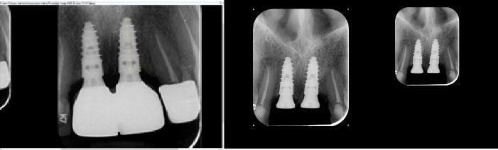
Figure 1.
Measurement of the Cervical Line of the Implant to the Alveolar Crest on the Mesial and Distal Aspects. Note. The greatest number was selected as bone loss.
.
Measurement of the Cervical Line of the Implant to the Alveolar Crest on the Mesial and Distal Aspects. Note. The greatest number was selected as bone loss.
All data were analyzed by SPSS software version 22.0, and descriptive statistics (i.e., number and percentage) were used for illustrating results.
Exclusion criteria were as follows:
-
Patients with any bone disease
-
Heavy smoker patients
-
Alcoholics
-
Pregnant or lactating women
-
Patients with long-term oral medication that interfere with bone remodeling
-
Patients who require guided bone regeneration (GBR) during implantation
-
Patients with parafunctional disorders including bruxism and clenching
-
Patients with poor hygiene with plaque index of more than 20%
-
Patients with pre-implantitis
-
Patients whose implants are placed as a fresh socket
-
Patients with adjacent implants inserted with a distance less than 3 mm or a distance less than 1.5 mm between the tooth and implant
-
Subcrestal implant placement greater than 1 mm
-
Food impaction in the implanted area
-
History of abscess during implant repair
-
Advanced osteoporosis with dual-energy X-ray absorptiometry (DEXA) less than -2.5
Results
Intra-lock implants, with their unique design, are claimed to have reduced torque during surgery and no damage to bone can reduce bone loss. The mean bone resorption rate among the 164 implants we studied was 1.87, with a standard deviation of 0.39, and out of 164 patients, 158 (96.3%) had permanent implants and 6 (3.7%) had missing implants. Of the 164 individuals, 80 (48.8%) were male and 84 (51.2%) were female. All implants were 9.5, 10.5 and 11.5 in length. Out of the 164 patients, the highest frequency belonged to the age range of 31 to 40 years (54 patients), and the lowest frequency was observed in patients under 20 years of age and above 70, respectively (Table 1).
Table 1.
Distribution of Implants in Different Age Groups
|
Age Group
|
Number
|
Percent
|
| Under 20 |
4 |
2.4 |
| 21-30 |
11 |
6.7 |
| 31-40 |
54 |
32.9 |
| 41-50 |
41 |
25 |
| 51-60 |
26 |
15.9 |
| 61-70 |
20 |
12.2 |
| Above 70 |
8 |
4.9 |
| Total |
164 |
100 |
Of all the implants, 3 (1.8%) were placed in the anterior maxilla, 76 (46.3%) in the posterior maxilla, and 85 (51.8%) in the posterior mandible. No implant was placed in the anterior mandible. out of 164 patients, 158 (96.3%) had permanent implants and 6 (3.7%) had implant loss. The mean bone loss among all 164 implants was 1.87 one year after loading the prosthesis, with the lowest rate of 1.04 and the highest rate of 2.90. Bone resorption was zero immediately after implant placement, and all selected cases had implants exactly at the crestal surface. In another part of our study, the relationship between patients’ age and bone resorption was examined. Although the lowest mean bone resorption was observed in the group age of below 20 years, there was no statistically significant difference in the mean bone resorption one year after loading intra-lock dental implants in patients of different age groups (P=0.11). This similarity is probably due to the low frequency of members in this age category in our study (4 out of 164).
The amount of bone resorption in the jaw was the next aspect to be studied. Except for the three implants which were placed in the maxillary anterior and in the canine area, the other implants were placed in the maxillary and mandibular premolars and molars. Additionally, 76 implants were placed in the posterior maxilla and 85 in the posterior mandible. The highest mean bone resorption was observed in the posterior maxilla.
One-way analysis of variance (ANOVA) was used to test the above-mentioned hypothesis and the results showed that there was no difference between mean bone resorption one year after loading intra-lock dental implants (P = 0.21).
The rate of bone resorption one year after loading intra-lock dental implants varies between male and female patients. To test this hypothesis, t test was used for comparing independent groups in terms of mean bone resorption one year after loading intra-lock dental implants among patients with a different gender. There were no statistically significant differences between them (P= 0.22).
Discussion
The highest bone resorption is observed in the first year after implant placement, and calculating it across different implant brands can provide a better and more reliable view of choosing an implant brand, which was calculated to be 1.87 ± 0.39 mm for this brand. Blossom design is a new architectural method to provide 180 degrees cutting surface which can cause absolute sharpness to protect bone from necrosis during the process of implantation. To investigate this claim, bone resorption around the implant was calculated one year after prosthetic loading using periapical radiographs by parallel technique. To minimize the bias on the part of the surgeon, all implants were implanted by a surgeon. Then, the rate of bone resorption in the jaws, different genders, and different age groups was studied.
Age was the first indicator studied. According to the groupings, people under the age of 20 showed the least amount of bone resorption. Moreover, people over 70 showed the highest bone resorption rate. Tissue and bone regeneration and turnover rates were much higher in healthy young people than in older adults. This speeds up osteointegration around the implant and thus reduces the rate of bone resorption in this age group. In a study by Simmons et al in 2017 (14), the durability of the 2 implant brands of OSP and OSPTX was investigated 6 weeks and 12 months after implant placement. The bone resorption rate in both groups was less than 0.5 mm and the implant survival rate was 93.3% after 1 year for both brands. Of the 30 implants placed, 2 failed.
In a study, Ho et al investigated 2 brands of ITI and Xive in 2016 and reported that there was less bone loss in the ITI brand. Initially, it was 0.10 mm, and 24 weeks after implant placement, this rate reached 0.16 mm. While in the Xive brand, it was initially 0.16 and after 24 weeks of placement, it reached 0.41 mm, which was significantly higher compared to the ITI brand (15).
In another study, Ebler et al studied 64 patients and 97 implants (54 Astra Tech OsseoSpeed implants and 43 Straumann Bone Level implants) and found that none of the implants were lost in one year. In the Astra brand, the bone resorption rate was 1.30 mm at the time of implant placement, and for the Straumann brand, it was 1.26 mm. In the one-year follow-up, the bone resorption rate for the above-mentioned two brands was 0.37 and 0.39, respectively, indicating no significant difference between the two brands (16).
Jaws have been under-studied and we were able to make a good comparison of bone resorption with respect to the location of the implants, indicating a relatively equal number of implants in the upper and lower jaws. The maxillary posterior showed more bone loss than the posterior maxilla. It could theoretically be justified by lower bone density and greater spongy bone mass in the posterior maxilla reduces osteosynthesis and enhances bone resorption. In a study conducted in 2007, Khayat and Milliez concluded that the success rate of implants was 98.6% in the maxilla and 98.8% in the mandible, but this difference was not statistically significant (17). In a study conducted in 1999, Cochran stated that “in general, implants placed in the mandible have a significantly higher success rate than maxillary implants” (18).
Gender was another factor analyzed in this study and it was found that there was no significant difference between men and women in term of bone resorption, indicating that gender could not be a factor in bone resorption.
Despite the fact that we only used periapical radiographs and this study was done in a short period of time, Intra lock brand was considered successful according to the criteria illustrated by Albrektsson et al in 1985 (8). In our study, the highest amount of mesial or distal bone loss was considered as the bone loss of that implant, while if the mesial and distal surfaces were examined separately, it could have given us more accurate results. Three-month radiographic examinations can show the amount of bone resorption before loading the prosthesis, and multi-year studies can show the pattern of bone resorption in this brand, which unfortunately was not included in our study. Moreover, by increasing the sample size, the average obtained from bone resorption can be more accurate and can also show better bone resorption in the anterior parts of the jaws which are important from the aesthetic point of view and the lowest rate of long-term bone resorption is highly required for patients.
Conclusions
Due to the limitations we had in this project, the amount of bone resorption in this brand due to the presence of a sufficient number of cases in Iran was normal according to previous studies. Because of its unique design, the brand can be suitable for patients.
Conflict of Interest Disclosures
The authors claim to have no financial interests, either directly or indirectly, in the products or information listed in the article.
Acknowledgments
This research was supported by Ardabil University of Medical Science, Dental School. We would like to thank Dr. Aryan (Prosthodontist, School of Dentistry, Ardabil University of Medical Sciences) for designing and doing all restorations as well as providing post-loading radiographs from his private archives.
Ethical Statement
Lack of available resources, insufficient funding to expand the research and limited information to a geographical area and lack of willingness of dental colleagues to participate in this project were the problems that troubled us in this research.
Authors’ Contribution
PS carried out the study and provided all data. ANB performed the analytic calculations and performed the numerical simulations. VK took the lead in writing the manuscript. All authors discussed the results and contributed to the final manuscript.
References
- Freitas AC Jr, Bonfante EA, Giro G, Janal MN, Coelho PG. The effect of implant design on insertion torque and immediate micromotion. Clin Oral Implants Res 2012; 23(1):113-8. doi: 10.1111/j.1600-0501.2010.02142.x [Crossref] [ Google Scholar]
- Branemark PI. Intravascular Anatomy of Blood Cells in Man. S. Karger; 1971.
- Brånemark PI. Rehabilitation with intra-osseous anchorage of dental prosthesis. Tandlakartidningen 1972; 844(20):662-3. [ Google Scholar]
- Adell R, Lekholm U, Rockler B, Brånemark PI. A 15-year study of osseointegrated implants in the treatment of the edentulous jaw. Int J Oral Surg 1981; 10(6):387-416. doi: 10.1016/s0300-9785(81)80077-4 [Crossref] [ Google Scholar]
- Tonetti MS, Schmid J. Pathogenesis of implant failures. Periodontol 2000 1994; 4:127-38. doi: 10.1111/j.1600-0757.1994.tb00013.x [Crossref] [ Google Scholar]
- Adell R, Lekholm U, Rockler B. Marginal tissue reactions at osseointegrated titanium fixtures (I) A 3-year longitudinal prospective study. Int J Oral Maxillofac Surg 1986; 15(1):39-52. doi: 10.1016/s0300-9785(86)80010-2 [Crossref] [ Google Scholar]
- Adell R, Lekholm U, Rockler B, Brånemark PI, Lindhe J, Eriksson B. Marginal tissue reactions at osseointegrated titanium fixtures (I) A 3-year longitudinal prospective study. Int J Oral Maxillofac Surg 1986; 15(1):39-52. doi: 10.1016/s0300-9785(86)80010-2 [Crossref] [ Google Scholar]
- Albrektsson T, Zarb G, Worthington P, Eriksson AR. The long-term efficacy of currently used dental implants: a review and proposed criteria of success. Int J Oral Maxillofac Implants 1986; 1(1):11-25. [ Google Scholar]
- Misch CE, Suzuki JB, Misch-Dietsh FM, Bidez MW. A positive correlation between occlusal trauma and peri-implant bone loss: literature support. Implant Dent 2005; 14(2):108-16. doi: 10.1097/01.id.0000165033.34294.db [Crossref] [ Google Scholar]
- Misch CE. Early crestal bone loss etiology and its effect on treatment planning for implants. Dental Learning Systems Co, Inc, Postgrad Dent 2: 1995.
- van Steenberghe D. A retrospective multicenter evaluation of the survival rate of osseointegrated fixtures supporting fixed partial prostheses in the treatment of partial edentulism. J Prosthet Dent 1989; 61(2):217-23. doi: 10.1016/0022-3913(89)90378-8 [Crossref] [ Google Scholar]
- Albrektsson T, Isidor F. Consensus report of session IV. In: Lang NP, Karring T, eds. Proceedings of the 1st European Workshop on Periodontology. London: Quintessence; 1993. p. 365-9.
- Wennstrom J, Palmer R. Consensus report session 3: clinical trials. In: Lang NP, Karring T, Lindhe J, eds. Proceedings of the 3rd European Workshop on Periodontology: Implant Dentistry. Berlin: Quintessence; 1999. p. 345-50.
- Simmons DE, Maney P, Teitelbaum AG, Billiot S, Popat LJ, Palaiologou AA. Comparative evaluation of the stability of two different dental implant designs and surgical protocols-a pilot study. Int J Implant Dent 2017; 3(1):16. doi: 10.1186/s40729-017-0078-2 [Crossref] [ Google Scholar]
- Ho KN, Salamanca E, Lin HK, Lee SY, Chang WJ. Marginal bone level evaluation after functional loading around two different dental implant designs. Biomed Res Int 2016; 2016:1472090. doi: 10.1155/2016/1472090 [Crossref] [ Google Scholar]
- Ebler S, Ioannidis A, Jung RE, Hämmerle CH, Thoma DS. Prospective randomized controlled clinical study comparing two types of two-piece dental implants supporting fixed reconstructions - results at 1 year of loading. Clin Oral Implants Res 2016; 27(9):1169-77. doi: 10.1111/clr.12721 [Crossref] [ Google Scholar]
- Khayat PG, Milliez SN. Prospective clinical evaluation of 835 multithreaded tapered screw-vent implants: results after two years of functional loading. J Oral Implantol 2007; 33(4):225-31. doi: 10.1563/1548-1336(2007)33[225:pceomt]2.0.co;2 [Crossref] [ Google Scholar]
- Cochran DL. A comparison of endosseous dental implant surfaces. J Periodontol 1999; 70(12):1523-39. doi: 10.1902/jop.1999.70.12.1523 [Crossref] [ Google Scholar]