Avicenna J Dent Res. 12(1):2-7.
doi: 10.34172/ajdr.2020.02
Original Article
Evaluation of Surface Preparations Combined With Different Generations of Bonding on the Bond Strength of Resin Composite Repair: An Original Article
Azam Valian 1  , Marzieh Nejatifard 2, * , Elham Moravej Salehi 3, Fatemeh Jamali 4
Author information:
1Associate Professor of Restorative Dentistry, School of Dentistry, Shahid Beheshti University of Medical Sciences, Tehran, Iran.
2Postgraduate student, Department of Operative Dentistry, School of Dentistry, Shahid Beheshti University of Medical Sciences, Tehran, Iran.
3Specialist in Operative Dentistry, Private practice, Tehran, Iran.
4DDS, Private practice.
Abstract
Background: This study evaluated the effect of different mechanical surface preparation methods, as well as different adhesives including universal bonding agents, on the shear bond strength of composite repairs.
Methods: This study was experimentally performed on 64 Z250 composite discs (3M, ESPE) with 6 mm diameter and 2 mm height. A total of 60 samples were randomly divided into 6 groups as follows: Group A) diamond milling + Adper Single Bond 2, group B) diamond milling + Single Bond Universal, group C) diamond milling + All Bond Universal, group D) sandblast + Adper Single Bond 2, group E) sandblast + Single Bond Universal, group F) sandblast + All Bond Universal. Then, the new composite was placed on the bonding layer, cured, and underwent aging again. The samples were assessed for shear bond strength by universal testing machine and their failure mode was investigated under the light microscope (20x and 100x). Finally, 4 remaining samples, which were surface-prepared by diamond milling and sandblasting, were evaluated for qualitative analysis of surface roughness using scanning electron microscopy (SEM) and atomic force microscopy (AFM). Data were analyzed by oneway ANOVA and Fisher’s exact tests.
Results: There was no statistically significant difference in shear bond strength and failure mode among the groups (P> 0.05). However, diamond milling + Single Bond Universal group showed the highest and Adper Single Bond 2 had the lowest bond strength.
Conclusions: There was no significant difference in bond strength using different methods. Therefore, diamond milling + Single Bond Universal was suggested as the best and most available method compared to sandblasting.
Keywords: Shear strength, Dental restoration repair, Dentin-bonding agents
Copyright and License Information
© 2020 The Author(s); Published by Hamadan University of Medical Sciences.
This is an open-access article distributed under the terms of the Creative Commons Attribution License (
http://creativecommons.org/licenses/by/4.0), which permits unrestricted use, distribution, and reproduction in any medium provided the original work is properly cited.
Citation: Valian A, Nejatifard N, Salehi M, Jamali F. Evaluation of Surface Preparations Combined With Different Generations of Bonding on the Bond Strength of Resin Composite Repair: An Original Article. Avicenna J Dent Res. 2020;12(1):2-7. doi: 10.34172/ajdr.2020.02.
Background
Highlights
-
Single bond universal had the best performance in composite repair.
-
Adhesive generation does not influence composite repair success.
-
Adhesive affinity to silica filler of composite may influence repair.
Composite resins had been introduced as an alternative restoration instead of amalgam in dentistry because of their better esthetics. Chemical and mechanical damages can destroy composite restorations during their service period. Clinical studies revealed 5%-45% failure rate during a 5-year period in this regard (1-3). It has been demonstrated in a clinically simulated study that more than twice as much tooth structure is lost when removing composite restorations than comparable amalgam restorations (4). The repair of composite restoration can reduce the cavity preparation size, preserving intact tooth structure, prevent pulpal damage, increase the longevity of the restoration and reduce costs (3-6). A successful repair cannot occur between the two layers without any surface preparation (chemical or mechanical) (7-9). One of the mechanical surface preparation techniques is increasing surface roughness, which improves bond strength by creating micro or macro interlock (8-13). Sandblasting by 50 µm alumina particles, milling by diamond bur (14-16), sandpaper, pumice abrasion (17), lasers irradiation (4) and using tribochemical silica coating (4,5,18) are some of the mechanical methods.
The adhesion between fresh and old composite surfaces is achieved by an oxygen-inhibited layer (5,9,10). The oxygen-inhibited layer has unreacted double covalent bonds (C=C) during the first 24 hours, that enable this layer to polymerize with new composite monomers. Aging of composite restoration with various agents in the oral environment removes this active layer (19); therefore, old composite surface preparation is critical for repairing restoration (20). This procedure removes the surface layer, exposes the fresh surface to higher energy, and increases the surface area by forming irregularities (18).
Chemical preparation methods include etching by hydrofluoric acid (19,21), phosphoric acid (4,22), 38% hydrogen peroxide (17), and silane (4,17,21,23,24).
Previous studies showed that the bond will be so weak if the adhesive is not applied after surface preparation (16,25). Despite many studies, there is still no agreement on the best composite repair protocol (2,11,26,27). Therefore, a combination of mechanical/chemical preparation and bonding agents has been used in this regard (28,29).
In recent decades, some new adhesive materials have been introduced called “universal” or “multipurpose” which can be used on different substrates including enamel, dentine, ceramic, metal, or composite. They make the bonding process easier and also reduce chair-time and technical sensitivity (30). These adhesives may contain MDP (methacryloyloxydecyl dihydrogen phosphate) as functional acidic monomers which improve the bond strength between different substrates such as metal and zirconia (31). Some of these universal bonds, such as Single Bond Universal, contain silane (5,24). Silane is a coupling agent that can bond to silica fillers in many of old composite matrices. Additionally, it may react as bi-functional molecules in order to increase surface wettability in organic materials such as composite or inorganic materials such as porcelain (4,24). There are a few studies on the composite repair by this bonding generation (3).
The aim of this study was to investigate the effect of different mechanical surface preparation methods and universal bonding agents on the bond strength of the repaired composites.
Materials and Methods
The present study was conducted using in-vitro experimental method. Table 1 shows the composite and bonding agents used in this study. Based on the pilot and similar studies, the number of groups was estimated to be 6 with 95% confidence interval (α = 0.05). The sample size was determined to be 10 samples for each group for multiple group comparisons using Minitab software. Therefore, 60 samples were investigated in this study. The samples were allocated to six groups randomly. In addition, 4 additional samples were provided for SEM and AFM to investigate the quality and quantity of surface roughness resulting from sandblasting and diamond milling, respectively.
Table 1.
The Ingredients of Materials Used in the Study
|
Material
|
Composition
|
Company
|
Composite Z250
(A1-A3) |
Bis-GMA, Bis-EMA, TEGDMA, Zirconia/Silica fillers
(filler volume: 60%)
(filler size: 0.01-3.5 µ) |
3M, ESPE, St Paul, MN, USA |
Schotchbond
Etching gel |
Phosphoric 35% |
3M, ESPE, St Paul, MN, USA |
| All Bond Universal |
10-MDP Phosphate monomer, HEMA, Bis-GMA. Ethanol |
BISCO, Schaumburg, IL, USA |
| Single Bond Universal |
HEMA, Dimethacrylates, MDP Phosphate monomer, Ethanol, initiators, water, Vitrebond copolymer, Silane |
3M, ESPE, St Paul, MN, USA |
| Adper Single Bond 2 |
HEMA, Bis-GMA, water, Ethanol, Dimethacrylates, novel photo initiator, polyitaconic acids, copolymer of polyacrylic, Vitrebond copolymer |
3M, ESPE, St Paul, MN, USA |
Z250 composite discs (shade A1/3M, ESPE ST, Paul, MN, USA) were prepared with a diameter of 1 mm and a height of 1 mm using a metal mold in one layer and a celluloid strip was placed on the packed composite. A glass slab was placed on the samples to plan the surface of the composite. Then, the glass slab was removed and the samples were cured from the upper surface by a Demetron Optilux 401 light curing device (Demetron/Kerr, Danbury, USA) for 40 seconds. The samples were stored in distilled water and placed in an incubator (Pars Azma Co, Iran) for 24 hours at 37°C. Afterwards, the samples underwent thermocycling (5000 cycles) at 5-55°C (TC_300, Vafaei Industrial, Iran) for 30 seconds in each bath with 10 seconds of transition time.
The samples were randomly divided into two groups of 32 and received one of the following surface preparations.
-
Surface grinding with coarse grit diamond bur (Fissure 008, Teeskavan, Iran): the bur was changed for each 5 samples to ensure that they are sharp.
-
The surface of the samples was sandblasted with an intraoral sandblasting device (Micro etcher Danvill, USA) using 50 μm particles of aluminum oxide (AL2O3) for 10 seconds at a constant distance of 5 mm with 3 bar pressure (Psi45) perpendicularly. The samples were then washed with water for 10 seconds and air-dried for 1 minute.
Two samples were separated from each group for surface roughness evaluation by SEM and AFM. Then, from both groups, 30 samples were randomly divided into 3 sub-groups (10 per each) and bonding was applied to each group as follow:
-
Adper single bond 2 (3M, ESPE ST, Paul MN, USA)
-
Single Bond Universal (3M, ESPE ST, Paul MN, USA(
-
All Bond Universal (BISCO, USA)
Light-curing was done for all six groups by Demetron Optilux 401 light curing device for 10 seconds with an intensity of 1200 mw/cm2.
Then, A3 shade of Z250 composite was used as 2 mm layers on the bonding surface of the samples in a transparent silicon tube (Tygon, Norton performance) with 3 mm diameter and 4 mm height and each layer from the occlusal surface was cured for 40 seconds. Afterwards, the composite cylinders were cured for an additional 120 seconds (from the occlusal surface and two arcs of irradiation from each side at a 45° angle). The plastic cylinders were cut with a sharp blade under a stereomicroscope (SMZ_10, SERIAL NO. 68100. Nikon, Japan). Then, the samples were placed in distilled water in the incubator at 37°C (Pars Azma Co, Iran) for 24 hours. After incubation, the samples were placed in a thermocycling machine again under 5000 cycles as before.
Shear Bond Strength Measurement
The samples were mounted in metal molds by cold-cure acrylic resin (ACROPARS 200, Iran). The mounted samples were then transferred to the Universal Testing Machine (Instron 5566, USA). The machine blade was applied to the interface of samples with 5N force and speed of 1 mm/cm2. Shear bond strength was recorded in MPa.
The failure mode of all samples was evaluated by stereomicroscope (SMZ_10, serial no.68100. Nikon, Japan) with a magnification of 20x and 100x, and they were classified into fracture groups of Mixed, Cohesive and Adhesive.
SEM and AFM were done on the 4 additional samples whose surfaces were prepared by sandblasting and diamond milling in order to evaluate surface roughness (Ra).
The collected data were analyzed by SPSS version 17.0 (IBM, USA).
In this study, descriptive-analytic statistical methods were used. To investigate the data distribution, Shapiro-Wilk test was used. Levene test was used to evaluate the equality of variances of the qualitative variables in groups. One-way ANOVA was used to compare the means of quantitative variables, such as bond strength, and Fisher’s test was used for comparing qualitative variables, such as failure mode of samples.
Results
Shapiro-Wilk test was used for evaluating data distribution. According to the probability values, the distribution of data in all six groups with a minimum probability value of P=0.489 was accepted. Table 2 shows the mean bond strength of each experimental group. Diamond milling + Single Bond Universal had the highest, while diamond milling + Adper Single Bond 2 group had the least bond strength.
Table 2.
Descriptive Data of the Experimental Groups
|
Group
|
Mean
|
Standard Deviation
|
Minimum
|
Maximum
|
| Diamond milling + Adper Single Bond 2 |
9.8710 |
0.65320 |
6.72 |
12.60 |
| Diamond milling + Single Bond Universal |
12.3930 |
0.83655 |
8.35 |
16.26 |
| Diamond milling + All Bond Universal |
12.1680 |
0.78795 |
8.41 |
15.82 |
| Sandblast + Adper Single Bond 2 |
10.9800 |
0.69599 |
7.10 |
14.09 |
| Sandblast + Single Bond Universal |
11.9470 |
0.71230 |
7.85 |
14.92 |
| Sandblast + All Bond Universal |
11.6630 |
0.67081 |
7.86 |
14.41 |
The similarity of the variance of the groups was evaluated by Levene test and this hypothesis was confirmed with P=0.864. Therefore, one-way ANOVA was used to compare the groups with each other. There was no significant difference in bond strength among the 6 groups (P=0.162).
Table 3 presents the distribution of the fracture types in each group. Given that this variable is qualitative, Fisher’s test was used with a probability value of P=0.07 for group comparison. There was no significant difference in fracture type among the 6 groups (P>0.05).
Table 3.
Distribution of Failure Modes in Experimental Groups
|
Group
|
Mixed Fracture
|
Cohesive Fracture
|
Adhesive Fracture
|
| Diamond milling + Adper Single Bond 2 |
80 |
20 |
0 |
| Diamond milling + Single Bond Universal |
100 |
0 |
0 |
| Diamond milling + All Bond Universal |
60 |
40 |
0 |
| Sandblast + Adper Single Bond 2 |
70 |
30 |
0 |
| Sandblast + Single Bond Universal |
80 |
20 |
0 |
| Sandblast + All Bond Universal |
40 |
60 |
0 |
Mixed and Cohesive failure modes were seen in the samples (Figures 1 and 2), and these samples were also evaluated by SEM (×20, ×100). Figure 3 illustrates the surface roughness through diamond milling and sandblasting by AFM.
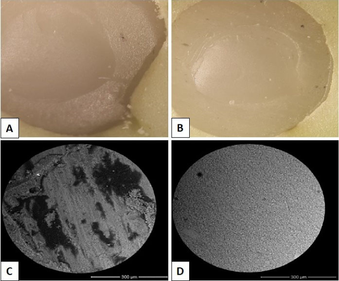
Figure 1.
Failure Mode Analysis by SEM; A) Mixed (×20 magnification) B) Cohesive (×20) C) Mixed (×100) D) Cohesive (×100).
It should be noted that there was not any adhesive fracture in this study.
.
Failure Mode Analysis by SEM; A) Mixed (×20 magnification) B) Cohesive (×20) C) Mixed (×100) D) Cohesive (×100).
It should be noted that there was not any adhesive fracture in this study.
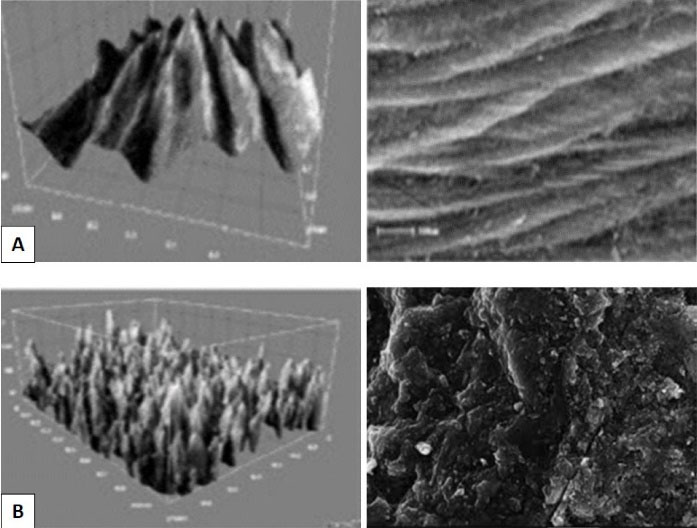
Figure 2.
Surface Roughness Analysis by SEM in ×100 magnification; A) Diamond Milling B) Sandblasting.
.
Surface Roughness Analysis by SEM in ×100 magnification; A) Diamond Milling B) Sandblasting.
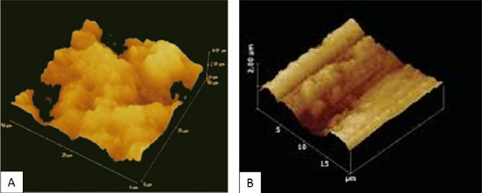
Figure 3.
Surface Roughness Analysis by AFM; A) Diamond milling B) Sandblasting.
.
Surface Roughness Analysis by AFM; A) Diamond milling B) Sandblasting.
Discussion
Old composite restorations do not have an oxygen-inhibited layer due to intraoral factors such as saliva, repeated temperature change, and also nutritional chemical alterations. Therefore, if restoration is damaged, we need chemomechanical methods to activate the surface of old restoration (23) in order to obtain the proper bond between the two composite layers (19).
We selected 5000 rpm thermocycling for our study in order to simulate oral conditions based on previous studies (22). Additionally, we decided to use the same composite for repaired composite discs in order to reduce factors affecting the bond strength (5,13,32).
Shahdad and Kennedy reported that using similar composites would not increase the bond strength between the two layers (13), and the chemical formula of the bonding adhesive used between the two composite layers is much more important than the chemical formula of composites (22). Nevertheless, in the present study, the same composites were used to minimize the effect of any dissimilarity. In the present study, Z250 composite was used due to its silica fillers which may interact with adhesives containing silane. One of our adhesives was Single Bond Universal that contained silane. Moreover, Adper Single Bond 2 contained nano-spherical silica particles (%10 weight) which were silane-treated to prevent agglomeration (33). Silane can react with the silica fillers in the composite and improve the bonding between the composite and the adhesive (17,34).
The bonding between the two layers of old and new composites is obtained by the following three mechanisms:
1. Through chemical bond with organic matrix
2. Through the chemical bond with the exposed fillers
3. Through micromechanical bond with the prepared surface (18).
Surface preparation can be done by mechanical or chemical approaches. Diamond milling (4,6,18,21-24) and sandblasting with 50 μm particles (15,18,21,24) are mechanical; however, etching with phosphoric acid gel 37% (6,17,24) or hydrofluoric acid (3,4,22,24) is a chemical method. In many studies, acid etchant is used before the adhesion. However, the acid invasion is not the same for all composites and it depends on the type of its filler particles (18). For example, hydrofluoric acid has no effect on a composite containing zirconia (17). According to Fawzy et al, acid alone does not affect the morphological deformation of the composite surface (34), and it is only used for cleaning. Therefore, we used these two mechanical methods in this study.
Applying adhesive after surface roughening has a great effect on the improvement of repair bond strength. This effect may be due to the penetration of adhesive into the surface layer and this micro-mechanical interlock has a positive effect on the repair bond strength (16).
Three adhesives have been used in this study. Single Bond Universal and All Bonds Universal are the two universal brands of multipurpose composite bonding used for enamel, dentin, metal alloy, amalgam, porcelain, and composite. The third brand is Adper Single Bond 2 which is a hydrophilic total etch adhesive (5th generation).
Diamond milling had slightly higher bond strength compared to sandblasting. However, there was no significant difference between the two methods. This result is similar to that found by Yesilyurt et al (22). Joulaei et al (17) revealed that diamond had better performance in improving bond strength; however, da Costa et al (21) and Hemadri et al (15) believed that sandblasting is more effective than diamond milling. Although a specific surface preparation may not have the same effect on different types of composite materials or various fillers, the use of diamond milling may provide greater bond strength. Surface roughness analysis by SEM showed that diamond milling provided rougher and more available exposed surface for the macro-mechanical bond compared to sandblasting. However, micro-retentive grooves were more irregular, with an average depth of 15 µm, in sandblasted samples and composite fillers or matrix structures were damaged greatly (16).
There were no significant statistical differences in the bond strength of experimental groups in the study. It is consistent with previous studies (6,16,18,23,24,35). It was found that adhesive generation is not important for having a successful composite repair. Single Bond Universal and Adper Single Bond 2 have silane in their structures and it is more likely to bond with silica filler of composite material. However, this was not confirmed statistically.
In order to recognize the type of failure mode, we used two different color composites (A1, A3) (7). The failure mode of the samples exhibited either cohesive (28.3%) or mixed fractures (71.7%). The adhesive pattern was not observed. There was no significant difference in the failure mode of different groups. Adhesive failure is the fracture in the adhesive layer between two composite layers while the cohesive fracture is a fracture within each composite matrix (8,9,36). The higher incidence of mixed fracture in the present study showed that the bond strength of composite repair is average.
One of the strong points of this study is the use of AFM along with SEM analysis. AFM data are more detailed compared to SEM because it contains information in three spatial dimensions. Therefore, AFM can exhibit irregularities better than SEM. SEM should be done under vacuum condition while AFM can be done in liquid. The samples may be more preserved in AFM methods; however, SEM may damage them greatly. In addition, AFM is less time-consuming (37,38).
Conclusion
There was no statistically significant difference in bond strength using diamond milling or sandblasting as surface preparation methods or using different generations of adhesives. However, two universal bonds had slightly better performance. Finally, diamond milling + Single Bond Universal is suggested as the best protocol related to the composite repair.
References
- Özcan M, Pekkan G. Effect of different adhesion strategies on bond strength of resin composite to composite-dentin complex. Oper Dent 2013; 38(1):63-72. doi: 10.2341/11-482-l [Crossref] [ Google Scholar]
- Ghavam M, Naeemi M, Hashemikamangar SS, Ebrahimi H, Kharazifard MJ. Repair bond strength of composite: effect of surface treatment and type of composite. J Clin Exp Dent 2018; 10(6):e520-e7. doi: 10.4317/jced.54030 [Crossref] [ Google Scholar]
- Gupta S, Parolia A, Jain A, Kundabala M, Mohan M, de Moraes Porto IC. A comparative effect of various surface chemical treatments on the resin composite-composite repair bond strength. J Indian Soc Pedod Prev Dent 2015; 33(3):245-9. doi: 10.4103/0970-4388.160402 [Crossref] [ Google Scholar]
- Eliasson ST, Tibballs J, Dahl JE. Effect of different surface treatments and adhesives on repair bond strength of resin composites after one and 12 months of storage using an improved microtensile test method. Oper Dent 2014; 39(5):E206-16. doi: 10.2341/12-429-l [Crossref] [ Google Scholar]
- Fornazari IA, Wille I, Meda EM, Brum RT, Souza EM. Effect of surface treatment, silane, and universal adhesive on microshear bond strength of nanofilled composite repairs. Oper Dent 2017; 42(4):367-74. doi: 10.2341/16-259-l [Crossref] [ Google Scholar]
- de Melo MA, Moysés MR, dos Santos SG, Alcântara CE, Ribeiro JC. Effects of different surface treatments and accelerated artificial aging on the bond strength of composite resin repairs. Braz Oral Res 2011; 25(6):485-91. doi: 10.1590/s1806-83242011000600003 [Crossref] [ Google Scholar]
- Cotes C, Cardoso M, Melo RM, Valandro LF, Bottino MA. Effect of composite surface treatment and aging on the bond strength between a core build-up composite and a luting agent. J Appl Oral Sci 2015; 23(1):71-8. doi: 10.1590/1678-775720140113 [Crossref] [ Google Scholar]
- Jafarzadeh Kashi TS, Erfan M, Rakhshan V, Aghabaigi N, Tabatabaei FS. An in vitro assessment of the effects of three surface treatments on repair bond strength of aged composites. Oper Dent 2011; 36(6):608-17. doi: 10.2341/10-386-l [Crossref] [ Google Scholar]
- Nassoohi N, Kazemi H, Sadaghiani M, Mansouri M, Rakhshan V. Effects of three surface conditioning techniques on repair bond strength of nanohybrid and nanofilled composites. Dent Res J (Isfahan) 2015; 12(6):554-61. doi: 10.4103/1735-3327.170575 [Crossref] [ Google Scholar]
- Craig RG. Craig’s Restorative Dental Materials. Mosby/Elsevier; 2006.
- Eliasson ST, Dahl JE. Effect of curing and silanizing on composite repair bond strength using an improved micro-tensile test method. Acta Biomater Odontol Scand 2017; 3(1):21-9. doi: 10.1080/23337931.2017.1301211 [Crossref] [ Google Scholar]
- Furuse AY, da Cunha LF, Benetti AR, Mondelli J. Bond strength of resin-resin interfaces contaminated with saliva and submitted to different surface treatments. J Appl Oral Sci 2007; 15(6):501-5. doi: 10.1590/s1678-77572007000600009 [Crossref] [ Google Scholar]
- Shahdad SA, Kennedy JG. Bond strength of repaired anterior composite resins: an in vitro study. J Dent 1998; 26(8):685-94. doi: 10.1016/s0300-5712(97)00044-4 [Crossref] [ Google Scholar]
- da Costa TR, Serrano AM, Atman AP, Loguercio AD, Reis A. Durability of composite repair using different surface treatments. J Dent 2012; 40(6):513-21. doi: 10.1016/j.jdent.2012.03.001 [Crossref] [ Google Scholar]
- Hemadri M, Saritha G, Rajasekhar V, Pachlag KA, Purushotham R, Reddy VK. Shear bond strength of repaired composites using surface treatments and repair materials: an in vitro study. J Int Oral Health 2014; 6(6):22-5. [ Google Scholar]
- Rathke A, Tymina Y, Haller B. Effect of different surface treatments on the composite-composite repair bond strength. Clin Oral Investig 2009; 13(3):317-23. doi: 10.1007/s00784-008-0228-2 [Crossref] [ Google Scholar]
- Joulaei M, Bahari M, Ahmadi A, Savadi Oskoee S. Effect of different surface treatments on repair micro-shear bond strength of silica- and zirconia-filled composite resins. J Dent Res Dent Clin Dent Prospects 2012; 6(4):131-7. doi: 10.5681/joddd.2012.027 [Crossref] [ Google Scholar]
- Rodrigues SA Jr, Ferracane JL, Della Bona A. Influence of surface treatments on the bond strength of repaired resin composite restorative materials. Dent Mater 2009; 25(4):442-51. doi: 10.1016/j.dental.2008.09.009 [Crossref] [ Google Scholar]
- Özcan M, Corazza PH, Marocho SM, Barbosa SH, Bottino MA. Repair bond strength of microhybrid, nanohybrid and nanofilled resin composites: effect of substrate resin type, surface conditioning and ageing. Clin Oral Investig 2013; 17(7):1751-8. doi: 10.1007/s00784-012-0863-5 [Crossref] [ Google Scholar]
- Imbery TA, Gray T, DeLatour F, Boxx C, Best AM, Moon PC. Evaluation of flexural, diametral tensile, and shear bond strength of composite repairs. Oper Dent 2014; 39(6):E250-60. doi: 10.2341/13-299-l [Crossref] [ Google Scholar]
- Costa TR, Ferreira SQ, Klein-Júnior CA, Loguercio AD, Reis A. Durability of surface treatments and intermediate agents used for repair of a polished composite. Oper Dent 2010; 35(2):231-7. doi: 10.2341/09-216-l [Crossref] [ Google Scholar]
- Yesilyurt C, Kusgoz A, Bayram M, Ulker M. Initial repair bond strength of a nano-filled hybrid resin: effect of surface treatments and bonding agents. J Esthet Restor Dent 2009; 21(4):251-60. doi: 10.1111/j.1708-8240.2009.00271.x [Crossref] [ Google Scholar]
- Hamano N, Chiang YC, Nyamaa I, Yamaguchi H, Ino S, Hickel R. Effect of different surface treatments on the repair strength of a nanofilled resin-based composite. Dent Mater J 2011; 30(4):537-45. doi: 10.4012/dmj.2010-202 [Crossref] [ Google Scholar]
- Zaghloul H, Elkassas DW, Haridy MF. Effect of incorporation of silane in the bonding agent on the repair potential of machinable esthetic blocks. Eur J Dent 2014; 8(1):44-52. doi: 10.4103/1305-7456.126240 [Crossref] [ Google Scholar]
- Staxrud F, Dahl JE. Role of bonding agents in the repair of composite resin restorations. Eur J Oral Sci 2011; 119(4):316-22. doi: 10.1111/j.1600-0722.2011.00833.x [Crossref] [ Google Scholar]
- Kanzow P, Wiegand A, Schwendicke F, Göstemeyer G. Same, same, but different? a systematic review of protocols for restoration repair. J Dent 2019; 86:1-16. doi: 10.1016/j.jdent.2019.05.021 [Crossref] [ Google Scholar]
- Lemos CA, Mauro SJ, de Campos RA, Dos Santos PH, Machado LS, Fagundes TC. Repairability of aged resin composites mediated by different restorative systems. Acta Odontol Latinoam 2016; 29(1):7-13. [ Google Scholar]
- Altinci P, Mutluay M, Tezvergil-Mutluay A. Repair bond strength of nanohybrid composite resins with a universal adhesive. Acta Biomater Odontol Scand 2018; 4(1):10-9. doi: 10.1080/23337931.2017.1412262 [Crossref] [ Google Scholar]
- Cuevas-Suárez CE, Nakanishi L, Isolan CP, Ribeiro JS, Moreira AG, Piva E. Repair bond strength of bulk-fill resin composite: Effect of different adhesive protocols. Dent Mater J 2020; 39(2):236-41. doi: 10.4012/dmj.2018-291 [Crossref] [ Google Scholar]
- Elkaffas AA, Hamama HHH, Mahmoud SH. Do universal adhesives promote bonding to dentin? a systematic review and meta-analysis. Restor Dent Endod 2018; 43(3):e29. doi: 10.5395/rde.2018.43.e29 [Crossref] [ Google Scholar]
- Carrilho E, Cardoso M, Marques Ferreira M, Marto CM, Paula A, Coelho AS. 10-MDP based dental adhesives: adhesive interface characterization and adhesive stability-a systematic review. Materials (Basel) 2019; 12(5). doi: 10.3390/ma12050790 [Crossref]
- Tezvergil A, Lassila LV, Vallittu PK. Composite-composite repair bond strength: effect of different adhesion primers. J Dent 2003; 31(8):521-5. doi: 10.1016/s0300-5712(03)00093-9 [Crossref] [ Google Scholar]
- Muñoz MA, Luque I, Hass V, Reis A, Loguercio AD, Bombarda NHC. Immediate bonding properties of universal adhesives to dentine. Journal of dentistry 2013; 41(5):404-411. doi: 10.1016/j.jdent.2013.03.001 [Crossref] [ Google Scholar]
- Fawzy AS, El-Askary FS, Amer MA. Effect of surface treatments on the tensile bond strength of repaired water-aged anterior restorative micro-fine hybrid resin composite. J Dent 2008; 36(12):969-76. doi: 10.1016/j.jdent.2008.07.014 [Crossref] [ Google Scholar]
- Hamano N, Ino S, Fukuyama T, Hickel R, Kunzelmann KH. Repair of silorane-based composites: microtensile bond strength of silorane-based composites repaired with methacrylate-based composites. Dent Mater J 2013; 32(5):695-701. doi: 10.4012/dmj.2013-129 [Crossref] [ Google Scholar]
- Rinastiti M, Özcan M, Siswomihardjo W, Busscher HJ. Effects of surface conditioning on repair bond strengths of non-aged and aged microhybrid, nanohybrid, and nanofilled composite resins. Clin Oral Investig 2011; 15(5):625-33. doi: 10.1007/s00784-010-0426-6 [Crossref] [ Google Scholar]
- Fischer NG, Dang J, Takamizawa T, Tsujimoto A, Barkmeier WW, Baruth AG. The role of spatial frequency analysis in correlating atomic force microscopy and optical profilometry with self-etch adhesive enamel bond fatigue durability. Microsc Res Tech 2019; 82(9):1419-29. doi: 10.1002/jemt.23294 [Crossref] [ Google Scholar]
- Piontek MC, Roos WH. Atomic force microscopy: an introduction. In: Peterman E, ed. Single Molecule Analysis. New York, NY: Humana Press; 2018. p. 243-58.10.1007/978-1-4939-7271-5_13.