Avicenna J Dent Res. 11(3):83-88.
doi: 10.34172/ajdr.2019.16
Original Article
Effect of Light Intensity and Curing Time on Color Stability of a Methacrylate-Based Composite Resin Using an LED Light-Curing Unit
Hosna Teimourian 1  , Negin Farahmandpour 1, Maryam Zali Moghadam 2, Hoda Pouyanfar 1, Narges Panahandeh 3, *
Author information:
1Department of Operative and Esthetic Dentistry, Faculty of Dentistry, Kermanshah University of Medical Sciences, Kermanshah, Iran
2Students Research Committee, Faculty of Dentistry, Kermanshah University of Medical Sciences, Kermanshah, Iran
3Dental Research Center, Research Institute of Dental Sciences, Shahid Beheshti University of Medical Sciences, Tehran, Iran
Abstract
Background: Despite improvements in the optical properties of composite resins, their color stability is still a matter of discussion. This study sought to assess the effect of light intensity and curing time by a light-emitting diode (LED) light-curing unit on color stability of a methacrylate-based composite resin.
Methods: In this in vitro study, 60 discs (8 × 2 mm) were fabricated of A2 shade of Z250 composite. Specimens were polished and divided into 4 groups (n=15) for curing for 20 or 40 seconds with a light intensity of 600 or 1200 mW/cm2. After immersion in distilled water at 37°C for 24 hours, colorimetry was performed using a spectroradiometer. The specimens were then immersed in tea solution for 7 days (3 times a day, each time for one hour) and were subjected to colorimetry again. Color change (∆E) was calculated and analyzed using two-way ANOVA.
Results: Significant color change was noted following an increase in curing time (P<0.05). No improvement in color stability was noted after increasing the light intensity (P>0.05). The interaction effect of light intensity and curing time on color change was significant (P<0.05).
Conclusions: Curing time is an important factor affecting the color stability of composite resin polymerized with LED light curing unit. On the other hand, increasing the light intensity over the standard threshold showed no significant effect.
Keywords: Color, Composite resin, Curing light, Light intensity, Time
Copyright and License Information
© 2019 The Author(s); Published by Hamadan University of Medical Sciences.
This is an open-access article distributed under the terms of the Creative Commons Attribution License (
http://creativecommons.org/licenses/by/4.0), which permits unrestricted use, distribution, and reproduction in any medium provided the original work is properly cited.
Citation: Teimourian H, Farahmandpour N, Zali Moghadam M, Pouyanfar H, Panahandeh N. Effect of Light Intensity and Curing Time on Color Stability of a Methacrylate-Based Composite Resin Using an LED Light-Curing Unit. Avicenna J Dent Res. 2019;11(3):83-88. doi: 10.34172/ajdr.2019.16.
Background
Highlights
Tooth-colored restorative materials should mimic the shade, translucency, shape and surface texture of natural teeth to blend in. They should also have adequate strength, optimal wear resistance, and good marginal adaptation and must be insoluble and biocompatible. They should also have color stability and mimic the external morphology of natural teeth.
Composite resins are direct tooth-colored restorative materials with optimal properties such as excellent esthetics and durability (1) and highly resemble the tooth structure (2). Due to the increased popularity of dental esthetic treatments and high patient demand for tooth-colored restorations, composite resin restorations constitute a large percentage of routine dental practice (3). However, these restorations have shortcomings as well, which include discoloration, wear, microleakage, and polymerization shrinkage (2).
Several intrinsic factors in the oral environment including chemical alterations in the structure of resin matrix, degree of polymerization, inorganic phase, inhibitors, activators, type of amine in resin and UV irradiation as well as several extrinsic factors such as nutrition and oral hygiene can cause color change in composite restorations (3). Many attempts have been made to improve the color stability of composite resins. However, their color stability is still a major problem (4).
Most previous studies on color stability of composite resins have focused on the effects of the type of light curing unit namely quartz tungsten halogen (QTH) or light emitting diode (LED) light-curing systems, type of coloring agent (comparison of colored drinks), methods of composite finishing and polishing, and composition of composite resins (in terms of the type of photo-initiator, or percentage and type of fillers) in this respect (2,5-8). However, studies evaluating the interaction effect of curing time and light intensity on color stability of composite resins are scarce. Therefore, this study aimed to assess the effect of light intensity and curing time by an LED light curing unit on the color stability of a methacrylate-based composite resin.
Materials and Methods
This in vitro experimental study was conducted on 60 composite discs, which were randomly divided into 4 groups of 15. The sample size was calculated to be 60 (n = 15 in each group) according to a study by Rüttermann et al (9), assuming the standard deviation of color change to be 0.8 and 0.5 after 20 seconds and 40 seconds of curing time, d = 0.8, alpha = 0.05 and power of 90%.
Preparation of Samples
A2 shade of Z250 composite (3M ESPE, St. Paul, MN, USA) was used for the fabrication of samples. Composite resin was applied to a prefabricated stainless steel mold, measuring 8 mm in internal diameter and 2 mm in height. The composite was packed in the mold using a medium-sized condenser. To prevent the formation of oxygen-inhibited layer during polymerization, the upper and lower surface of the composite were covered with a transparent celluloid strip. Polymerization was performed using an LED light curing unit (DB-685, Coxo, China). The tip of the light-curing unit had no distance from the composite surface. The diameter of the light guide tip was 8 mm, which matched the diameter of the mold. Before photo-curing, the light intensity was measured using a radiometer (LM-100; Monitex, DigiRate, Taiwan).
Composite discs were divided into 4 groups of 15 as follows:
-
Group 1: Radiation time of 20 seconds, light intensity of 600 mW/cm2
-
Group 2: Radiation time of 40 seconds, light intensity of 600 mW/cm2
-
Group 3: Radiation time of 20 seconds, light intensity of 1200 mW/cm2
-
Group 4: Radiation time of 40 seconds, light intensity of 1200 mW/cm2
All samples were then finished and polished. First, a 600-grit silicon carbide abrasive paper was used (991A; Starcke, Germany) for 20 seconds to polish the surface. Next, medium to superfine polishing discs (TOR VM, Stem Polishing Discs, Russia) were used with a hand-piece operating at 1000 rpm for 30 seconds. The samples were rinsed for 10 seconds between the polishing steps.
The samples in each group were then coded from 1 to 15 and placed in microtubes containing 2 cc of distilled water for 24 hours and incubated at 37°C (10). After 24 hours, the samples were subjected to primary colorimetry using a spectroradiometer (CS2000/2000A; Konica Minolta, Japan).
Colorimetry
The spectroradiometer used for this purpose had a wavelength spectrum of 380 to 780 nm with a wavelength accuracy of 1 nm. The light source illuminated the samples at 45° angle. White Leneta paper was used as a background according to the manufacturer’s instructions (11). The spectroradiometer was adjusted perpendicular to the surface of samples at 70 cm distance. The measuring angle was 0.2° (yielding a circular surface area with 2.4 mm diameter at the center of samples). Measurements were made in triplicate for each sample and the mean of the values was calculated and reported. Colorimetry was performed at 25°C.
After initial color assessment, tea was used for the aging process. For this purpose, one tea bag (Mahmood Tea) was placed in 100 cc of boiling water at 95°C for 3 minutes. After reaching 70°C, 2 cc of the tea solution was poured on each sample and they were stored in an oven at 37°C for 1 hour. This was repeated three times a day and a fresh tea solution was prepared for each time of testing as described earlier. At the end of the three experiments per day, the samples were stored in distilled water in an oven at 37°C until the next day and this was continued for 7 days.
The standard time for immersion in tea solution was considered to be 21 hours (3 hours a day for 7 days). For colorimetry, excess moisture of the samples was eliminated by a paper towel and they were then subjected to spectroradiometry as explained earlier.
The color parameters under D65/20 visual conditions were calculated by CS-S10W software (12,13). Color change (∆E) was calculated using the following formula:
Where the a* axis represents the green-red component, the b* axis indicates the blue-yellow component and L* indicates lightness.
Statistical Analysis Data were analyzed using descriptive and inferential statistics. The mean and standard deviation values were reported for descriptive data and tables and diagrams were drawn accordingly. In inferential statistics, the normal distribution of data was checked using the Shapiro-Wilk test. Data were then analyzed using two-way ANOVA. All statistical analyses were carried out using SPSS version 18.0 (SPSS Inc., IL, USA) at a significance level of 0.05.
Results
The results of Shapiro-Wilk test revealed that the color change data were normally distributed in all 4 groups (P> 0.05). Table 1 shows the color change values in the 4 groups. As demonstrated, the greatest color change occurred after irradiation of low intensity light (600 mW/cm2) for 20 seconds.
Table 1.
Descriptive Data for the Color Change in the 4 Groups According to the Intensity of Light and Duration of Curing
|
|
|
|
|
Count
|
Mean
|
SD
|
Min
|
Max
|
Q1
|
Med
|
Q3
|
| Intensity |
Low |
Time |
20 s |
15 |
11.17 |
1.68 |
8.42 |
13.92 |
9.43 |
11.54 |
12.38 |
| 40 s |
15 |
9.46 |
1.27 |
7.54 |
11.96 |
8.34 |
9.40 |
10.39 |
| Total |
30 |
10.31 |
1.70 |
7.54 |
13.92 |
8.96 |
10.14 |
11.77 |
| High |
Time |
20 s |
15 |
10.28 |
1.25 |
7.84 |
12.65 |
10.01 |
10.19 |
11.25 |
| 40 s |
15 |
10.07 |
1.07 |
7.32 |
11.82 |
9.22 |
10.34 |
10.54 |
| Total |
30 |
10.18 |
1.15 |
7.32 |
12.65 |
9.92 |
10.33 |
10.74 |
| Total |
Time |
20 s |
30 |
10.72 |
1.52 |
7.84 |
13.92 |
10.01 |
10.67 |
11.77 |
| 40 s |
30 |
9.77 |
1.19 |
7.32 |
11.96 |
9.17 |
10.00 |
10.41 |
| Total |
60 |
10.25 |
1.44 |
7.32 |
13.92 |
9.21 |
10.26 |
11.22 |
Note. SD: standard deviation; Min: minimum; Max: maximum; Q1: first quartile; Med: median;
Q3: third quartile.
Table 2 shows the results of two-way ANOVA. As presented, the light intensity had no significant effect on color change (P = 0.697) but curing time significantly affected the color change (P = 0.007), indicating that the mean color change was less after 40 seconds of curing than after 20 seconds of curing. Moreover, the interaction effect of light intensity and duration of curing on color change was statistically significant (P = 0.034), indicating that color change depended on light intensity at different curing times. Using low light intensity, the color change significantly decreased from 20 seconds of curing to 40 seconds of curing but this reduction was not significant with the use of high intensity light (Figure 1).
Table 2.
Results of Two-way ANOVA
|
Source
|
Type III Sum of Squares
|
df
|
Mean Square
|
F
|
P
Value
|
Partial Eta Squared
|
Observed Power
b
|
| Corrected model |
22.392a |
3 |
7.464 |
4.191 |
0.010 |
0.183 |
0.831 |
| Intercept |
6298.447 |
1 |
6298.447 |
3536.646 |
0.000 |
0.984 |
1.000 |
| Severity |
0.272 |
1 |
0.272 |
0.153 |
0.697 |
0.003 |
0.067 |
| Time |
13.727 |
1 |
13.727 |
7.708 |
0.007 |
0.121 |
0.779 |
| Severity * time |
8.393 |
1 |
8.393 |
4.713 |
0.034 |
0.078 |
0.569 |
| Error |
99.731 |
56 |
1.781 |
|
|
|
|
| Total |
6420.570 |
60 |
|
|
|
|
|
| Corrected total |
122.123 |
59 |
|
|
|
|
|
a R squared = 0.183 (adjusted R squared = 0.140).
b Computed using alpha = 0.05.
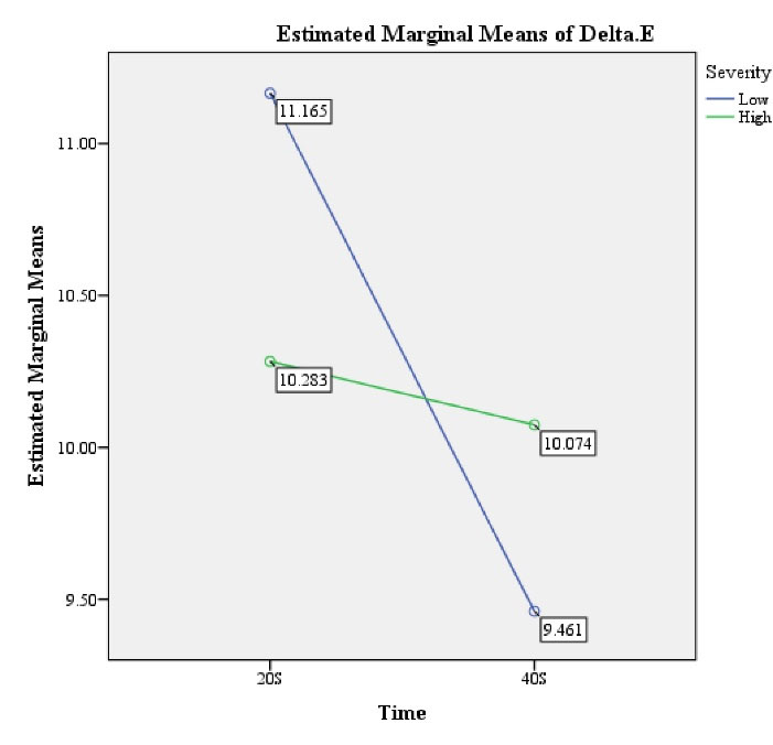
Figure 1.
Mean Color Change Depending on the Light Intensity and Duration of Curing.
.
Mean Color Change Depending on the Light Intensity and Duration of Curing.
Discussion
This in vitro experimental study assessed the effect of intensity and duration of light curing by an LED light curing unit on color stability of a methacrylate-based composite resin. The results showed significant color change following an increase in irradiation time (P < 0.05). No improvement in color stability was noted after increasing the light intensity (P > 0.05). The interaction effect of light intensity and curing time on color change was significant (P< 0.05).
The use of tea as the coloring agent in our study was attributed to the popularity of this drink in the Iranian community. It contains coloring agents that can cause significant discoloration of composite resins (14). All samples were stored in tea solution for 21 hours (3 hours a day for 7 days). Assuming that an individual averagely drinks three cups of tea per day and the teeth are exposed to tea solution for 15 minutes when drinking a cup of tea (15), a 21-hour immersion time simulates tea consumption for 28 days. To better simulate the oral environment, the temperature of tea was adjusted to 70°C. The samples were stored at 37°C simulating body temperature for the rest of the day.
The CIE L*a*b* color space was used for color assessment in our study. This system has the approval of the American Dental Association and is the most accurate and commonly applicable color system for colorimetry (14). In our study, spectroradiometer was used for the measurement of color parameters since (a) it is reliable, (b) when using spectroradiometer, edge-loss effect does not happen and therefore, shadows are not seen on the target object (16), and (c) it can analyze mathematical data with high accuracy and according to visual perception. In other words, the difference between mathematical data and visual perception is small in this device (17). Spectroradiometer is preferred to other colorimetric tools such as spectrophotometer for colorimetry of translucent samples, and in general, conditions can be better controlled in spectroradiometry. Spectroradiometer has been investigated in different studies (18,19).
In our study, the L*, a*, and b* values were measured before and after the immersion of samples in the coloring agent. To minimize measurement errors, colorimetry was repeated 3 times for each sample and the mean value was calculated and reported.
The current findings regarding the effect of light intensity on color stability of a methacrylate-based composite revealed that increasing the light intensity over the standard value (600 mW/cm2) had no significant effect on color stability of composite samples (P > 0.05); however, changing the curing time significantly affected the color stability of the tested methacrylate-based composite (P < 0.05).
Stress cracks in polymer matrix and relative debonding of filler from resin due to hydrolysis increase the opacity and changes the appearance of composite resin. Color change also occurs due to oxidation and is the result of the interaction of water with the polymer matrix as well as the interaction effect of unreacted polymers and the residual initiators or accelerators (5).
Polymerization shrinkage can be divided into two phases of pregel and postgel. In pregel shrinkage phase, composite can still flow and the internal stresses are released. The duration of this phase depends on the speed of reaction, which depends on the light intensity and concentration of initiator molecules (20). In postgel shrinkage phase, the composite can no longer flow and the generated stresses cannot be released. This phase of shrinkage can create stresses at the tooth-restoration interface (21). Therefore, the higher the intensity of light irradiated to the composite surface, or in other words, the faster the composite is polymerized, the shorter the viscoplastic phase would be. Therefore, the polymerization stress and consequently the microleakage of restoration would increase.
Yoshikawa et al (22) evaluated the effect of 20, 270 and 600 mW/cm2 light intensities and showed that higher light intensity can create greater gaps and increase the microleakage. Discacciati et al (23) indicated that 200 and 400 mW/cm2 light intensities had no significant effect on volumetric shrinkage of composite resins. Davidson-Kaban et al (24) demonstrated that higher light intensity (i.e., 700 mW/cm2) increases shrinkage and can cause marginal gap. The effect of low and high intensity light such as ramp curing on volumetric shrinkage of composite resin has been previously confirmed (25). Although composite curing with high intensity light is associated with superior mechanical properties, no linear correlation exists in this respect. In other words, higher intensity of energy is converted to a higher level of stress, which can increase the staining of dental composites and compromise their color stability (26).
Higher color stability after 40 seconds of curing can be due to a higher rate of polymerization during longer curing time. During longer curing time, higher number of photons reach composite resin and a higher number of camphorquinone molecules are excited. This leads to the generation of a higher number of free radicals and consequently greater polymerization of composite resin (27).
Inadequate polymerization is correlated to a low degree of conversion and higher number of double bonds, which decrease physical properties and increase water sorption, solubility and discoloration of composite (28).
With an increase in output of irradiated light by increasing the curing time or light intensity, the degree of conversion of double bonds to single bonds and consequently the polymerization of composite increase, which eventually improve color stability of samples (8).
Our study showed significant interaction effect of light intensity and curing time on the color stability, which means that color change that occurs following different curing times depends on the intensity of light. Poorsattar Bejeh Mir and Poorsattar Bejeh Mir (29) evaluated the effect of curing time on the color change of composite resins and found different results from ours. They demonstrated that color change was significantly greater after a longer curing time (20 seconds) than after shorter curing time (10 seconds). This finding may be due to the immediate measurement of color change without aging.
Rüttermann et al (9) evaluated the effect of curing time by LED and QTH light curing units on color stability of composites during 180 days and found that by increasing the curing time to 60 seconds, color stability did not improve in any group. This difference in the results may be due to different aging times and different aging conditions (water storage and xenon lamp). Our study showed that increasing the light intensity over the standard level had no significant effect on color stability of methacrylate-based composite samples. Zamarripa et al (30) found no significant association between the level of received energy and color stability of composites. They evaluated the effect of energy density (which is the result of light intensity and time and is expressed in mJ/cm2) on the color stability of composite resins. Both time and light intensity are involved in the calculation of energy density. Pires-de-Souza Fde et al (31) assessed the color stability of three composite resins cured with LED (320 mW/cm2) and QTH (500 mW/cm2) light curing units and found no significant difference in the color change of light shade composites irrespective of the type of light-curing unit and the intensity of light in the three composite resins. However, the dark shade of Tetric C3 showed greater color change when QTH light-curing unit was used instead of LED. This finding was in agreement with our result because we also used a light shade of composite.
Increased curing time, particularly with high-intensity light generates excessive heat, which is dangerous for the tooth and the supporting structures (1). Our findings suggest standard light intensity for curing composite resins because by the two-fold increase in the light intensity, no change occurred in color stability of composites. However, the generated heat at higher light intensity may compromise the vitality of the tooth. On the other hand, the standard curing time with most dental curing lights is 20 seconds. Assuming that the light source is completely adjacent to the restoration surface, this time is often sufficient for curing a light shade resin with depths of 2 to 2.5 mm. The tooth anatomy often prevents close contact of the light source with the restoration surface. A curing time of 40 seconds improves the degree of curing at all depths and is necessary for adequate curing of darker shades of composites (5). Similarly, we witnessed greater color stability after this time period in our study.
Since color change of tooth-colored restorations such as methacrylate-based composites in the oral environment is affected by several factors, which cannot be simulated in vitro, future in vivo studies are required to obtain more reliable results. Further studies are also recommended to assess longer curing times, different composites, different types of LED light-curing units and their effect on color stability of methacrylate-based composites.
Conclusions
Within the limitations of this study, the results showed that change in light intensity of LED light-curing unit had no effect on color stability of a methacrylate-based composite but changing the curing time with an LED light-curing unit significantly affected the color stability of composite. The induced color change following different curing times depended on the intensity of light.
Conflict of Interest Disclosures
The authors declared that they have no conflict of interests.
Acknowledgements
The authors gratefully acknowledge the Research Council of Kermanshah University of Medical Sciences for financial support.
Ethical Statement
The results of this study have been partly presented in a dissertation (code: 95627) for the degree of DDS in the Faculty of Dentistry, Kermanshah University of Medical Sciences.
Authors’ Contribution
HT and NF conceived the presented idea and designed the study. MZM carried out the experiment. HP contributed to sample preparation and performed the analytic calculations. NP wrote the manuscript.
References
- Hilton TJ, Ferracane JL, Broome JC. Summitt’s Fundamentals of Operative Dentistry: A Contemporary Approach. Quintessence Publishing Company Incorporated; 2013.
- Falkensammer F, Arnetzl GV, Wildburger A, Freudenthaler J. Color stability of different composite resin materials. J Prosthet Dent 2013; 109(6):378-83. doi: 10.1016/s0022-3913(13)60323-6 [Crossref] [ Google Scholar]
- Barutcigil Ç, Yildiz M. Intrinsic and extrinsic discoloration of dimethacrylate and silorane based composites. J Dent 2012; 40 Suppl 1:e57-63. doi: 10.1016/j.jdent.2011.12.017 [Crossref] [ Google Scholar]
- Sabatini C. Color stability behavior of methacrylate-based resin composites polymerized with light-emitting diodes and quartz-tungsten-halogen. Oper Dent 2015; 40(3):271-81. doi: 10.2341/14-080-l [Crossref] [ Google Scholar]
- Sakaguchi RL, Powers JM. Craig’s Restorative Dental Materials-E-Book. Elsevier Health Sciences; 2012.
- de Oliveira DC, Rocha MG, Gatti A, Correr AB, Ferracane JL, Sinhoret MA. Effect of different photoinitiators and reducing agents on cure efficiency and color stability of resin-based composites using different LED wavelengths. J Dent 2015; 43(12):1565-72. doi: 10.1016/j.jdent.2015.08.015 [Crossref] [ Google Scholar]
- Lepri CP, Palma-Dibb RG. Surface roughness and color change of a composite: influence of beverages and brushing. Dent Mater J 2012; 31(4):689-96. doi: 10.4012/dmj.2012-063 [Crossref] [ Google Scholar]
- Yazici AR, Celik C, Dayangaç B, Ozgünaltay G. The effect of curing units and staining solutions on the color stability of resin composites. Oper Dent 2007; 32(6):616-22. doi: 10.2341/07-3 [Crossref] [ Google Scholar]
- Rüttermann S, Suyoun K, Raab WH, Janda R. Effect of exposure time on the color stability of resin-based restorative materials when polymerized with quartz-tungsten halogen and LED light. Clin Oral Investig 2010; 14(5):599-605. doi: 10.1007/s00784-009-0316-y [Crossref] [ Google Scholar]
- Tekçe N, Tuncer S, Demirci M, Serim ME, Baydemir C. The effect of different drinks on the color stability of different restorative materials after one month. Restor Dent Endod 2015; 40(4):255-61. doi: 10.5395/rde.2015.40.4.255 [Crossref] [ Google Scholar]
- Hashemi Kamangar S, Kiakojoori K, Mirzaii M, Kharazifard M. Comparison of color change of silorane and methacrylate-based composites due to bleaching. J Islam Dent Assoc Iran 2015; 27(2):77-84. [ Google Scholar]
- Dozić A, Kleverlaan CJ, El-Zohairy A, Feilzer AJ, Khashayar G. Performance of five commercially available tooth color-measuring devices. J Prosthodont 2007; 16(2):93-100. doi: 10.1111/j.1532-849X.2007.00163.x [Crossref] [ Google Scholar]
- Johnston WM. Color measurement in dentistry. J Dent 2009; 37 Suppl 1:e2-6. doi: 10.1016/j.jdent.2009.03.011 [Crossref] [ Google Scholar]
- Arman M, Khalilak Z, Rajabi M, Esnaashari E, Saati K. In vitro spectrophotometry of tooth discoloration induced by tooth-colored mineral trioxide aggregate and calcium-enriched mixture cement. Iran Endod J 2015; 10(4):226-30. doi: 10.7508/iej.2015.04.003 [Crossref] [ Google Scholar]
- Ertaş E, Güler AU, Yücel AC, Köprülü H, Güler E. Color stability of resin composites after immersion in different drinks. Dent Mater J 2006; 25(2):371-6. [ Google Scholar]
- Bayindir F, Kuo S, Johnston WM, Wee AG. Coverage error of three conceptually different shade guide systems to vital unrestored dentition. J Prosthet Dent 2007; 98(3):175-85. doi: 10.1016/s0022-3913(07)60053-5 [Crossref] [ Google Scholar]
- Ghinea R, Ugarte-Alvan L, Yebra A, Pecho OE, Paravina RD, Perez Mdel M. Influence of surface roughness on the color of dental-resin composites. J Zhejiang Univ Sci B 2011; 12(7):552-62. doi: 10.1631/jzus.B1000374 [Crossref] [ Google Scholar]
- Gozalo-Diaz DJ, Lindsey DT, Johnston WM, Wee AG. Measurement of color for craniofacial structures using a 45/0-degree optical configuration. J Prosthet Dent 2007; 97(1):45-53. doi: 10.1016/j.prosdent.2006.10.013 [Crossref] [ Google Scholar]
- Wee AG, Lindsey DT, Kuo S, Johnston WM. Color accuracy of commercial digital cameras for use in dentistry. Dent Mater 2006; 22(6):553-9. doi: 10.1016/j.dental.2005.05.011 [Crossref] [ Google Scholar]
- Stanford CM, Fan PL, Leung RL, Knoeppel R, Stanford JW. Polymerization of composites by sequential and continuous irradiation with visible light. Oper Dent 1986; 11(2):51-4. [ Google Scholar]
- Yap AU, Wang HB, Siow KS, Gan LM. Polymerization shrinkage of visible-light-cured composites. Oper Dent 2000; 25(2):98-103. [ Google Scholar]
- Yoshikawa T, Burrow MF, Tagami J. A light curing method for improving marginal sealing and cavity wall adaptation of resin composite restorations. Dent Mater 2001; 17(4):359-66. doi: 10.1016/s0109-5641(00)00095-6 [Crossref] [ Google Scholar]
- Discacciati JAC, Neves AD, Oréfice RL, Pimenta FJGS, Sander HH. Effect of light intensity and irradiation time on the polymerization process of a dental composite resin. Mater Res 2004; 7(2):313-8. doi: 10.1590/S1516-14392004000200015 [Crossref] [ Google Scholar]
- Davidson-Kaban SS, Davidson CL, Feilzer AJ, de Gee AJ, Erdilek N. The effect of curing light variations on bulk curing and wall-to-wall quality of two types and various shades of resin composites. Dent Mater 1997; 13(6):344-52. doi: 10.1016/s0109-5641(97)80105-4 [Crossref] [ Google Scholar]
- Silikas N, Eliades G, Watts DC. Light intensity effects on resin-composite degree of conversion and shrinkage strain. Dent Mater 2000; 16(4):292-6. doi: 10.1016/s0109-5641(00)00020-8 [Crossref] [ Google Scholar]
- Dennison JB, Yaman P, Seir R, Hamilton JC. Effect of variable light intensity on composite shrinkage. J Prosthet Dent 2000; 84(5):499-505. doi: 10.1067/mpr.2000.110494 [Crossref] [ Google Scholar]
- Moein N, Darabi F, Davalloo R, Tavangar M, Hasanzade E. The effect of standard and extended curing time in different distances on composite’s degree of conversion. Journal of Dentomaxillofacial Radiology, Pathology and Surgery 2013; 2(1):22-7. doi: 10.18869/acadpub.3dj.2.1.4 [Crossref] [ Google Scholar]
- Danesh G, Davids H, Reinhardt KJ, Ott K, Schäfer E. Polymerisation characteristics of resin composites polymerised with different curing units. J Dent 2004; 32(6):479-88. doi: 10.1016/j.jdent.2004.03.003 [Crossref] [ Google Scholar]
- Poorsattar Bejeh Mir A, Poorsattar Bejeh Mir M. The effect of different curing time regimens on immediate postpolymerization color changes of resin composites. J Contemp Dent Pract 2012; 13(4):472-5. doi: 10.5005/jp-journals-10024-1171 [Crossref] [ Google Scholar]
- Zamarripa E, Ancona AL, D’Accorso NB, Macchi RL, Abate PF. Effect of energy density on color stability in dental resin composites under accelerated aging. Acta Odontol Latinoam 2008; 21(1):11-5. [ Google Scholar]
- Pires-de-Souza Fde C, Garcia Lda F, Hamida HM, Casemiro LA. Color stability of composites subjected to accelerated aging after curing using either a halogen or a light emitting diode source. Braz Dent J 2007; 18(2):119-23. doi: 10.1590/s0103-64402007000200006 [Crossref] [ Google Scholar]