Avicenna J Dent Res. 11(3):94-97.
doi: 10.34172/ajdr.2019.18
Original Article
Detection of Radiopaque Dental Materials From Lateral Cephalometric Images Using Canny Edge Detection and Nonlinear Gamma Correction
Shiva Gheibi 1  , Morteza Yadekar 2, * , Mahrokh Gheibi 3
Author information:
1Department of Oral and Maxillofacial Radiology, Faculty of Dentistry, Dental and Periodontal Research Center, Tabriz University of Medical Sciences, Tabriz, Iran
2Department of Electronic Engineering, Seraj Higher Education Institute, Tabriz, Iran
3Department of Biotechnology, Urmia Branch, Islamic Azad University, Urmia, Iran
Abstract
Background: One of the most critical issues of cone-beam computed tomographic (CBCT) imaging is the presence of high-density subjects such as metal implants and dental fillings that consequently waste the benefit information in imaging due to their artifact developments. Given that 3D images of CBCT consist of a series of orthogonal images (images similar to lateral cephalometric images), the aim of this study was to offer an algorithm for detection of radiopaque dental materials from lateral cephalometric images to remove cupping artifacts from CBCT images.
Methods: In this paper, lateral cephalometric images of Planmeca Scara 3 were used, each of which with 1259×1674 resolution in JPEG format. A total of 35 images of patients with radiopaque dental materials were selected. To decrease the time and image detection steps and for the purpose of acceleration we used Robinson edge detection and nonlinear gamma correction instead of filtering stage and noise reduction.
Results: After applying the proposed algorithm to images and comparison with the acquired images, it was observed that the proposed algorithm was able to successfully detect radiopaque dental materials on lateral cephalometric images with high accuracy.
Conclusions: Comparison of the reconstructed images and Table showed that the proposed algorithm was successful in detecting radiopaque dental materials on latral cephalometric images without reduction in image quality.
Keywords: Lateral cephalometric images, Canny edge detector, Cupping artifact, Radiopaque dental materials, Nonlinear gamma correction
Copyright and License Information
© 2019 The Author(s); Published by Hamadan University of Medical Sciences.
This is an open-access article distributed under the terms of the Creative Commons Attribution License (
http://creativecommons.org/licenses/by/4.0), which permits unrestricted use, distribution, and reproduction in any medium provided the original work is properly cited.
Citation: Gheibi S, Yadekar M, Gheibi M. Detection of Radiopaque Dental Materials From Lateral Cephalometric Images Using Canny Edge Detection and Nonlinear Gamma Correction. Avicenna J Dent Res. 2019;11(3):94-97. doi: 10.34172/ ajdr.2019.18.
Background
Cone-beam computed tomographic (CBCT) imaging is consist of multiple sequential planar projection images which are obtained while the x-ray source and detector move through an arc of 180 to 360°. Basis images appear similar to cephalometric radiographic images except that each is slightly offset from the next. There are usually several hundred two-dimensional basis images from which the image volume is calculated and constructed.
As an x-ray beam passes through a dense object, lower energy photons are absorbed in preference to higher energy photons. This phenomenon, called beam hardening, results in two types of artifact: 1) Cupping artifact, and 2) streaks and dark bands.
In clinical practice, it is advised that the field size should be reduced, patient position be modified, or the dental arches be separated to avoid scanning regions susceptible to beam hardening (e.g., metallic restorations, dental implants) (1-3).
To improve these artifacts, various methods, including software-based and hardware-based algorithms, have been proposed. Among the methods mentioned above, algorithm-based methods, with high-speed operation, low cost, and no need for additional equipment are preferable to other methods.
Various methods have been used to determine the exact location of different objects. Of all these methods, we can mention accurate caries lesion detection with segmentation and boundary points (4,5), automatic detection of pulmonary tuberculosis by watershed segmentation (6,7), extraction of cervical vertebrae from panoramic images used Tomosynthesis method (8,9), and so on.
We assume that if the proposed algorithm can detect radiopaque dental materials on raw orthogonal images and import resultant images before the reconstruction stage, we can correct cupping artifacts on CBCT images. Therefore, we proposed an algorithm to detect radiopaque dental materials on lateral cephalometric images.
Materials and Methods
In this study, lateral cephalometric images of Planmeca Scara 3 were used. Each image had a resolution of 1259×1674 in the JPEG format. A total of 35 images of patients with radiopaque dental materials were selected. The images belonged to patients with an age range of 20–50 years. Figure 1 shows a sample of lateral cephalometric images belonging to the patients with radiopaque dental materials.

Figure 1.
Lateral Cephalometric Image of Patient with Radiopaque Dental Materials.
.
Lateral Cephalometric Image of Patient with Radiopaque Dental Materials.
One of the problems of radiographic images is mottle noise, which impairs radiographic images and influences their distinction and interpretation. Therefore, filtering and reducing noise is an important stage before the extraction and recognition of medical image features. In this study, Canny edge detection and nonlinear gamma correction was used instead of filtering stage and noise reduction in order to decrease the time and image detection steps and for the purpose of acceleration. Figure 2 presents the proposed algorithm.
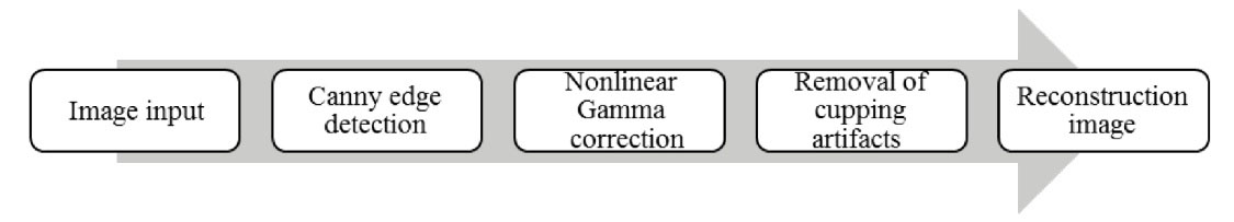
Figure 2.
The Proposed Algorithm.
.
The Proposed Algorithm.
Canny Edge Detector
This method, at the beginning, softens the image to omit the effect of noise. Then, it gives the gradient information to find the areas with high variation. Then, the algorithm moves along these areas to nullify every pixel whose gradient magnitude is not maximum.
In other words, there should be a connection between this pixel and edge pixels and if the pixel value is above the threshold, that pixel is selected as edge. Therefore, the edge detection process using the Canny algorithm is classified into steps as follows:
-
Softening and reducing noise
-
Calculating gradient magnitude based on image
-
Detecting the direction of probable edges
-
Detecting real edges
-
Thresholding
Figure 3 shows the result of Canny edge detection.
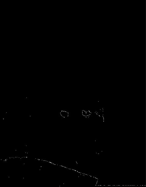
Figure 3.
Canny Edge Detection Result.
.
Canny Edge Detection Result.
Nonlinear Gamma Correction
Nonlinear gamma correction, or often simply gamma, is the name of a nonlinear operation used to encode and decode luminance in fixed image systems or definition of tristimulus values in video. Nonlinear gamma correction is, in the simplest cases, defined by the following power-law expression (10).
Iout = A Iinγ (1)
In the above relation, where Iin and Iout are the input and output image intensities, respectively, A is the constant value, and γ is nonlinear Gamma correction value, the computation of which is shown in equation 1. In the common case of A = 1, inputs and outputs are typically in the 0–1 range.
A γ value of <1 is sometimes called an encoding gamma, and the process of encoding with this compressive power-law nonlinearity is called gamma compression; conversely, a γ value of >1 is called a decoding gamma and the application of the expansive power-law nonlinearity is called gamma expansion.
The result of nonlinear gamma correction is shown in Figure 4.
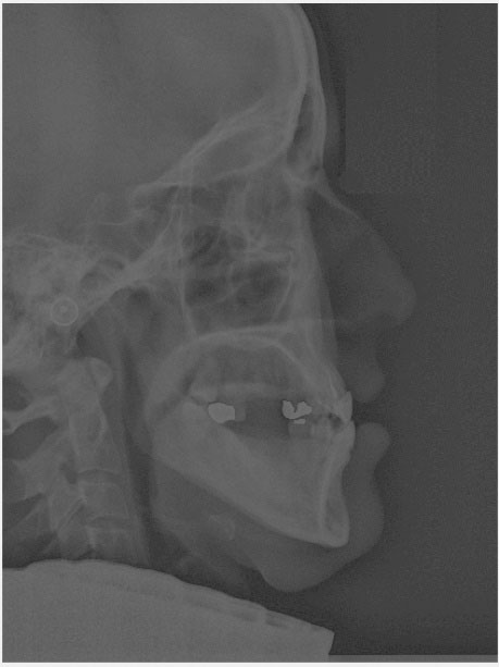
Figure 4.
Nonlinear Gamma Correction Result.
.
Nonlinear Gamma Correction Result.
Radiopaque Dental Materials Detection and Image Reconstruction
After applying Canny edge detector and nonlinear gamma correction, density difference of lateral cephalometric images in the basis image and the image produced by nonlinear Gamma correction is computed in outcome areas of Canny. The points at which this density difference is beyond a definite limit are considered image radiopaque dental materials boundary and are detected from the basis image. The result of such a procedure is shown in Figure 5.
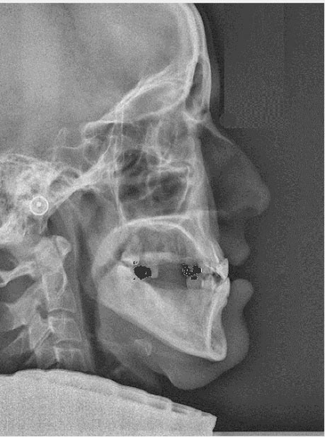
Figure 5.
Lateral Cephalometric Image Without Cupping Artifacts.
.
Lateral Cephalometric Image Without Cupping Artifacts.
Evaluation Criteria
Three techniques are used to evaluate the method used for noise reduction of radiographic images and compare it with the most common methods used as follows:
1. Median to Standard Deviation Ratio (MSR)
In (3), 𝜂d and 𝜎𝑑 are median and standard deviation of the region of interest, respectively.
2. Contrast to Noise Ratio
Contrast-to-noise ratio (CNR) is also an important quality evaluation method for medical imaging, represented as follows:
In (4), 𝑑 and 𝜎𝑑 are median and standard deviation of the region of interest, respectively.
3. Peak Signal-to-Noise Ratio
For evaluation of the efficiency of the new filter used in this study, we computed peak signal-to-noise ratio (PSNR)as image quality evaluation method.
Peak signal-to-noise relation used in medical x-ray images is defined as follows:
In (5), (𝑥,y)ε𝐷 is high-dose x-ray image, 𝐻∗𝐿 is pixel content, and 𝑜(𝑥,𝑦)ε𝑂 is the final image.
In Table 1, the results of evaluation algorithm processes by max, variance, median, PSNR, MSR, and CNR are presented.
Table 1.
Results of the Evaluation Criteria in Axial Images
|
|
Max
|
Variance
|
Median
|
PSNR
|
MSR
|
CNR
|
| Nonlinear gamma conversion |
117 |
209.7 |
83.03 |
33.6275 |
5.73 |
1.07 |
| Final image |
255 |
2.09e+03 |
118.93 |
18.1201 |
2.601 |
0.009 |
Conclusion
Currently, the use of computer systems to determine the exact location of objects on radiographic images has attracted the attention of researchers since they increase assessment accuracy and reduce response time. The aim of this study was to determine the exact location of radiopaque objects, including metal implants, amalgam fillings, post and cores, and so forth on lateral cephalometric images. One of the most important advantages of this study was the use of Canny edge detection and nonlinear Gamma correction instead of filtering stage and noise omission to decrease the number of procedural steps and accelerate image detection. Figure 5 and Table 1 showed that the proposed algorithm decreased median, variance, maximum value of the image, CNR, MSR, and PSNR.
Comparison of the final images showed that the proposed algorithm successfully detected radiopaque dental materials on lateral cephalometric images with high accuracy. If we can do this algorithm on raw orthogonal images of CBCT, we can remove cupping artifacts from CBCT images.
Conflict of Interest Disclosures
The authors declare that they have no conflict of interests.
Ethical Statement
The study uses archival images, so all ethical considerations are taken into account.
Authors’ Contribution
Shiva Gheibi: Preparing images and contributing to the editing of the article.
Mahrokh Gheibi: Review of previous research.
References
- William C. Cone-Beam Computed Tomography: Volume Acquisition. In: White SC, Pharoah MJ, eds. Oral Radiology. 7th ed. Canada: Elsevier; 2014. p. 193-97.
- Mengko TL, Adiono T, Setyawan H, Setiadarma R. Design and implementation of object detection and classification system based on deformable template algorithm. Chiangmai, Thailand: IEEE. APCCAS 1998. 1998 IEEE Asia-Pacific Conference on Circuits and Systems. Microelectronics and Integrating Systems. Proceedings (Cat. No.98EX242); 1998. p. 311-4. 10.1109/APCCAS.1998.743755.
- Leymarie F, Levine MD. Tracking deformable objects in the plane using an active contour model. IEEE Trans Pattern Anal Mach Intell 1993; 15(6):617-34. doi: 10.1109/34.216733 [Crossref] [ Google Scholar]
- Datta S, Chaki N. Detection of dental caries lesion at early stage based on image analysis technique. Bhubaneswar, India: 2015 IEEE International Conference on Computer Graphics, Vision and Information Security (CGVIS); 2015. p. 89-93. 10.1109/CGVIS.2015.7449899.
- Amit Y, Kong A. Graphical templates for model registration. IEEE Trans Pattern Anal Mach Intell 1996; 18(3):225-36. doi: 10.1109/34.485529 [Crossref] [ Google Scholar]
- Poornimadevi CS, Halen Sulochana C. Automatic detection of pulmonary tuberculosis using image processing techniques. Chennai, India: 2016 International Conference on Wireless Communications, Signal Processing and Networking (WiSPNET); 2016. p. 798-802. 10.1109/WiSPNET.2016.7566243.
- Staib LH, Duncan JS. Boundary finding with parametrically deformable models. IEEE Trans Pattern Anal Mach Intell 1992; 14(11):1061-75. doi: 10.1109/34.166621 [Crossref] [ Google Scholar]
- Yamamoto J, Yanase M, Ogawa K, Katsumata A. Extraction of cervical vertebrae from panoramic X-ray images. Seoul, South Korea: 2013 IEEE Nuclear Science Symposium and Medical Imaging Conference; 2013. p. 1-2. 10.1109/NSSMIC.2013.6829282.
- Jolly MPD, Lakshmanan S, Jain AK. Vehicle segmentation and classification using deformable templates. IEEE Trans Pattern Anal Mach Intell 1996; 18(3):293-308. doi: 10.1109/34.485557 [Crossref] [ Google Scholar]
- Rahman S, Rahman MM, Abdullah-Al-Wadud M, Al-Quaderi GD, Shoyaib M. An adaptive gamma correction for image enhancement. EURASIP Journal on Image and Video Processing 2016; 2016(1):35. doi: 10.1186/s13640-016-0138-1 [Crossref] [ Google Scholar]