Avicenna J Dent Res. 11(2):76-78.
doi: 10.34172/ajdr.2019.14
Case Report
The Effect of Mandibular Symphysis Fracture on the Position of the Permanent Incisor Tooth Germ
Mohammad Esmaelzadeh 1  , Ali Aghajani 2, * , Arghavan Afshar 2, Fahimeh Daneshyar 2
Author information:
1Pedodontist, Hamadan, Iran
2Postgraduate Student, Department of Pediatric Dentistry, School of Dentistry, Hamadan University of Medical Sciences, Hamadan, Iran
*
Correspondence to Ali Aghajani, Department of Pediatric Dentistry, School of Dentistry, Hamadan University of Medical Sciences, Hamadan, Iran Email:
Ali_dnt.2000@yahoo.com
Abstract
Background: Among facial bones in children, mandible bone has the highest fracture rate, 8.4% of which is related to Symphysis fractures.
Case Presentation: A 3.5-year-old girl with complaints about the mobility of the mandibular left primary central incisor without a history of recent dental trauma and caries referred to the Department of Pediatric Dentistry of Hamadan University. After radiographic evaluation, occlusal displacement of lower left permanent central incisor tooth germ and root resorption of lower left primary central incisor were seen.
Conclusions: This case report implies that because of the close proximity of the root of the primary tooth to its developing permanent successor, jaw fracture and especially mandibular fracture combined with dental injuries (e.g., intrusion, avulsion, and extrusive luxation) can cause damages and significant displacement in permanent successor tooth germs.
Keywords: Fractures, Dentition, Deciduous
Copyright and License Information
© 2019 The Author(s); Published by Hamadan University of Medical Sciences.
This is an open-access article distributed under the terms of the Creative Commons Attribution License (
http://creativecommons.org/licenses/by/4.0), which permits unrestricted use, distribution, and reproduction in any medium provided the original work is properly cited.
Citation: Esmaelzadeh M, Aghajani A, Afshar A, Daneshyar F. The Effect of Mandibular Symphysis Fracture on the Position of the Permanent Incisor Tooth Germ. Avicenna J Dent Res. 2019;11(2):76-78. doi: 10.15171/ajdr.2019.14.
Background
Highlights
Among facial bones in children, mandible bone has the highest fracture rate, 8.4% of which is related to symphysis fractures. Generally, 7.7% of mandible fractures occur in children under 16 years old and 2.9% in children under the age of 10 years. The incidence of fractures increases with age (1). There are several causes for symphysis fractures, the most important of which are conflicts, traffic accidents and falls (2). Considering the fact that only the incisors have erupted in infants, lack of support in the lateral regions can imply that forceful occlusion resulting from trauma to the chin can result in fracture of the anterior portion of the alveolar process (3). Because of the close proximity of the root of the primary tooth to its developing permanent successor, injuries to the primary dentition can cause considerable damages to the permanent tooth germ (4).
Injuries to the primary dentition are common and occur with a significantly higher annual incidence compared to the permanent dentition (5). One difference between injuries in the primary and permanent dentition appears to be the difference in the mechanical properties of the supporting periodontium. In primary dentition, the surrounding bone is less dense and less mineralized, which implies that a tooth damaged by trauma can easily be displaced instead of fractured. This difference in the trauma pattern favoring luxations rather than fractures has been found to be typical for the primary dentition (6). Disturbances in permanent tooth germs are latent complications that may be seen following all types of primary tooth injuries especially intrusion, avulsion, and alveolar bone fracture (4).
In particular, children between 18 to 30 months are very prone to accidents. Poor motor coordination and the child’s inability to evaluate potential risks are relevant conditions to be considered for a close supervision at this early age. As falls are the predominant cause for dental injuries in young age groups, the incidence of trauma is usually equally high in both genders. It is not until early school age (considering the eruption of permanent teeth) that boys become more prone to dental trauma (7). The radiographic study of symphysis fracture should include extra-oral and intraoral radiographs. Generally, extra-oral radiographs, especially panoramic methods help a lot in determining the direction and location of the fracture line (8). The management of pediatric mandible fractures is substantially different from that of the adult injury. This is due primarily to the presence of multiple tooth buds throughout the substance of the mandible and probability of damage to them (1). The treatment of symphysis fractures in children is by minimal interference in skeletal position with putting the parts together and fixing them (9).
Case Report
The case of the study was a 3.5-year-old girl who referred to the Department of Pediatric Dentistry of Hamadan University with complaints about the mobility of mandibular left primary central incisor without a history of recent dental trauma and caries. During the intraoral examination, severe mobility of mandibular left primary central incisor (7) was seen and there were no signs of the mobility of primary dentition in other parts of the mouth. In examining the medical history of the patient, it was determined that the child had a history of trauma and mandibular symphysis fracture due to an accident she had when she was one year old. According to the observation, panoramic radiography was performed for a complete evaluation of the teeth and the skeletal jaw and also periapical radiography was used for a more accurate examination of the area of interest (Figure 1). After radiographic evaluation, occlusal displacement of lower left permanent central incisor tooth germ and root resorption of lower left primary central incisor were seen. For preventing damage to permanent tooth germ, the bone plate was used instead of wire in the treatment of symphysis fractures. Due to the severe mobility of the teeth and the possibility of aspiration in the child, the decision was to extract this tooth and follow up the eruption and root development of mandibular first left permanent central incisor. After one year follow up, the complete eruption of the tooth with grade 2 mobility during the intraoral evaluation and lack of root development in the radiographic examination were observed (Figure 2). Due to the poor prognosis of the tooth and the possibility of aspiration, the decision was made to extract the tooth.
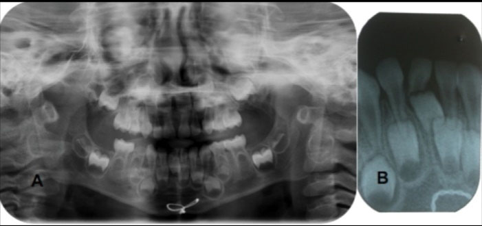
Figure 1.
Radiographic Examination of a 3.5 Year Old Girl at First Appointment (A: Panoramic View, B: Periapical View (Anterior Mandible))
.
Radiographic Examination of a 3.5 Year Old Girl at First Appointment (A: Panoramic View, B: Periapical View (Anterior Mandible))
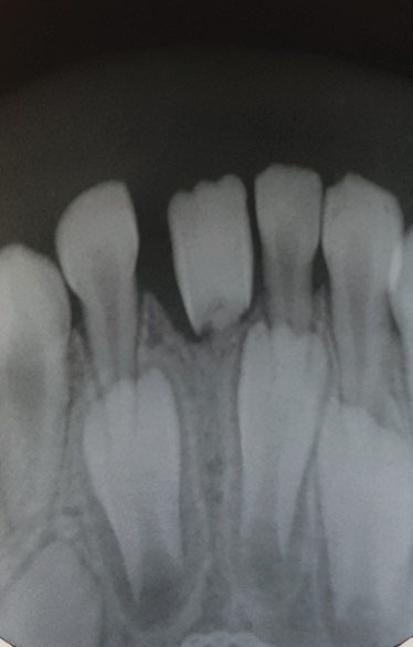
Figure 2.
Radiographic Examination after A Follow-up of One Year (Periapical View)
.
Radiographic Examination after A Follow-up of One Year (Periapical View)
Discussion
Facial fractures in the pediatric age group generally account for about 5% of all fractures and this percentage drops considerably in those less than 5 years of age. Their incidence rises as children begin school and also peaks during puberty and adolescence (10). Pediatric fractures are unusual when compared with fractures in adults (11). The reasons for this statement are based primarily on social and anatomical factors. Most often, children are in protected environments, under the supervision of parents and thus are less exposed to major trauma, occupational accidents or interpersonal violence, which are common causes of facial fractures in adults. Regarding maxillofacial fractures, a low incidence is due to the early stage of development of the facial skeleton and the sinuses, leading to a craniofacial disproportion. Moreover, the flexibility of the facial skeleton and the relative protection offered by existing fat in the subcutaneous tissue around the bones of the face contribute to the reduction of the incidence of fractures, especially maxillofacial fractures (12). Approximately half of all pediatric facial fractures involve the mandible and boys are more commonly affected than girls with a ratio of 2:1 (13). In another study, falls and traffic accidents were two causes of the fractured mandible in children (8). Unrestrained children who are seated or standing often hit the dashboard or windshield when the car is stopped suddenly (14). Considering that only the incisors have erupted in infants, lack of support in the lateral regions can imply that forceful occlusion resulting from trauma to the chin can result in fracture of the anterior portion of the alveolar process (2). An injury to the chin may produce a sudden forceful closure of the mandibular teeth with their maxillary counterparts. As a result, a wide range of injuries may affect molar teeth. These injuries include minor enamel fractures, fractures with dentin exposure that may imitate a carious lesion on the radiograph, crown-root fractures with or without pulp exposure, and injuries to the PDL. In addition, fracture of the mandible may occur mainly in the symphysis, mental, and subcondylar areas. These injuries have also been correlated with cervical spine fractures (15). Because of the close proximity of the root of the primary tooth to its developing permanent successor, injuries to the primary dentition can cause considerable damages to the permanent tooth germ (3).
In this case, after the examination of medical history, it was determined that the child had a history of trauma and mandibular symphysis fracture due to an accident she had when she was one year old. After radiographic evaluation, occlusal displacement of lower left permanent central incisor tooth germ and root resorption of lower left primary central incisor were seen. For preventing damage to permanent tooth germ, the bone plate was used instead of wire in the treatment of symphysis fractures. Due to the severe mobility of the teeth and the possibility of aspiration in the child, the decision was to extract this tooth and follow up the eruption and root development of mandibular first left permanent central incisor. After one year follow up, the complete eruption of the tooth with grade 2 mobility during the intraoral evaluation and lack of root development in the radiographic examination were observed. Due to the poor prognosis of the tooth and the possibility of aspiration, the decision was made to extract the tooth.
Conclusions
This case report implies that because of the close proximity of the root of the primary tooth to its developing permanent successor, jaw fracture and especially mandibular fracture combined with dental injuries can cause damages and significant displacement in permanent successor tooth germs.
Authors’ Contribution
The authors had same contribution in this study.
Ethical Statement
Informed consent was obtained from the parents’ patient for publication of this study.
Conflict of Interest Disclosures
The authors declare that they have no conflict of interests.
References
- Iida S, Matsuya T. Paediatric maxillofacial fractures: their aetiological characters and fracture patterns. J Craniomaxillofac Surg 2002; 30(4):237-41. doi: 10.1054/jcms.2002.0295 [Crossref] [ Google Scholar]
- Ghosh R, Gopalkrishnan K, Anand J. Pediatric facial fractures: a 10-year study. J Maxillofac Oral Surg 2018; 17(2):158-63. doi: 10.1007/s12663-016-0965-8 [Crossref] [ Google Scholar]
- Kramer PF, Onetto J, Flores MT, Borges TS, Feldens CA. Traumatic Dental Injuries in the primary dentition: a 15-year bibliometric analysis of Dental Traumatology. Dent Traumatol 2016; 32(5):341-6. doi: 10.1111/edt.12262 [Crossref] [ Google Scholar]
- Sleiter R, von Arx T. [Developmental disorders of permanent teeth after injuries of their primary predecessors A retrospective study]. Schweiz Monatsschr Zahnmed 2002; 112(3):214-9. [ Google Scholar]
- Demianczuk AN, Verchere C, Phillips JH. The effect on facial growth of pediatric mandibular fractures. J Craniofac Surg 1999; 10(4):323-8. doi: 10.1097/00001665-199907000-00007 [Crossref] [ Google Scholar]
- Glendor U. On dental trauma in children and adolescents Incidence, risk, treatment, time and costs. Swed Dent J Suppl 2000; 140:1-52. [ Google Scholar]
- Ferreira PC, Amarante JM, Silva PN, Rodrigues JM, Choupina MP, Silva AC. Retrospective study of 1251 maxillofacial fractures in children and adolescents. Plast Reconstr Surg 2005; 115(6):1500-8. doi: 10.1097/01.prs.0000160268.20294.fd [Crossref] [ Google Scholar]
- Hariharan VS, Rayen R. Case report: management of crown-root fracture in lower first primary molar caused by injury to the chin: report of an unusual case. Eur Arch Paediatr Dent 2012; 13(4):217-20. [ Google Scholar]
- Srinivasan I, Kumar MN, Kumaran PS, Bhandari A, Udhya J. Management of symphysis fracture in a 3-year-old child with prefabricated acrylic splint and circum-mandibular wiring. J Dent Child (Chic) 2013; 80(1):36-40. [ Google Scholar]
- Eggensperger Wymann NM, Holzle A, Zachariou Z, Iizuka T. Pediatric craniofacial trauma. J Oral Maxillofac Surg 2008; 66(1):58-64. doi: 10.1016/j.joms.2007.04.023 [Crossref] [ Google Scholar]
- Abdullah WA. The use of a single titanium microplate in displaced pediatric parasymphysial mandibular fractures. Saudi Dent J 2009; 21(2):95-100. doi: 10.1016/j.sdentj.2009.07.007 [Crossref] [ Google Scholar]
- Ferreira PC, Amarante JM, Silva AC, Pereira JM, Cardoso MA, Rodrigues JM. Etiology and patterns of pediatric mandibular fractures in Portugal: a retrospective study of 10 years. J Craniofac Surg 2004; 15(3):384-91. doi: 10.1097/00001665-200405000-00008 [Crossref] [ Google Scholar]
- Qadri GW, Mokhtar SM. Paediatric mandibular fractures: report of a case. Dent Traumatol 2008; 24(6):e67-70. doi: 10.1111/j.1600-9657.2008.00700.x [Crossref] [ Google Scholar]
- Hurt TL, Fisher B, Peterson BM, Lynch F. Mandibular fractures in association with chin trauma in pediatric patients. Pediatr Emerg Care 1988; 4(2):121-3. doi: 10.1097/00006565-198806000-00009 [Crossref] [ Google Scholar]
- Holan G. Traumatic injuries to the chin: a survey in a paediatric dental practice. Int J Paediatr Dent 1998; 8(2):143-8. doi: 10.1046/j.1365-263x.1998.00071.x [Crossref] [ Google Scholar]