Avicenna J Dent Res. 17(3):167-171.
doi: 10.34172/ajdr.2350
Original Article
Effect of Image Processing on the Diagnosis of Root Fractures in Digital Periapical Radiography
Bahareh Hekmat Conceptualization, Formal analysis, Investigation, Methodology, Project administration, Resources, Supervision, Visualization, Writing – original draft, Writing – review & editing, 1, * 
Farhad Aghmasheh Investigation, Methodology, Resources, Supervision, Visualization, 1
Yasaman Moghadam Far Data curation, Funding acquisition, Validation, Visualization, 2
Author information:
1Department of Oral and Maxillofacial Radiology, School of Dentistry, Zanjan University of Medical Sciences, Zanjan, Iran
2Dentist, Private Practitioner, Zanjan, Iran
Abstract
Background: Two-dimensional digital radiography is the most common diagnostic tool for detecting root fractures. Contrast, sharpness, and colorization are tools used to enhance radiographic images. Any activity aimed at improving, restoring, analyzing, or altering a digital image in any manner is referred to as image processing. This study aimed to evaluate the effects of utilizing image editing features on the diagnosis of root fractures in digital radiographs.
Methods: A total of 70 single-rooted teeth were examined in this cross-sectional study. The teeth were initially endodontically treated, and the coronal third of the gutta-percha was removed to induce fractures. The crowns of the teeth were then sectioned, and the samples were randomly divided into two groups. In the first group, controlled fractures were induced using gentle hammer blows, while the teeth in the second group remained intact. The teeth were placed in the bovine rib bone, and red wax was used to simulate the gingiva. Digital periapical radiographs of all teeth were captured using a size 2 sensor. Modifications were then made to the sharpness, contrast, and colorization of the radiographic images to investigate the impact of these alterations on the precision of diagnosing root fractures. The obtained data were analyzed using SPSS.
Results: The results showed that the area under the receiver operating characteristic curve was higher when the sharpness (62.8%) and colorization (65.71%) of the images were altered compared to other cases. Considering that this value is an appropriate criterion for selecting the optimal point for sensitivity and specificity, it appears that enhancing images in terms of sharpness and colorization can improve the accuracy of specialists in diagnosing root fractures.
Conclusion: Enhancing images through adjustments in sharpness and colorization can improve the diagnostic accuracy for root fractures.
Keywords: Processing, Root, Fracture, Radiography
Copyright and License Information
© 2025 The Author(s); Published by Hamadan University of Medical Sciences.
This is an open-access article distributed under the terms of the Creative Commons Attribution License (
http://creativecommons.org/licenses/by/4.0), which permits unrestricted use, distribution, and reproduction in any medium provided the original work is properly cited.
Please cite this article as follows: Hekmat B, Aghmasheh F, Moghadam Far Y. Effect of image processing on the diagnosis of root fractures in digital periapical radiography. Avicenna J Dent Res. 2025;17(3):167-171. doi:10.34172/ajdr.2350
Background
Between 5% and 7% of dental injuries are related to root fractures. Compared to other dental traumas, root fractures in permanent teeth are relatively rare and often lead to tooth extraction (1-3). Root fractures are among the most common causes of root canal treatment failure (4,5). Patients may complain of pain and tenderness during chewing; however, the fracture line is usually subtle and difficult to detect on radiographs. The clinical and radiographic diagnosis of root fractures is often challenging and requires meticulous care in dental management and treatment (6).
Depending on the direction of the fracture line relative to the tooth’s longitudinal axis, root fractures can be classified as horizontal or vertical. Diagnosing most undisplaced fractures and fractures occurring in the mesiodistal plane is particularly challenging and needs radiographs from various angles. Sometimes, the fracture line is invisible, and the only diagnostic indicator is localized widening of the periodontal ligament space adjacent to the fracture site (7).
Several factors affect the prognosis of the damaged tooth, including the stage of root development, the patient’s age, the degree of displacement and mobility of the coronal segment, and the extent of separation between the fractured segments (8,9). In root fractures, tooth sensitivity is temporarily lost but normally returns to normal within six months (10).
In horizontal root fractures (HRFs), the fracture plane extends horizontally or obliquely across the longitudinal axis of the root and often occurs in the maxillary central incisors as a result of direct forces (10). HRFs typically occur in fully erupted teeth with complete root formation. These fractures are more commonly found in the middle third of the root, but they can occur at any level of the root and in one or all roots of multi-rooted teeth (11).
HRFs in the coronal third of the root have a poor prognosis and are typically treated by extraction. Horizontal fractures in the middle and apical thirds have a more favorable prognosis and are usually managed by stabilizing the tooth with a splint (1,3,6).
In vertical root fractures (VRFs), the fracture plane extends longitudinally from the crown toward the apex and passes through the facial or lingual surfaces of the root. Vertical fractures most commonly occur in molars and premolars that have undergone root canal treatment. They may also be iatrogenic, caused by the placement of retentive pins or screws within the tooth or by excessive occlusal forces, especially in teeth with large restorations. One of the most common recurring endodontic conditions is VRFs, with an incidence of 8.8–13.4% in endodontically treated teeth (10).
The prognosis of teeth with VRFs is poor, and once diagnosed, the affected tooth should be extracted to minimize bone loss and ensure that subsequent implant placement is not compromised. Early diagnosis of root fractures is crucial to prevent extensive damage to the supporting tissues, determine the prognosis of each tooth, and select appropriate treatment (12).
One of the current challenges in dentistry is the radiographic diagnosis of root fractures. Two-dimensional digital radiography remains the most common diagnostic tool for detecting root fractures. Analog imaging methods have been replaced by digital imaging systems due to their lower radiation doses and faster imaging times. More recently, high-resolution, small field-of-view cone-beam computed tomography imaging (CBCT) has been used to examine root fractures, although with varying true positive and true negative rates (13).
To detect a root fracture, the X-ray beam must pass directly through the fracture line. A root fracture may appear radiographically as a single radiolucent line with clearly defined margins, a single line with unclear margins, or as two separate lines converging on the mesial or distal surfaces of the root. To enhance the visual quality of diagnostic images, post-processing is performed using image enhancement tools (14). Inverse contrast is an electronic image processing tool that generates both a negative radiographic image and a positive radiographic image. Considering that humans can distinguish colors more effectively than shades of gray, converting grayscale values of a digital image into various colors may improve diagnostic ability. Noise refers to random intensity variations and is often categorized as high-frequency or low-frequency type. The purpose of sharpening and smoothing filters is to improve image quality by eliminating noise or blurring (15).
The results of image enhancement tools are visually more appealing. However, there is no scientific evidence to suggest that they can enhance diagnostic value (16).
This study seeks to investigate the effects of using image editing features on the diagnosis of root fractures in digital images.
Materials and Methods
Overall, 70 single-rooted teeth extracted due to severe periodontal issues or for orthodontic purposes were evaluated in this laboratory study. For this reason, the protocol of the study was thoroughly approved by the Ethics Committee of Zanjan University of Medical Sciences, Zanjan, Iran (IR.ZUMS.REC.1401.256). Teeth with multiple roots, pre-existing fractures, or root caries were excluded from the analysis. All samples were disinfected with a 2% sodium hypochlorite solution before use. Endodontic procedures utilized NiTi rotary files (Dentsply, Ballaigues, Switzerland) and filled canals with size 40 gutta-percha cones (Pumadent Company, Ltd., Tianjin, China). The upper third of the root canal filling was removed using Gates–Glidden drills (size 2, Mani Inc., Japan). Any tooth that fractured during preparation was eliminated from further analysis. The teeth were randomly assigned to two equal groups (n = 35). In the experimental group, fractures were intentionally created by placing a wedge inside the canal and gently striking it with a hammer. The control group remained unaltered. Each sample of the tooth was placed in a segment of bovine rib bone, with dimensions of 15 × 10 × 20 mm, simulating the human alveolar bone. Red wax was applied to mimic soft gingival tissue (Figure 1).
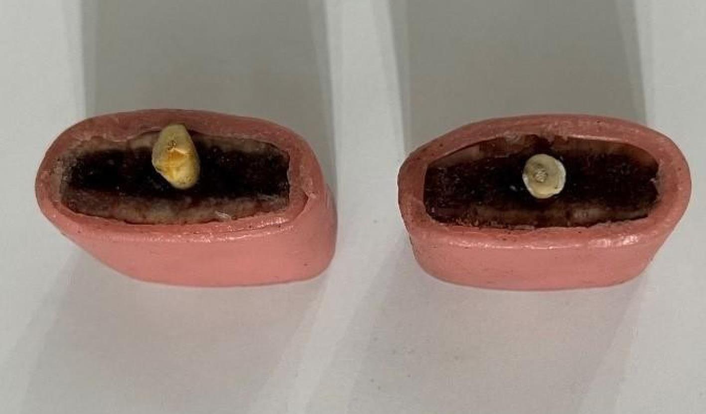
Figure 1.
Teeth Placement
.
Teeth Placement
Digital periapical radiographs were acquired using size 2 photostimulable phosphor plates (Optime®, Soredex, Helsinki, Finland) and a Minray X-ray unit set at 70 kVp, 10 mA, and 0.16 seconds exposure. The parallel technique was employed to standardize image acquisition.
Three types of image enhancements—sharpening, inverse contrast, and colorization—were applied to the radiographs. Two oral and maxillofacial radiologists, each with five years of experience, independently evaluated both raw and processed images in a double-blind manner, using a 20-inch monitor (200P; LG Corporation, Seoul, South Korea) in a semi-dark room. Observations were recorded in a checklist, noting the presence or absence of root fractures (Figures 2 and 3).
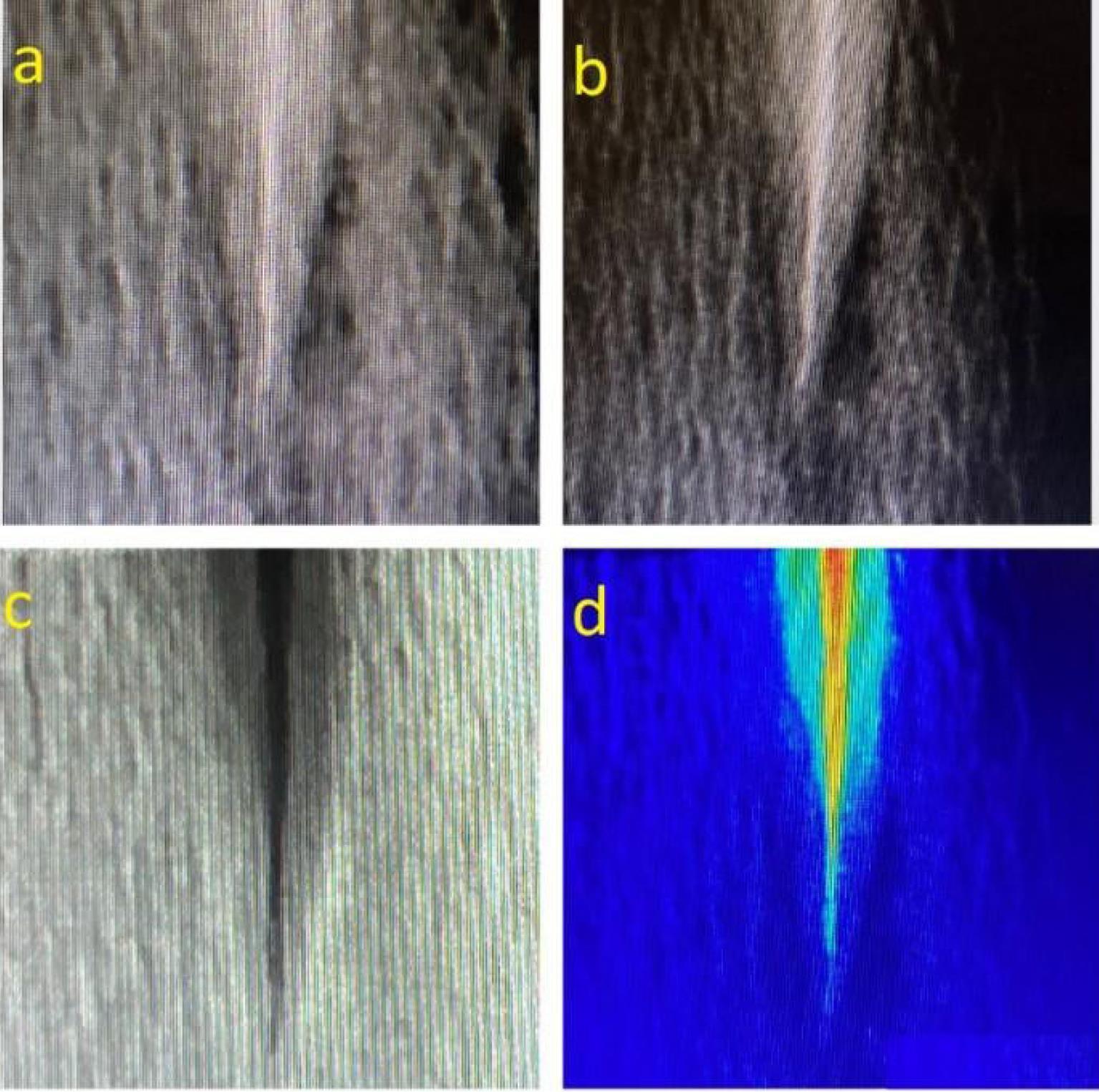
Figure 2.
Digital Periapical Radiograph of a Tooth with a Vertical Root Fracture (a) Row Images, (b) Sharpness, (c) Inverse Contrast, and (d) Colorization
.
Digital Periapical Radiograph of a Tooth with a Vertical Root Fracture (a) Row Images, (b) Sharpness, (c) Inverse Contrast, and (d) Colorization
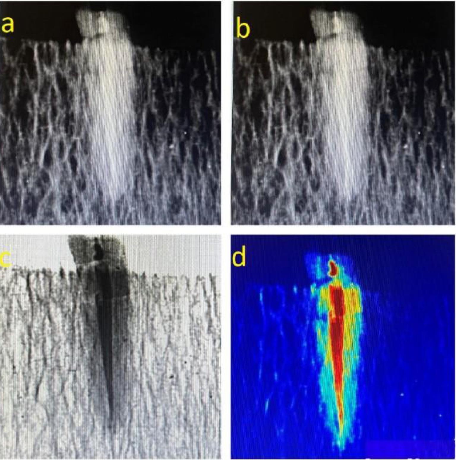
Figure 3.
Digital Periapical Radiograph of a Tooth With a Horizontal Root Fracture: (a) Row Images, (b) Sharpness, (c) Inverse Contrast, and (d) Colorization
.
Digital Periapical Radiograph of a Tooth With a Horizontal Root Fracture: (a) Row Images, (b) Sharpness, (c) Inverse Contrast, and (d) Colorization
The data were entered into SPSS (version 26, IBM Corporation, Armonk, NY, USA). The chi-square test was used for comparisons, and inter-observer agreement was measured using Cohen’s kappa coefficient. A P-value below 0.05 was considered statistically significant.
Results
The analysis of inter-observer agreement using Cohen’s kappa coefficient revealed a high level of consistency between the two radiologists across all enhancement modalities. Specifically, kappa values were 0.91 for the sharpening filter and 0.96 for both inverse contrast and colorization, indicating substantial to almost perfect agreement (Table 1).
Table 1.
Inter-Observer Agreement for Different Processing Enhancement Tools Determined by Calculating the Cohen’s κCoefficient
|
Processing Enhancement Tools
|
Conflict of Agreement
|
| Sharpening |
0.910 |
| Inverse contrast |
0.960 |
| Colorization |
0.960 |
The evaluation of diagnostic performance metrics for each enhancement method demonstrated notable variations. For images processed with inverse contrast, sensitivity reached 85.7%, while specificity was substantially lower at 17.1%. Correspondingly, false positive and false negative rates were 54.5% and 50.8%, respectively.
Images modified with sharpening filters yielded a sensitivity of 68.6% and specificity of 57.1%, with false positives at 64.5% and false negatives at 61.5%. In comparison, the application of colorization resulted in a sensitivity of 60% and a specificity of 71.4%, alongside 67.7% false positives and 64.1% false negatives.
Regarding the area under the receiver operating characteristic curve, colorized images produced the highest area under the curve at 65.71%, followed by sharpening at 62.86% and inverse contrast at 61.40%. Statistically significant differences in diagnostic accuracy were observed for sharpening and colorization techniques (P < 0.001), whereas the performance of inverse contrast images did not reach statistical significance (P = 0.06), the details of which are provided in Table 2.
Table 2.
Sensitivity, Specificity, and False Positive and False Negative Values for the Three Processing Enhancement Tools Assessed in This Study
|
Processing Enhancement Tools
|
Sensitivity (%)
|
Specificity (%)
|
False Negative
(%)
|
False
Positive (%)
|
SE (AUC)
|
95% CI (AUC)
|
P
Value
|
| Inverse contrast |
85.7 |
17.1 |
50.8 |
54.5 |
61.40 |
(39–63) |
0.06 |
| Sharpness |
68.6 |
57.1 |
61.5 |
64.5 |
62.86 |
(50–74) |
< 0.001 |
| Colorization |
60 |
71.4 |
67.7 |
64.1 |
65.71 |
(65.4–76.53) |
< 0.001 |
Note. SE: Standard error; AUC: Area under the curve; CI: Confidence interval.
Discussion
Intraoral digital radiography software typically provides operators with image processing tools, such as brightness and contrast adjustments and sharpness enhancement. However, improper use of some of these tools may interfere with clinical diagnosis. The key point in using these images is that they should be considered an adjunctive method alongside other diagnostic techniques. Therefore, determining their accuracy in correctly identifying teeth with root fractures and distinguishing them from healthy teeth has always been a concern in dentistry (17).
This study evaluated the impact of image editing on the diagnosis of root fractures in digital periapical radiographs.
The results of this study revealed that enhancing radiographic images through sharpness adjustments increased sensitivity and specificity in the diagnosis of root fractures. The findings of Nascimento et al regarding the effect of sharpness on the accuracy of root fracture diagnosis are consistent with our results. They examined the diagnostic accuracy of radiographs using digital filters for detecting VRFs and reported that the sharpness filter resulted in better diagnostic outcomes compared to the original image and other filtered images (18).
In contrast to the present study, Gaêta-Araujo et al, who enhanced images in terms of sharpness, reported no significant difference in the diagnostic accuracy of VRFs between the test groups (19). They investigated 15 single-rooted human teeth in the control group and 15 teeth in the enhanced image group for VRFs. It seems that the difference in sample size may explain the discrepancy in the results of the two studies.
Enhancing radiographic images through colorization adjustments also increased sensitivity and specificity in the diagnosis of root fractures. In line with our findings regarding colorization, Mikrogeorgis et al found that pseudo-coloring techniques assisted in the diagnosis of VRFs (20). Similarly, in the study by Kal et al, brightness adjustments were mentioned as one of the image enhancement tools that increased the accuracy of root fracture diagnosis (21).
In contrast to our results, the findings of Tofangchiha et al revealed that the diagnostic accuracy of VRFs in radiographic images decreased with the use of colorized images (22). They compared enhanced images of 100 single-rooted teeth with VRFs to those of another 100 teeth. The discrepancy in sample size and the fact that they exclusively examined vertical fractures may explain the conflicting results. Additionally, Tofangchiha et al used a charge-coupled device sensor (Trophy Radiologie, Vincennes, France), which differed from the equipment utilized in the present study. This difference in device and sensor type could be another reason for the variation in findings.
The study by Ghazizadeh et al, contrary to our findings, demonstrated that color filters do not enhance the diagnosis of vertical and HRFs. They examined 25 mandibular premolars and 25 maxillary incisors for vertical and HRFs, with imaging conditions set at 70 kVp and 8 mA (23). Differences in sample size and imaging conditions may explain the discrepancies between their results and those of the present study.
In the current study, the inverse contrast filter did not improve the diagnosis of root fractures, which aligns with the findings of previous studies (9,24-26). However, this result contradicts the findings of Soares et al, confirming that post-processing of radiographic images, including geometric adjustments, noise reduction, inverse contrast, and gamma correction, could enhance sensitivity in detecting VRFs (26). This discrepancy suggests that in the above-mentioned study, changes in image contrast were combined with geometric adjustments, noise reduction, and gamma correction, indicating a potential synergistic effect of contrast enhancement alongside other image processing techniques (21).
It appears that enhancing radiographic images in terms of sharpness and colorization can be beneficial in improving the diagnostic accuracy of specialists for detecting root fractures.
Currently, CBCT imaging with a small field of view and high resolution is widely used for evaluating teeth with root fractures. CBCT provides multiplanar views of the teeth, overcoming the limitations associated with the orientation of X-ray beams. Additionally, it offers the superior visualization of the surrounding periradicular bone and supporting alveolar process.
Conclusion
Applying image enhancement techniques, especially those that improve sharpness and apply colorization, can lead to better diagnostic accuracy when identifying root fractures in digital periapical radiographs. Although these enhancements do not replace advanced imaging methods, such as CBCT, they can serve as valuable adjunctive tools in clinical settings, particularly during the early diagnostic stage.
To further improve diagnostic reliability, future research should focus on developing more advanced image processing methods, including artificial intelligence-based algorithms that can adaptively highlight potential fracture lines and reduce observer variability.
Acknowledgments
We would like to extend our sincere gratitude to Dr. Kamyar Mansouri for their invaluable guidance and support throughout this study.
Competing Interests
The authors have no financial interest in the companies whose materials were included in this study.
Ethical Approval
This study was approved by the Ethics Committee of Zanjan University of Medical Sciences, Zanjan, Iran (IR.ZUMS.REC.1401.256).
Funding
This research received no specific grant from funding agencies in the public, commercial, or not-for-profit sectors.
References
- Mortazavi S, Hasanpour S, Anbiaee N, Rouhani A, Naghavi N, Babazadeh S. Evaluating the effectiveness of cone-beam computed tomography reverse contrast images in the diagnosis of vertical root fractures: an in vitro pilot study. Saudi Endod J 2025; 15(1):31-7. doi: 10.4103/sej.sej_92_24 [Crossref] [ Google Scholar]
- Arkhipova A, Bovanova N, Lastovichek D, Ramonova A, Generalov E, Byakova S. Radiographic criteria for differential diagnosis between vertical root fracture and apical periodontitis in single-rooted endodontically treated premolars using cone-beam computed tomography. J Endod 2024; 50(3):329-35. doi: 10.1016/j.joen.2023.12.008 [Crossref] [ Google Scholar]
- Saati S, Eskandarloo A, Falahi A, Tapak L, Hekmat B. Evaluation of the efficacy of the metal artifact reduction algorithm in the detection of a vertical root fracture in endodontically treated teeth in cone-beam computed tomography images: an in vitro study. Dent Med Probl 2019; 56(4):357-63. doi: 10.17219/dmp/109902 [Crossref] [ Google Scholar]
- Edlund M, Nair MK, Nair UP. Detection of vertical root fractures by using cone-beam computed tomography: a clinical study. J Endod 2011; 37(6):768-72. doi: 10.1016/j.joen.2011.02.034 [Crossref] [ Google Scholar]
- Martins JN, Canta JP, Coelho A, Baharestani M. Vertical root fracture diagnosis of crowned premolars with root canal treatment–two case reports. Rev Port Estomatol Med Dent Cir Maxilofac 2014; 55(1):60-4. doi: 10.1016/j.rpemd.2013.11.002 [Crossref] [ Google Scholar]
- Farman AG, Farman TT. A comparison of 18 different x-ray detectors currently used in dentistry. Oral Surg Oral Med Oral Pathol Oral Radiol Endod 2005; 99(4):485-9. doi: 10.1016/j.tripleo.2004.04.002 [Crossref] [ Google Scholar]
- Lai JY, Basrani B. Radiographic analysis of periodontal and endodontic lesions. In: Basrani B, ed. Endodontic Radiology. 2nd ed. John Wiley & Sons; 2012. p. 166-76. doi: 10.1002/9781119421689.ch11.
- Bornstein MM, Wölner-Hanssen AB, Sendi P, Von Arx T. Comparison of intraoral radiography and limited cone beam computed tomography for the assessment of root-fractured permanent teeth. Dent Traumatol 2009; 25(6):571-7. doi: 10.1111/j.1600-9657.2009.00833.x [Crossref] [ Google Scholar]
- Kamburoğlu K, Murat S, Pehlivan SY. The effects of digital image enhancement on the detection of vertical root fracture. Dent Traumatol 2010; 26(1):47-51. doi: 10.1111/j.1600-9657.2009.00841.x [Crossref] [ Google Scholar]
- White SC, Pharoah MJ. White and Pharoah’s Oral Radiology: Principles and Interpretation. Elsevier; 2019. p. 672.
- Sheikhnezami M, Shahmohammadi R, Jafarzadeh H, Azarpazhooh A. Long-term outcome of horizontal root fractures in permanent teeth: a retrospective cohort study. J Endod 2024; 50(5):579-89. doi: 10.1016/j.joen.2024.02.002 [Crossref] [ Google Scholar]
- Nair MK, Nair UP. Digital and advanced imaging in endodontics: a review. J Endod 2007; 33(1):1-6. doi: 10.1016/j.joen.2006.08.013 [Crossref] [ Google Scholar]
- Kambungton J, Janhom A, Prapayasatok S, Pongsiriwet S. Assessment of vertical root fractures using three imaging modalities: cone beam CT, intraoral digital radiography and film. Dentomaxillofac Radiol 2012; 41(2):91-5. doi: 10.1259/dmfr/49798768 [Crossref] [ Google Scholar]
- White SC, Pharoah MJ. The evolution and application of dental maxillofacial imaging modalities. Dent Clin North Am 2008; 52(4):689-705. doi: 10.1016/j.cden.2008.05.006 [Crossref] [ Google Scholar]
- Gormez O, Yilmaz HH. Image post-processing in dental practice. Eur J Dent 2009; 3(4):343-7. doi: 10.1055/s-0039-1697455 [Crossref] [ Google Scholar]
- Mol A. Image processing tools for dental applications. Dent Clin North Am 2000; 44(2):299-318. doi: 10.1016/s0011-8532(22)01304-0 [Crossref] [ Google Scholar]
- Fulton A, Fiani N. Diagnostic imaging in veterinary dental practice Dentigerous cyst with secondary infection. J Am Vet Med Assoc 2011; 238(4):435-7. doi: 10.2460/javma.238.4.435 [Crossref] [ Google Scholar]
- Nascimento HA, Ramos AC, Neves FS, de-Azevedo-Vaz SL, Freitas DQ. The ‘Sharpen’ filter improves the radiographic detection of vertical root fractures. Int Endod J 2015; 48(5):428-34. doi: 10.1111/iej.12331 [Crossref] [ Google Scholar]
- Gaêta-Araujo H, Nascimento EL, Oliveira-Santos N, Queiroz PM, Oliveira ML, Freitas DQ. Effect of digital enhancement on the radiographic assessment of vertical root fractures in the presence of different intracanal materials: an in vitro study. Clin Oral Investig 2021; 25(1):195-202. doi: 10.1007/s00784-020-03353-x [Crossref] [ Google Scholar]
- Mikrogeorgis G, Eirinaki E, Kapralos V, Koutroulis A, Lyroudia K, Pitas I. Diagnosis of vertical root fractures in endodontically treated teeth utilising digital subtraction radiography: a case series report. Aust Endod J 2018; 44(3):286-91. doi: 10.1111/aej.12240 [Crossref] [ Google Scholar]
- Kal BI, Baksi BG, Dündar N, Sen BH. Effect of various digital processing algorithms on the measurement accuracy of endodontic file length. Oral Surg Oral Med Oral Pathol Oral Radiol Endod 2007; 103(2):280-4. doi: 10.1016/j.tripleo.2006.06.001 [Crossref] [ Google Scholar]
- Tofangchiha M, Bakhshi M, Shariati M, Valizadeh S, Adel M, Sobouti F. Detection of vertical root fractures using digitally enhanced images: reverse-contrast and colorization. Dent Traumatol 2012; 28(6):478-82. doi: 10.1111/j.1600-9657.2012.01120.x [Crossref] [ Google Scholar]
- Ghazizadeh M, Sheikhi M, Aminian M, Ghazizadeh MM. Accuracy of digital image enhancement in detection of vertical and horizontal root fracture. Dent Res J (Isfahan) 2020; 17(4):266-72. [ Google Scholar]
- Barayan M, Nasseh I, Geha H, Noujeim M. The effects of imaging enhancement tools in the detection of horizontal root fractures. J Clin Diagn Res 2017; 11(8):ZC98-101. doi: 10.7860/jcdr/2017/26775.10490 [Crossref] [ Google Scholar]
- Mehralizadeh S, Mehrvarzfar P, Taghizadeh S, Edalat M, Mohebi M. Reverse contrast enhancement in digital radiography in detection of vertical root fracture (in vitro). J Dent Med 2015;28(2):115-21. [Persian].
- Soares LE, Freitas DQ, de Lima KL, Silva LR, Yamamoto-Silva FP, da Costa Vieira MA. Application of image processing techniques to aid in the detection of vertical root fractures in digital periapical radiography. Clin Oral Investig 2021; 25(8):5077-85. doi: 10.1007/s00784-021-03820-z [Crossref] [ Google Scholar]