Avicenna J Dent Res. 17(3):146-153.
doi: 10.34172/ajdr.2135
Original Article
Evaluation of Morphology of Maxillary Premolar Roots and Canals Using Cone-Beam Computed Tomography in Northeastern Iran
Atie Safaee Conceptualization, Funding acquisition, Methodology, Supervision, 1 
Hamid Jafarzadeh Bakooei Data curation, Methodology, Supervision, 2, 3
Ali Bagherpour Formal analysis, Methodology, 1
Seyed Amir Abas Noorbakhsh Writing – original draft, Writing – review & editing, 4
Mohamadreza Zare Investigation, 5
Masoumeh Soheyli Writing – original draft, Writing – review & editing, 6, *
Author information:
1Department of Oral and Maxillofacial Radiology, School of Dentistry, Mashhad University of Medical Sciences, Mashhad, Iran
2Department of Endodontics, Faculty of Dentistry, Mashhad University of Medical Sciences, Mashhad, Iran
3Department of Endodontics, School of Dentistry, West Virginia University, Morgantown, West Virginia, USA
4Immunology of Infectious Diseases Research Center, Research Institute of Basic Medical Sciences, Rafsanjan University of Medical Sciences, Rafsanjan, Iran
5Dentist, Mashhad, Iran
6Department of Oral & Maxillofacial Radiology, School of Dentistry, Rafsanjan University of Medical Sciences, Rafsanjan, Iran
Abstract
Background: Understanding the morphology of the root canal is crucial for successful root canal treatment (CBCT), which is the most effective method for detecting canal anatomy and morphology. This study aimed to investigate the root canal morphology of maxillary premolars using CBCT.
Methods: Overall, 71 CBCT images, comprising 97 maxillary first and 94 maxillary second premolars, were analyzed in the current study. Various sections of the CBCT images were examined to gather information on the roots, canals per root, and canal types, categorized using Vertucci’s classification. Statistical analysis was conducted using independent t-tests and Mann-Whitney tests (P<0.05).
Results: The first premolars with a single canal had a frequency of 51.5%, while those with two canals had a frequency of 87.6%. For the second premolars, the occurrence of a single canal was over 85%, and for two canals, it was 69.1%. In maxillary first and second premolars, the predominant canal type was type I as per Vertucci’s classification. In terms of root and canal symmetry, about 83.7% and 91.89% of the first premolars exhibited symmetry, with 93.1% and 72.41% of second premolars displaying comparable levels of symmetry.
Conclusion: The findings indicated that single-rooted second and double-rooted first premolars were more common in the studied Iranian population. However, the occurrence of three-rooted maxillary first premolars is very low. In terms of Vertucci’s classification, the prevailing canal types were type III, I, and IV in maxillary first premolars and type III, I, and II in maxillary second premolars.
Keywords: Cone-beam computed tomography, Maxilla, Root canal
Copyright and License Information
© 2025 The Author(s); Published by Hamadan University of Medical Sciences.
This is an open-access article distributed under the terms of the Creative Commons Attribution License (
http://creativecommons.org/licenses/by/4.0), which permits unrestricted use, distribution, and reproduction in any medium provided the original work is properly cited.
Please cite this article as follows: Safaee A, Jafarzadeh Bakooei H, Bagherpour A, Noorbakhsh SAA, Zare M, Soheyli M. Evaluation of Morphology of Maxillary Premolar Roots and Canals Using Cone-Beam Computed Tomography in Northeastern Iran. Avicenna J Dent Res. 2025;17(3):146-153. doi:10.34172/ajdr.2135
Background
The effectiveness of dental procedures can be improved if dentists have detailed knowledge of the internal anatomy of the teeth. This knowledge helps perform root canal therapy with accurate orifice identification, enabling proper cleaning, shaping, and successful irrigation of the root canals to avoid treatment complications (1). The endodontic treatment of maxillary premolars is complex due to the varying number of roots, canals, and morphology of the pulp chamber. Genetic factors and ethnicity may affect the variability in root and canal numbers, with an increased incidence of extra roots and canals found in Chinese, Australian, and African populations (2).
The widely used Vertucci classification defines eight different canal configurations. Type I has a single canal from the pulp chamber to the apex. Type II has two converging canals in the apical region. Type III comprises a single canal that branches from the pulp chamber, reunites, and converges at the apex. Type IV consists of two separate canals from the chamber to the apex. Type V shows one canal with branches near the apex. Type VI has two canals that unite in the middle of the root and then separate at the apex. Type VII has one canal that branches both in the ventricle and in the apical region. Type VIII consists of three canals that extend from the chamber to the apex (3).
Radiographs, which are commonly utilized to assess the internal anatomy of teeth, have limited value for diagnosis due to the superimposition of different structures in two-dimensional (2D) images (4). Periapical radiographs are frequently employed in clinical practices to examine dental anatomy. These images are usually taken before, during, and after root canal treatments to gather information about teeth with endodontic problems (5). The reliability related to the diagnosis of the number of canals is limited by the overlap and poor quality of periapical radiographs (6).
Cone-beam computed tomography (CBCT) is a 3D radiographic imaging technique that has gained considerable popularity in dental radiology over the past two decades. CBCT overcomes the limitations of 2D imaging and provides a precise image of the jawbone and surrounding tissue. One of the key strengths of CBCT imaging is its ability to generate various 3D images and format volumetric image data on multiple planes (7). Studies have demonstrated that the number and configuration of root canals in maxillary premolars exhibit significant variability influenced by factors such as ethnicity, age, gender, and the study methodology (in vivo versus in vitro). The root canal anatomy and morphology of maxillary premolars can be remarkably complex and diverse. Considering these factors and the inherent advantages of CBCT in evaluating root canal anatomy and endodontic treatment planning, this study seeks to investigate the root and canal anatomy and morphology of maxillary premolars in a northeast Iranian population using CBCT imaging.
Materials and Methods
The present descriptive cross-sectional study was approved by the Ethics Committee of the School of Dentistry, Mashhad University of Medical Sciences (Ethical code IR.MUMS.SD.REC.1394.305).
CBCT scans obtained using Classic Planmeca Promax (Finland, Helsinki) with an 8 × 8-inch field of view and a power of 54 to 84 kVp were randomly selected from patients referred to the Radiology Department of the Mashhad School of Dentistry between 2016 and 2017 for diagnostic purposes per physician’s referral. The samples meeting the inclusion criteria included those showing developed and visible roots on at least one side of the maxillary first or second premolars, with adequate image quality for precise diagnosis. On the other hand, the samples were excluded from the study if they exhibited poor image definition (because of misinterpretation of the root canal anatomy), undeveloped roots (it can result in atypical canal morphology, which is not representative of the general population), or root canal calcification (it can obscure the canal space, making it difficult to accurately assess the canal configuration). Moreover, they were excluded if they represented post/core restoration (it can block the view of the root canal system, leading to incomplete or inaccurate assessments) or internal and external resorption (resorptive defects can alter the natural anatomy, making it difficult to apply the classification system). The maxillary first and second premolars were examined in different sections of the CBCT images using Planmeca Romexis Viewer 3.8.0.R software on a personal computer.
CBCT images were assessed by a postgraduate student of oral and maxillofacial radiology under the supervision of an oral and maxillofacial radiologist and an endodontist in axial planes from the pulp chamber bottom to the root apex. The examination involved scrolling through these planes. If uncertainty arose regarding the pulp chamber’s position, alternative planes were utilized to determine its exact location. Data on root number, canal count per root, and canal classification were recorded according to Vertucci’s system (Figure 1). The analysis included the number of roots and canals and the type of canals in each tooth, as well as an assessment of symmetry between the left and right sides when maxillary premolars were present bilaterally.
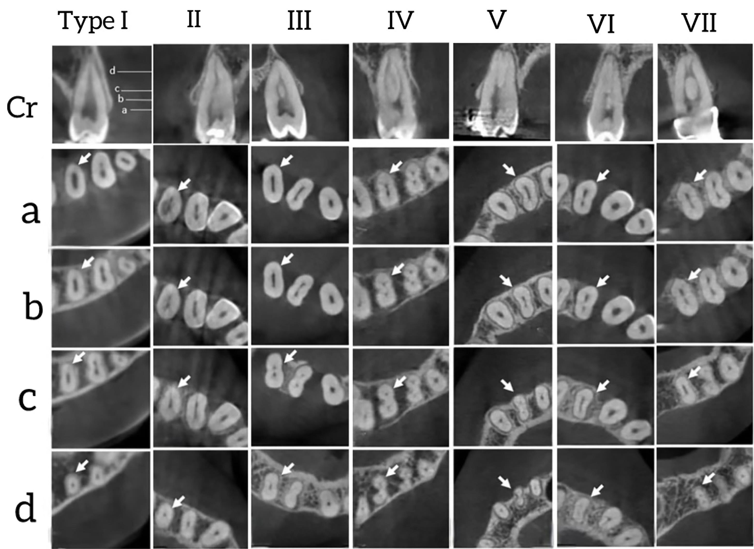
Figure 1.
Vertucci’s Classification of Root Canal Morphology in Samples Studied at Different Axial Sections (From a to d): Near the Floor of the Pulp Chamber (a), Slightly Below the Pulp Chamber (b), Around the Middle of the Root (c), Near the Root End (d), and The Coronal Section of Each Premolar (Cr)
.
Vertucci’s Classification of Root Canal Morphology in Samples Studied at Different Axial Sections (From a to d): Near the Floor of the Pulp Chamber (a), Slightly Below the Pulp Chamber (b), Around the Middle of the Root (c), Near the Root End (d), and The Coronal Section of Each Premolar (Cr)
The obtained data were statistically analyzed using SPSS software (version 20). Descriptive statistics were utilized to describe the frequency and distribution of each canal type. In addition, cross-tabulations were employed to assess the concordance in the number of roots, the number of root canals, and root canal morphology between the left and right sides. The Mann-Whitney test was also applied to compare the prevalence of the different types of canals between the two sides and between the two genders. A significance level of less than 0.05 was taken into consideration.
Results
This study included 71 CBCT images of 71 patients with at least one maxillary first or second premolar. The study population consisted of 34 males with a mean age of 41.91 ± 16.13 years and 36 females with a mean age of 39.86 ± 13.16 years and one male patient of unknown age.
Based on the results (Table 1), the frequencies of single-rooted teeth in the first and second premolar groups were 51.5% and more than 85%, respectively.
Table 1.
Frequency of Number of Roots by Tooth Type and Gender
|
Tooth Type
|
Gender
|
Number of Roots
|
Frequency (%)
|
P
Value
|
| Number of roots of maxillary first premolar |
Male |
Single |
21 (47.7) |
0.05 |
| Double |
20 (45.5) |
| Triple |
3 (6.8) |
| Female |
Single |
29 (54.7) |
| Double |
24 (45.3) |
| Triple |
0 (0) |
| Number of roots of maxillary second premolar |
Male |
Single |
36 (87.8) |
0.39 |
| Double |
5 (12.2) |
| Triple |
0 (0) |
| Female |
Single |
44 (83.0) |
| Double |
9 (17.0) |
| Triple |
0 (0) |
The results (Tables 1 and 2) showed no significant difference in the number of roots and canals between the two genders in all the first and second premolars.
Table 2.
Frequency of Number of Root Canals by Tooth Type and Gender
|
Tooth Type
|
Gender
|
Number of Root Canals
|
Frequency (%)
|
P
Value
|
| Number of root canals of the maxillary first premolar |
Male |
Single |
2 (4.5) |
0.06 |
| Double |
38 (86.4) |
| Triple |
4 (9.1) |
| Female |
Single |
4 (7.5) |
| Double |
47 (88.7) |
| Triple |
2 (3.8) |
| Number of root canals of the maxillary second premolar |
Male |
Single |
14 (34.1) |
0.50 |
| Double |
27 (65.9) |
| Triple |
0 (0) |
| Female |
Single |
15 (28.3) |
| Double |
38 (71.7) |
| Triple |
0 (0) |
Frequency of Canal Morphology
Table 3. Morphology of Root Canals by Gender and Tooth Type
Table 3.
provides the data related to the canal morphology according to tooth and root type. Among the examined samples, three maxillary first premolars exhibited three roots, and their canals were classified as Vertucci type I.
|
Tooth Type
|
Gender
|
Root Canal Morphology
|
Frequency (%)
|
| Single root maxillary first premolar |
Male |
Vertucci 1 |
3 (14.3) |
| Vertucci 2 |
4 (19.0) |
| Vertucci 3 |
7 (33.3) |
| Vertucci 4 |
4 (19.0) |
| Vertucci 5 |
3 (14.3) |
| Female |
Vertucci 1 |
3 (10.3) |
| Vertucci 2 |
4 (13.8) |
| Vertucci 3 |
6 (20.7) |
| Vertucci 4 |
9 (31.0) |
| Vertucci 5 |
6 (20.7) |
| Vertucci 6 |
1 (3.4) |
| Single root maxillary second premolar |
Male |
Vertucci 1 |
14 (38.9) |
| Vertucci 2 |
8 (22.2) |
| Vertucci 3 |
8 (22.2) |
| Vertucci 5 |
2 (5.6) |
| Vertucci 7 |
4 (11.1) |
| Female |
Vertucci 1 |
15 (34.1) |
| Vertucci 2 |
3 (6.8) |
| Vertucci 3 |
20 (45.5) |
| Vertucci 4 |
1 (2.3) |
| Vertucci 5 |
4 (9.4) |
| Vertucci 7 |
1 (2.3) |
Double root maxillary first premolar
(buccal root) |
Male |
Vertucci 1 |
18 (90.0) |
| Vertucci 5 |
2 (10.0) |
| Female |
Vertucci 1 |
22 (91.7) |
| Vertucci 5 |
1 (4.2) |
| Vertucci 6 |
1 (4.2) |
Double root maxillary second premolar
(buccal root) |
Male |
Vertucci 1 |
5 (100.0) |
| Female |
Vertucci 1 |
9 (100.0) |
Double root maxillary first premolar
(palatal root) |
Male |
Vertucci 1 |
20 (100.0) |
| Female |
Vertucci 1 |
24 (100.0) |
Double root maxillary second premolar
(palatal root) |
Male |
Vertucci 1 |
5 (100.0) |
| Female |
Vertucci 1 |
9 (100.0) |
The findings (Table 3) indicated that the most prevalent root canal morphology in single-rooted first premolars was type III in males and type IV in females. In single-rooted second premolars, the most common canal morphology was type I in both males and females, followed by type III. The most prevalent canal morphology observed in the buccal roots of maxillary first and second premolars with two roots was Vertucci type I, regardless of gender.
According to the results (Table 4), the morphology of all palatal roots of maxillary premolars that had two roots was Vertucci type I. The most common canal morphology in the maxillary first and second premolars was Vertucci type I. Types VII and VIII were not observed in the first premolars. Types VI and VIII were also not detected in the second premolars.
Table 4.
Frequency of Canal Morphology Types Regardless of Gender
|
Tooth Type
|
Canal Morphology
|
Frequency in Single Roots
|
Frequency in Double Roots
|
Frequency in Triple Roots
|
Total (%)
|
| Maxillary first premolar |
Vertucci 1 |
6 |
40 |
3 |
49 (50.5) |
| Vertucci 2 |
8 |
0 |
0 |
8 (8.2) |
| Vertucci 3 |
13 |
0 |
0 |
13 (13.4) |
| Vertucci 4 |
13 |
0 |
0 |
13 (13.4) |
| Vertucci 5 |
9 |
3 |
0 |
12 (12.3) |
| Vertucci 6 |
1 |
1 |
0 |
2 (2.0) |
| Vertucci 7 |
0 |
0 |
0 |
0 (0) |
| Total |
50 |
44 |
3 |
97 (100) |
| Maxillary second premolar |
Vertucci 1 |
29 |
14 |
0 |
43 (45.7) |
| Vertucci 2 |
11 |
0 |
0 |
11 (11.7) |
| Vertucci 3 |
28 |
0 |
0 |
28 (29.8) |
| Vertucci 4 |
1 |
0 |
0 |
1 (1.0) |
| Vertucci 5 |
6 |
0 |
0 |
6 (6.4) |
| Vertucci 6 |
0 |
0 |
0 |
0 (0) |
| Vertucci 7 |
5 |
0 |
0 |
5 (5.3) |
| Total |
80 |
14 |
0 |
94 (100) |
Evaluation of the Symmetry Between the Right and Left Teeth in the Examined Samples
A total of 191 teeth from 71 patients underwent assessment. Among these patients, 44 had matching maxillary premolars on both sides, suitable for similarity analysis. Cross-tabulations were used to study root number, root canal number, and root canal morphology similarity.
Comparison of the Symmetry of the Number of Roots of the First and Second Premolars in the Maxilla on the Left and Right Side in Both Genders
According to Table 5, symmetry was observed in 12 out of 16 upper first premolars in males (75%) and in 19 out of 21 in females (90%). In addition, it was found in all 11 pairs of maxillary second premolars in males (100%) and in 16 out of 18 pairs in females (88%), suggesting that symmetry in the number of maxillary second premolars roots is more common in males.
Table 5.
Cross-Tabulations of the Symmetry of the Number of Roots in the Right and Left First and Second Premolars in Both Genders
|
Tooth Type
|
Gender
|
Number of Roots
|
Number of Maxillary Left Premolar Roots (First or Second)
|
Total
|
|
Single Roots
|
Double Roots
|
Triple Roots
|
| The right maxillary first premolar |
Male |
Single roots |
5 |
2 |
0 |
7 |
| Double Roots |
1 |
6 |
1 |
8 |
| Triple roots |
0 |
0 |
1 |
1 |
| Total |
6 |
8 |
2 |
16 |
| Female |
Single roots |
9 |
1 |
0 |
10 |
| Double Roots |
1 |
10 |
0 |
11 |
| Total |
10 |
11 |
0 |
21 |
| The right maxillary second premolar |
Male |
Single roots |
10 |
0 |
0 |
10 |
| Double Roots |
0 |
1 |
0 |
1 |
| Total |
10 |
1 |
0 |
11 |
| Female |
Single roots |
13 |
1 |
0 |
14 |
| Double roots |
1 |
3 |
0 |
4 |
| Total |
14 |
4 |
0 |
18 |
Comparison of Symmetry in the Number of Root Canals of the Left and Right First and Second Premolars in Both Genders
Symmetry in the number of root canals in maxillary first premolars was observed in 15 out of 16 cases in males (93%) and in 19 out of 21 cases in females (90%). Similarly, symmetry in the number of root canals in the maxillary second premolars was found in 7 out of 11 cases in males (63%) and in 14 out of 18 cases in females (77%), demonstrating that symmetry in the number of root canals in the maxillary second premolars is more prevalent in females (Table 6).
Table 6.
Symmetrical Cross-Tabulation of the Number of Root Canals of the Right and Left Premolars in Both Genders
|
Tooth Type
|
Gender
|
Number of Root Canals
|
Number of Maxillary Left Premolar Root Canal (First or Second)
|
Total
|
|
One Root Canal
|
Two Root Canals
|
Three Root Canals
|
| The right maxillary first premolar |
Male |
Two root canals |
0 |
13 |
0 |
13 |
| Three root canals |
0 |
1 |
2 |
3 |
| Total |
0 |
14 |
2 |
16 |
| Female |
One root canal |
1 |
0 |
0 |
1 |
| Two root canals |
0 |
18 |
1 |
19 |
| Three root canals |
0 |
1 |
0 |
1 |
| Total |
1 |
19 |
1 |
21 |
| The right maxillary second premolar |
Male |
One root canal |
1 |
2 |
0 |
3 |
| Two root canals |
2 |
6 |
0 |
8 |
| Total |
3 |
8 |
0 |
11 |
| Female |
One root canal |
3 |
3 |
0 |
6 |
| Two root canals |
1 |
11 |
0 |
12 |
| Total |
4 |
14 |
0 |
18 |
Comparison of the Symmetry of the Morphology of the Root Canals of the Left and Right First and Second Premolars in Both Genders
Given the analogous morphology of all root canals in the palatal roots of multi-rooted teeth, only the buccal roots were subjected to comparative analysis.
Based on the data (Table 7), 11 out of 16 male cases (68%) and 15 out of 21 female cases (71%) showed symmetry in maxillary first premolars’ root canal morphology. Furthermore, for maxillary second premolars, 6 out of 11 male cases (54%) and 11 out of 18 female cases (61%) exhibited symmetry, indicating a higher prevalence of this trait in females.
Table 7.
Symmetry Cross-table of Root Canal Morphology in the Right and Left First Premolars in Both Genders
|
Tooth Type
|
Gender
|
Canal Morphology
|
Canal Morphology of Maxillary Left Premolar (First or Second)
|
Total
|
|
Vertucci 1
|
Vertucci 2
|
Vertucci 3
|
Vertucci 4
|
Vertucci 5
|
Vertucci 6
|
Vertucci 7
|
| The right maxillary first premolar |
Male |
Vertucci 1 |
7 |
1 |
1 |
0 |
1 |
0 |
0 |
10 |
| Vertucci 2 |
0 |
1 |
0 |
0 |
0 |
0 |
0 |
1 |
| Vertucci 3 |
0 |
0 |
2 |
0 |
0 |
0 |
0 |
2 |
| Vertucci 4 |
0 |
0 |
0 |
1 |
1 |
0 |
0 |
2 |
| Vertucci 5 |
1 |
0 |
0 |
0 |
0 |
0 |
0 |
1 |
| Total |
8 |
2 |
3 |
1 |
2 |
0 |
0 |
16 |
| Female |
Vertucci 1 |
10 |
0 |
0 |
0 |
1 |
0 |
0 |
11 |
| Vertucci 2 |
0 |
1 |
1 |
0 |
0 |
0 |
0 |
2 |
| Vertucci 3 |
0 |
1 |
1 |
0 |
0 |
0 |
0 |
2 |
| Vertucci 4 |
0 |
0 |
0 |
1 |
1 |
1 |
0 |
3 |
| Vertucci 5 |
1 |
0 |
0 |
2 |
0 |
0 |
0 |
3 |
| Total |
11 |
2 |
2 |
1 |
4 |
1 |
0 |
21 |
| The right maxillary second premolar |
Male |
Vertucci 1 |
2 |
0 |
0 |
0 |
1 |
0 |
1 |
4 |
| Vertucci 2 |
1 |
2 |
1 |
0 |
0 |
0 |
0 |
4 |
| Vertucci 3 |
0 |
0 |
1 |
0 |
0 |
0 |
0 |
1 |
| Vertucci 5 |
1 |
0 |
0 |
0 |
0 |
0 |
0 |
1 |
| Vertucci 7 |
0 |
0 |
0 |
0 |
0 |
0 |
1 |
1 |
| Total |
4 |
2 |
2 |
0 |
1 |
0 |
2 |
11 |
| Female |
Vertucci 1 |
7 |
0 |
2 |
0 |
1 |
0 |
0 |
10 |
| Vertucci 3 |
1 |
1 |
3 |
0 |
0 |
0 |
1 |
6 |
| Vertucci 5 |
0 |
0 |
1 |
0 |
1 |
0 |
0 |
2 |
| Total |
8 |
1 |
6 |
0 |
2 |
0 |
1 |
18 |
Discussion
In this study, single-rooted first premolars had a frequency of 51.5%, while second premolars were more than 85% single-rooted. The occurrence of two canals was 87.6% in first premolars and 69.1% in second premolars. Only 3 three-root first premolars were found, all with Vertucci type I canals. In maxillary first premolars, males and females commonly had types III and IV canals, respectively, and the overall canal was of type I. For second premolars, males and females typically had type I and type III canals, respectively, and type I was the overall canal. Symmetry in the number of roots and canals was detected in 83.7% and 91.89% of the first premolars and 93.1% and 72.41% of the second premolars, respectively.
Root canal access is the cornerstone of successful endodontic treatment, and unfamiliarity with root canal anatomy can be one of the main reasons for treatment failure (8). Given the complex anatomy of the maxillary premolars, this study investigated the anatomy of these teeth in northeast Iran. CBCT imaging provides a useful diagnostic tool by offering clinicians 3D information on root morphology and diversity. Images from axial, sagittal, and coronal sections are obtained to minimize the superimposition of anatomical structures, aiding clinicians in comprehending root canal morphology (7).
In recent years, numerous studies have been conducted on the anatomy and shape of the dental pulp. It is important to note that each study has its own set of limitations and challenges. Conversely, these limitations result in significant discrepancies regarding the internal anatomy of teeth. These contradictions can be attributed to the subject’s race, gender, age, and the nature of the study (in vivo/in vitro) (9).
In a study on premolars from a German population, Bürklein et al (10) showed that most maxillary first premolars had two roots (62.4%) and were mainly two-canal (88.4%). The majority of maxillary second premolars had a single root (82.6%). The occurrence of two-canal roots (56.3%) was higher than single-canal roots (43.1%), consistent with the findings of the current study.
Tafakhori and Rafiei investigated the root canal morphology of maxillary premolars in southeastern Iran. According to Vertucci’s classification, types II and IV were more common in southeastern Iran. The results of the present study demonstrated that types I, III, and IV are more common in the root canal of maxillary first premolars. Similar results were found in the prevalence of type IV in both studies, but the difference in the prevalence of the other types was due to the racial difference between the southeastern and northeastern Iranian populations (11).
A study conducted by Alqedairi et al revealed that most first premolars in a Saudi population displayed two roots (75.1%), while a greater occurrence of single roots was noted in second premolars (85.2%), which conforms to the findings of the current study. Type IV Vertucci was predominant in the first premolars (69.1%), whereas type I was more prevalent in the second premolars (49.4%). Various canal types were identified in maxillary premolars, except for type VII, which was absent in second premolars (12). In the study performed by Li et al on the Chinese population, the most common anatomy of maxillary first and second premolars included a single root with two canals (58%) and a single root with one canal (50.3%), respectively, which is in line with the results of the present study. The predominant canal morphology observed in maxillary first and second premolars was of type IV (42.7%) and type I (50.3%), respectively, aligning with the present study’s results. The study’s authors highlighted that single-rooted maxillary premolars displayed more diversity in canal morphology compared to multi-rooted teeth (13).
Celikten et al, studying the Turkish Cypriot population, found that types I and IV canals were respectively predominant in the maxillary first and second premolars, according to Vertucci’s classification, which corroborates the results of the current study. Similar to the present study, just two maxillary first premolars and one maxillary second premolar represented three roots, all displaying Vertucci type I canal morphology (14).
De Lima et al, investigating the Brazilian population, reported a higher prevalence of two roots in maxillary first premolars and a higher prevalence of one root in maxillary second premolars. However, our findings showed nearly equal prevalence of one and two roots in maxillary first premolars, with a higher prevalence of one root than that of two roots in maxillary second premolars. Unlike our study, male patients in their study had a higher prevalence of two roots compared to female patients. They concluded that types IV and I were more common in maxillary first and second premolars, respectively (15), which matches our results.
In their study on the Egyptian population, Saber et al found that more than half of the maxillary first premolars exhibited two roots, while the majority of maxillary second premolars displayed a single root (16). In contrast to the present study, the most common canal type in maxillary first and second premolars, according to Vertucci’s classification, was type IV in the aforementioned study.
Studies conducted in China (13), Saudi Arabia (12), Egypt (16), Turkey (14), and northeastern Iran (the present study) have consistently demonstrated a predominantly single-rooted, single-canal morphology for maxillary second premolars, suggesting a notable degree of anatomical similarity for this tooth across diverse populations.
Conversely, maxillary first premolars represent greater inter-population variability in root and canal configurations. For instance, two-rootedness is a more prevalent characteristic in populations from China (13) and northeastern Iran, whereas studies in Saudi Arabia (12) and Egypt (16) have reported Vertucci type IV canal configurations to be more common. These differences may be attributed to a complex interplay of racial, genetic, and geographical factors.
Across the majority of studies, with the notable exceptions of those conducted in Saudi Arabia (12) and Egypt (16), Vertucci type I canal configuration has been identified as the most frequent, indicating a degree of shared root canal anatomy among certain populations.
Martins et al, in their comprehensive, worldwide cross-sectional study, analyzed the root and root canal characteristics of 26,400 maxillary premolars utilizing CBCT and found a general trend of fewer roots and root canals in Asian populations compared to a higher prevalence in European populations (17).
Collectively, these comparative analyses revealed that while certain anatomical patterns are conserved across populations, significant variations also exist, underscoring the importance of considering population-specific anatomical data in the context of endodontic treatment planning.
Differences in the study results may stem from a variety of factors, such as race, genetics, and how root canal morphology is documented. Some studies classify two-rooted teeth with two canals as type IV (10,12,13,18), unlike Vertucci’s definition, where type IV refers to two canals within one root. Considering one canal in each root as Type I may yield similar results to those of other studies. The absence of Vertucci type VIII in this study contrasts with the results of previous studies(10,12,13,18), possibly due to variations in applying Vertucci’s classification system. For instance, Abella et al, studying the Spanish population, classified three-rooted teeth with three canals as type VIII (18), whereas our study categorized them as type I.
Our findings confirmed that the highest root and root canal number symmetry in maxillary first premolars was observed in females. Conversely, in maxillary second premolars, the greatest root number symmetry was found in males, with females exhibiting the highest symmetry in root canal numbers. The maxillary first and second premolars of females showed the greatest symmetry in root canal morphology. Based on the results, symmetry was detected in 31, 34, and 26 pairs of maxillary first premolars for the number of roots, number of root canals, and canal morphology, respectively. It was also observed in 27, 21, and 17 pairs of 29 pairs of maxillary second premolars. A few studies have focused on the symmetry of root number and root canal type. In the study conducted by Tian et al on the Chinese population, 38 out of 59 pairs of maxillary first premolars displayed symmetrical anatomical patterns for both root number and root canal type (5). Similarly, Li et al, exploring root and root canal symmetry in maxillary premolars, reported 80.2% symmetry in both canal and root numbers in maxillary first premolars. The symmetry rates were 81.8% for the maxillary second premolars (13). Our research found that the symmetry in root and canal numbers was 83.8% and 72.4% for the first and second maxillary premolars, indicating a similarity between the Chinese and Iranian populations in root and canal symmetry, likely due to their Asian ancestry. The observed symmetry in maxillary premolar anatomy may offer clinicians a predictive tool for anticipating the contralateral tooth’s morphology, potentially minimizing the necessity for additional imaging. However, practitioners should exercise caution against undue reliance on this symmetry, as significant bilateral variations can occur.
These findings underscore the importance of employing advanced imaging modalities, such as CBCT, for the accurate diagnosis and treatment of complex root canal systems, especially in populations exhibiting significant variability in dental anatomy. Clinicians should be cognizant of the potential influence of ethnic and genetic factors on root canal morphology, as these variations may impact treatment efficacy and prognosis.
Conclusion
The study highlights that endodontists and dentists can gain valuable insights into the anatomy of maxillary premolar canals using CBCT scans. It is crucial to focus on the precise details of maxillary premolars’ anatomy during dental procedures. The varied anatomy of these root canals can be better addressed with CBCT technology, overcoming the constraints of 2D images in root canal treatments. Considering how race impacts the diversity of root canal configurations, utilizing such imaging may decrease failures in endodontic procedures.
Acknowledgments
The authors express their gratitude for the support of the Vice-Chancellor for Research at Mashhad University of Medical Sciences for funding this research project.
Competing Interests
None declared.
Ethical Approval
The proposal for this research was approved by Mashhad University of Medical Sciences (Ethics code: IR.MUMS.SD.REC.1394.305)
Funding
This study was financially supported by Mashhad University of Medical Sciences (grant number: 4012177).
References
- Datta P, Zahir S, Kundu GK, Dutta K. Different methods of studying root canal morphology of human tooth: a review. Bangladesh J Dent Res Educ 2015; 5(2):59-63. doi: 10.3329/bjdre.v5i2.24718 [Crossref] [ Google Scholar]
- Liu X, Gao M, Ruan J, Lu Q. Root canal anatomy of maxillary first premolar by microscopic computed tomography in a Chinese adolescent subpopulation. Biomed Res Int 2019; 2019:4327046. doi: 10.1155/2019/4327046 [Crossref] [ Google Scholar]
- Vertucci FJ. Root canal morphology and its relationship to endodontic procedures. Endod Topics 2005; 10(1):3-29. doi: 10.1111/j.1601-1546.2005.00129.x [Crossref] [ Google Scholar]
- Shah N, Bansal N, Logani A. Recent advances in imaging technologies in dentistry. World J Radiol 2014; 6(10):794-807. doi: 10.4329/wjr.v6.i10.794 [Crossref] [ Google Scholar]
- Tian YY, Guo B, Zhang R, Yu X, Wang H, Hu T. Root and canal morphology of maxillary first premolars in a Chinese subpopulation evaluated using cone-beam computed tomography. Int Endod J 2012; 45(11):996-1003. doi: 10.1111/j.1365-2591.2012.02059.x [Crossref] [ Google Scholar]
- Yapp KE, Suleiman M, Brennan P, Ekpo E. Periapical radiography versus cone beam computed tomography in endodontic disease detection: a free-response, factorial study. J Endod 2023; 49(4):419-29. doi: 10.1016/j.joen.2023.02.001 [Crossref] [ Google Scholar]
- Kaasalainen T, Ekholm M, Siiskonen T, Kortesniemi M. Dental cone beam CT: an updated review. Phys Med 2021; 88:193-217. doi: 10.1016/j.ejmp.2021.07.007 [Crossref] [ Google Scholar]
- Olczak K, Pawlicka H, Szymański W. Root form and canal anatomy of maxillary first premolars: a cone-beam computed tomography study. Odontology 2022; 110(2):365-75. doi: 10.1007/s10266-021-00670-9 [Crossref] [ Google Scholar]
- Karobari MI, Parveen A, Mirza MB, Makandar SD, Nik Abdul Ghani NR, Noorani TY. Root and root canal morphology classification systems. Int J Dent 2021; 2021:6682189. doi: 10.1155/2021/6682189 [Crossref] [ Google Scholar]
- Bürklein S, Heck R, Schäfer E. Evaluation of the root canal anatomy of maxillary and mandibular premolars in a selected German population using cone-beam computed tomographic data. J Endod 2017; 43(9):1448-52. doi: 10.1016/j.joen.2017.03.044 [Crossref] [ Google Scholar]
- Tafakhori Z, Rafiei M. Root canal morphology assessment of the maxillary first premolar using cone-beam computed tomography (CBCT) imaging in a selected Iranian population. Caspian J Dent Res 2021; 10(2):30-7. doi: 10.22088/cjdr.10.2.30 [Crossref] [ Google Scholar]
- Alqedairi A, Alfawaz H, Al-Dahman Y, Alnassar F, Al-Jebaly A, Alsubait S. Cone-beam computed tomographic evaluation of root canal morphology of maxillary premolars in a Saudi population. Biomed Res Int 2018; 2018:8170620. doi: 10.1155/2018/8170620 [Crossref] [ Google Scholar]
- Li YH, Bao SJ, Yang XW, Tian XM, Wei B, Zheng YL. Symmetry of root anatomy and root canal morphology in maxillary premolars analyzed using cone-beam computed tomography. Arch Oral Biol 2018; 94:84-92. doi: 10.1016/j.archoralbio.2018.06.020 [Crossref] [ Google Scholar]
- Celikten B, Orhan K, Aksoy U, Tufenkci P, Kalender A, Basmaci F. Cone-beam CT evaluation of root canal morphology of maxillary and mandibular premolars in a Turkish Cypriot population. BDJ Open 2016; 2:15006. doi: 10.1038/bdjopen.2015.6 [Crossref] [ Google Scholar]
- de Lima CO, de Souza LC, Devito KL, do Prado M, Campos CN. Evaluation of root canal morphology of maxillary premolars: a cone-beam computed tomography study. Aust Endod J 2019; 45(2):196-201. doi: 10.1111/aej.12308 [Crossref] [ Google Scholar]
- Saber SE, Ahmed MH, Obeid M, Ahmed HM. Root and canal morphology of maxillary premolar teeth in an Egyptian subpopulation using two classification systems: a cone beam computed tomography study. Int Endod J 2019; 52(3):267-78. doi: 10.1111/iej.13016 [Crossref] [ Google Scholar]
- Martins JN, Ensinas P, Chan F, Babayeva N, von Zuben M, Berti L. Worldwide assessment of the root and root canal characteristics of maxillary premolars - a multi-center cone-beam computed tomography cross-sectional study with meta-analysis. J Endod 2024; 50(1):31-54. doi: 10.1016/j.joen.2023.10.009 [Crossref] [ Google Scholar]
- Abella F, Teixidó LM, Patel S, Sosa F, Duran-Sindreu F, Roig M. Cone-beam computed tomography analysis of the root canal morphology of maxillary first and second premolars in a Spanish population. J Endod 2015; 41(8):1241-7. doi: 10.1016/j.joen.2015.03.026 [Crossref] [ Google Scholar]