Avicenna J Dent Res. 17(1):1-7.
doi: 10.34172/ajdr.1794
Original Article
Comparative Evaluation of Compressive Bond Strength Between Denture Base and Two Types of Iranian Artificial and Vita Artificial Teeth
Samaneh Abbasi Conceptualization, Data curation, Formal analysis, Investigation, Methodology, Resources, Validation, Visualization, Writing – original draft, Writing – review & editing, 1 
Zahra Bagheri Conceptualization, Data curation, Formal analysis, Investigation, Methodology, Project administration, Resources, Supervision, Validation, Visualization, Writing – original draft, Writing – review & editing, 1, *
Mobina Dalvandi Formal analysis, Methodology, Resources, Visualization, 2
Erfan Ayubi Formal analysis, Methodology, Resources, Visualization, 3
Author information:
1Department of Prosthodontics, Faculty of Dentistry, Hamadan University of Medical Sciences, Hamadan, Iran
2Dentist, Hamadan University of Medical Sciences, Hamadan, Iran
3Social Determinants of Health Research Center, Health Sciences & Technology Research Institute, Hamadan University of Medical Sciences, Hamadan, Iran
Abstract
Background: One of the challenges of complete prosthetics is to achieve the ideal compressive bond strength (BS) between the denture base and artificial teeth and reduce the possibility of teeth separation. Due to the increase in the price of foreign artificial teeth, it is reasonable to utilize high-quality Iranian brands. Therefore, this study aimed to compare the BS between the denture base and two types of Iranian artificial teeth and Vita artificial teeth with a combination of chemical and mechanical surface treatments with orthophosphoric acid.
Methods: In this study, 99 maxillary artificial posterior teeth from Vita, Stone, and Classic brands were evaluated in 9 groups. Mechanical surface treatment using diamond burs and chemical treatment with orthophosphoric acid were used at the junction of the tooth and the denture base. They were mounted in heat-cured acrylic blocks, and then the compressive strength was evaluated by applying point pressure to each of the samples. The results were analyzed using an ANOVA statistical test with SPSS software.
Results: The mean compressive strength of Vita teeth was 14.09, 16.09, and 17.90 in the control group, diamond bur treatment, and orthophosphoric acid treatment, respectively. This value was measured as 10.72, 13.18, and 12.54 in Stone teeth, as well as 13.27, 14.81, and 15.63, respectively, in Classic teeth. Diamond burr and orthophosphoric acid treatments significantly increased compressive strength in Vita (P<0.001), Stone (P=0.002), and Classic (P=0.008) teeth.
Conclusion: Based on the findings, the surface treatments of diamond burr and orthophosphoric acid could noticeably increase the compressive strength of Classic, Vita, and Stone teeth. However, the physical treatment of diamond burr was more effective in Stone teeth. Orthophosphoric acid treatment significantly increased compressive strength in Vita and Classic teeth.
Keywords: Acrylic resins, Denture bases, Dentures, Artificial teeth
Copyright and License Information
© 2025 The Author(s); Published by Hamadan University of Medical Sciences.
This is an open-access article distributed under the terms of the Creative Commons Attribution License (
https://creativecommons.org/licenses/by/4.0), which permits unrestricted use, distribution, and reproduction in any medium provided the original work is properly cited.
Please cite this article as follows: Abbasi S, Bagheri Z, Dalvandi M, Ayubi E. Comparative evaluation of compressive bond strength between denture base and two types of Iranian artificial and vita artificial teeth. Avicenna J Dent Res. 2025;17(1):1-7. doi:10.34172/ajdr.1794
Background
Oral and dental diseases such as caries, periodontal diseases, and tooth loss indicate the impact of poor oral hygiene on tooth loss and reduced quality of life (1). The main goal of prosthetic treatment for missing teeth is to restore lost oral function and improve appearance (2). Complete denture treatment is widely used to restore oral function in completely edentulous patients (3). This treatment is one of the most popular and common prosthetic treatment options for patients who have systemic, anatomical, or economic limitations (4). Prostheses are exposed to considerable stress in the oral environment and during chewing (5). One of the most frequent issues or problems with dental prostheses is the artificial tooth detachment from the prosthesis base. Stress or exhaustion may cause the artificial tooth detachment from the denture base. Numerous studies were undertaken to assess the causes of bond failure and strengthen the bond between the prosthesis base and the artificial teeth (6). Jain et al emphasized that failures that occur due to cracks and expansion of cracks in load-bearing areas, in addition to wasting the dentist’s time and effort, impose more costs and discomfort on the patient (7). Approximately 26%–33% of artificial teeth are repaired due to bond loss (8).
To produce a cross-linked polymer and strengthen the bond between the denture base and artificial teeth, both polymers must be mutually soluble. The physical connection between the denture base resin and the artificial tooth resin, as well as the ensuing chemical reaction that creates an interwoven polymer network, has a substantial impact on the bond between the two components. The adhesion of two components is one of the most important elements in achieving the prosthesis’s best performance and long-term durability (9). The most frequent causes of debonding are the smaller ridge lap area gained for bonding, chewing stresses, incompatible surface conditions, or the presence of impurities, such as low monomer during polymerization and residual wax on the surface of the connection between the teeth and the prosthesis base (10). Implant-supported prostheses put more force on the teeth by improving the chewing function. They thereby stand a greater chance of debonding. Furthermore, the attachment in the anterior region of prostheses based on implants may cause the acrylic resin there to be too thin, which may cause the teeth to separate from the base (11).
To increase the prosthesis’s effectiveness for the patient and decrease the need for restoration in the future, studies have investigated the effect of various methods, including chemical and mechanical treatments, on the compressive bond strength (BS) between the denture base and the teeth, which have obtained different and occasionally contradictory results. Therefore, more investigations are necessary. Due to the increasing trend in the price of foreign artificial teeth, it is reasonable to use high-quality Iranian brands. Thus, Iranian artificial and Vita artificial teeth with a combination of chemical and mechanical surface treatments using orthophosphoric acid were compared for compressive BS in the present research.
Materials and Methods
In general, 99 maxillary posterior molar teeth from three brands of Stone and Classic (Azindandan, Hamadan, Iran) and Vita (Vita, Bad Säckingen, Germany) teeth were used in the current research. The Stone and Classic teeth utilized in this study were produced in Hamadan Science and Technology Park and have the national standard mark of Iran, CE, and ISO 13485:2016 certificate, along with approval from Hamadan Dental School. Vita teeth were made of polymer-infiltrated-feldspathic ceramic-network material and produced at VITA-Zahnfabrik Company, Germany. None of the three types of teeth in the control group had their surface treatment. In the mechanical treatment group, the connection surface was roughened with a diamond bur; in this way, first, guiding grooves were cut with a depth cut bur to a depth of 0.5 mm (Meisinger, 834 Depth Cutter), and then the islands between the grooves were removed with a flat bur (Crosstech Multi-Use Diamond Burs 859/012F (a head size of 1.2 mm and head length of 10.0 mm; Friction Grip Shank, Fine, Flats). In the chemical treatment groups, the samples were etched with orthophosphoric acid 85% (EMSURE® ACS, ISO, Reag. Ph Eur) for 30 seconds at the tooth-to-base junction, and then the teeth were washed and dried.
Therefore, the samples were divided into 9 groups, and each group consisted of 11 samples as follows:
-
Classic teeth
-
Classi teeth + orthophosphoric acid
-
Classic teeth + roughening of the connection surface with a diamond bur
-
Vita teeth
-
Vita teeth + orthophosphoric acid
-
Vita teeth + roughening of the connection surface with a diamond bur
-
Stone teeth
-
Stone teeth + orthophosphoric acid teeth
-
Stone teeth + roughening of the connection surface with a diamond bur (Figure 1).
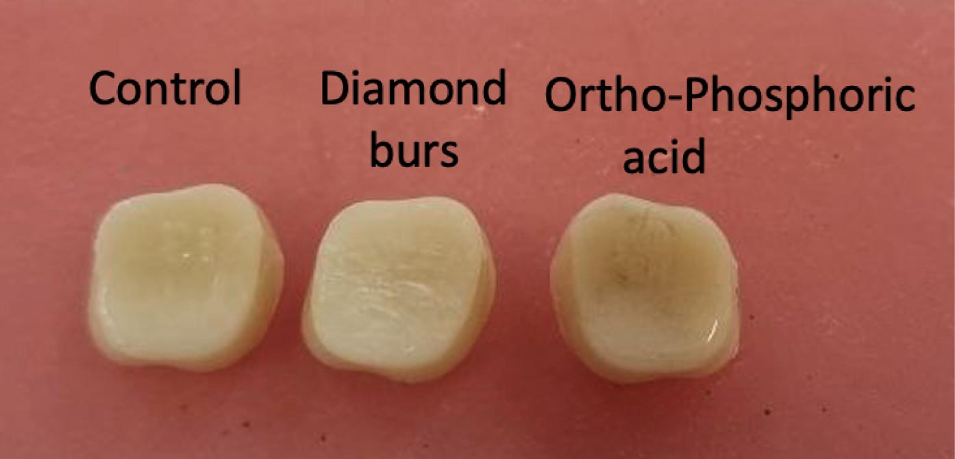
Figure 1.
Control Group, Mechanical and Chemical Treatment Groups
.
Control Group, Mechanical and Chemical Treatment Groups
The teeth were placed in wax blocks in such a way that the longitudinal axis of the teeth was placed at an angle of 45 degrees to the base block (Figure 2).
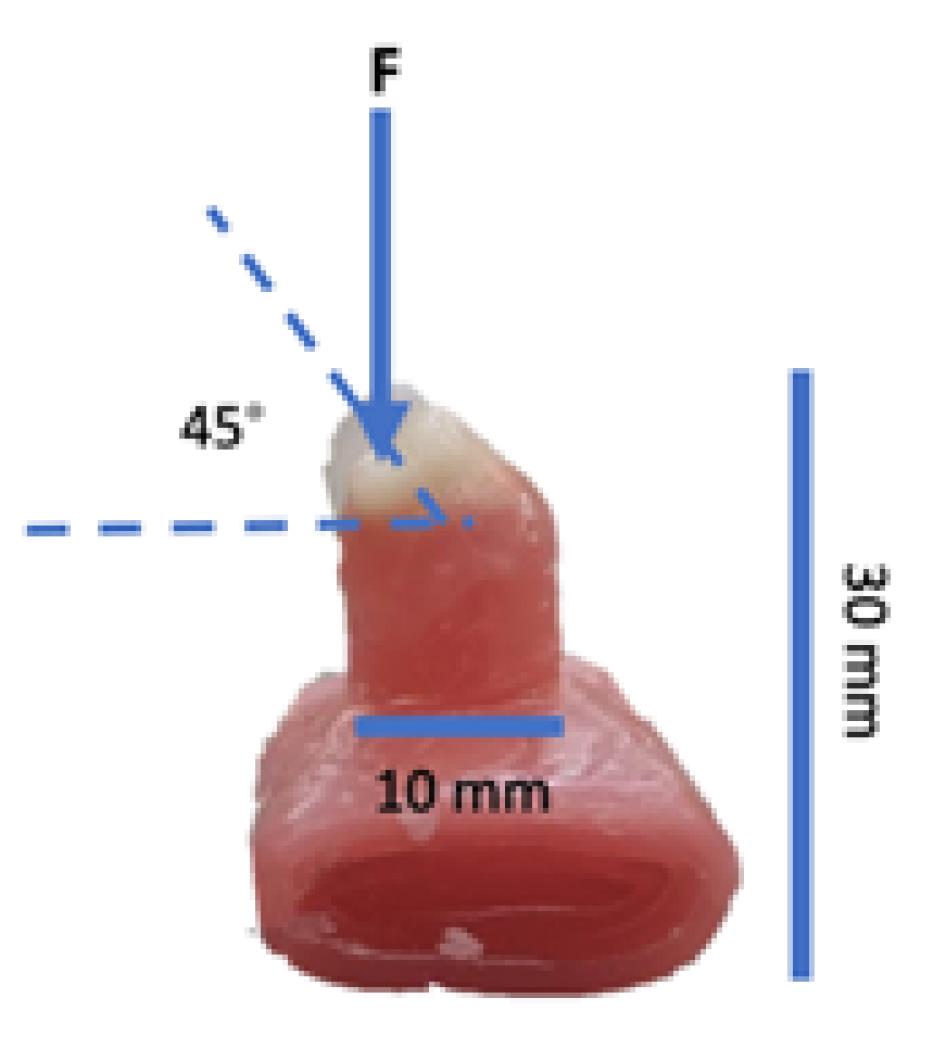
Figure 2.
The Teeth Mounted in the Wax Block and the Schematic Diagram of How the Force is Applied
.
The Teeth Mounted in the Wax Block and the Schematic Diagram of How the Force is Applied
Plaster type 2 and dental stone type 4 were mixed in equal proportions, the wax blocks were flasked, and burnout was performed. Then, the flasks were opened, and heat cured acrylic resin was mixed based on the manufacturer’s instructions (Acropars, Tehran, Iran), packed inside the flasks, and pressed twice under 1.5 bar pressure. Finally, the acrylic additions were removed in each step and then heated (Figure 3).
To simulate oral conditions, the samples were subjected to 10 000 cycles of thermocycling in distilled water between 5 and 55 °C (12), which is equivalent to placing the samples in the patient’s mouth for 100 days. Point compressive load was applied using a universal mechanical testing machine (Model 4301, Instron, Canton, MA, USA; Figure 4).
Figure 4.
Universal Mechanical Testing Machine
.
Universal Mechanical Testing Machine
The compression point on the palatal surface of the teeth measured 1 mm and pointed in the direction of the fracture. Each sample was removed after failure, and the maximum load at failure was measured in Newtons (N). The compressive stress (in MPa) at the maximum compressive load was computed using the formula S = Force/Area, where F is the maximum load necessary to cause failure and A denotes the test specimen’s interface area, measured in mm2.
For analysis, all data were entered into SPSS software, version 24. Inferential statistics and appropriate statistical tests such as independent t-test, analysis of variance (ANOVA), and post hoc tests (including LSD) were used. It should be mentioned that the Shapiro-Wilks and Kolmogorov-Smirnov tests were initially utilized to verify the assumption of data normality; if the normality assumption was not established, the non-parametric equivalent of the mentioned parametric tests was considered to use appropriate transformations to normalize the data. The significance level of the test was considered 0.05.
Results
Table 1 and Figure 5 show the compressive BS in all the studied groups. The highest compressive strength was related to the Vita artificial teeth group with orthophosphoric acid chemical treatment, while the lowest compressive strength was related to the Stone artificial teeth group without treatment. Based on the results (Table 1), in the groups where the surface treatment was performed either mechanically or chemically on the Iranian Classic brand, the average compressive BS with denture base resin was similar to or more than Vita artificial teeth, which were much more expensive.
Table 1.
Compressive BS Score Distribution According to Study Groups
|
Groups
|
Mean
|
Standard Deviation
|
Minimum
|
Maximum
|
| Vita |
14.09 |
1.04 |
13 |
16 |
| Vita under mechanical treatment (diamond bur) |
16.09 |
1.04 |
14 |
17 |
| Vita under chemical treatment (orthophosphoric acid) |
17.90 |
1.97 |
14 |
20 |
| Stone |
10.72 |
1.67 |
8 |
13 |
| Stone under mechanical treatment (diamond bur) |
13.18 |
1.60 |
11 |
15 |
| Stone under chemical treatment (orthophosphoric acid) |
12.54 |
1.21 |
10 |
14 |
| Classic |
13.27 |
2.05 |
11 |
18 |
| Classic under mechanical treatment (diamond bur) |
14.81 |
1.32 |
13 |
17 |
| Classic under chemical treatment (orthophosphoric acid) |
15.63 |
1.56 |
14 |
18 |
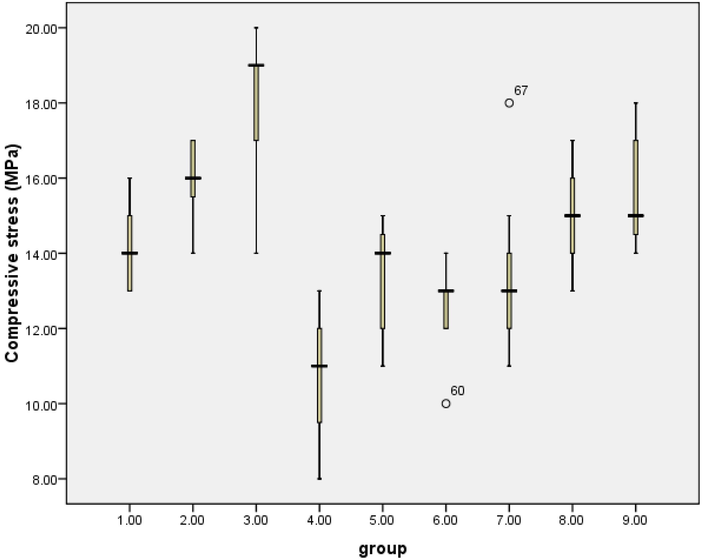
Figure 5.
Boxplot Diagram of Compressive Stress Score Distribution According to Study Groups
.
Boxplot Diagram of Compressive Stress Score Distribution According to Study Groups
Considering the normality of the distribution of the compressive stress value, ANOVA analysis was employed to check the difference in the average value of the compressive stress according to the groups.
According to the findings (Table 2), the compressive BS between the teeth and the base of the acrylic prosthesis in all three types of artificial teeth (Vita, Stone, and Classic) showed significant differences in the control groups and the groups that were treated with diamond burs and orthophosphoric acid.
Table 2.
Comparison of Compressive BS of Control Groups and Chemical and Mechanical Treatments in Each of the Brands
|
Groups
|
Mean
|
Standard Deviation
|
P
Value
|
| Vita |
14.09 |
1.04 |
< 0.001 |
| Vita under mechanical treatment (diamond bur) |
16.09 |
1.04 |
| Vita under chemical treatment (orthophosphoric acid) |
17.90 |
1.97 |
| Stone |
10.72 |
1.67 |
0.002 |
| Stone under mechanical treatment (diamond bur) |
13.18 |
1.60 |
| Stone under chemical treatment (orthophosphoric acid) |
12.54 |
1.21 |
| Classic |
13.27 |
2.05 |
0.008 |
| Classic under mechanical treatment (diamond bur) |
14.81 |
1.32 |
| Classic under chemical treatment (orthophosphoric acid) |
15.63 |
1.56 |
The results (Table 3) further revealed a remarkable distinction in the compressive BS between the teeth and acrylic prosthesis base in Vita, Stone, and Classic teeth in the control groups (P< 0.001) and in the groups where the mechanical treatment was performed by roughening the connection surface with a diamond bur (P < 0.001). Moreover, there was a remarkable distinction in the compressive BS between the teeth and the acrylic prosthesis base in Vita, Stone, and Classic teeth in the groups that underwent chemical treatment using orthophosphoric acid (P < 0.001).
Table 3.
Comparison of Compressive BS of Control Groups From Different Brands and Mechanical and Chemical Treatments From Different Brands
|
Groups
|
Mean
|
Standard Deviation
|
P
Value
|
| Vita |
14.09 |
1.04 |
< 0.001 |
| Stone |
10.72 |
1.67 |
| Classic |
13.27 |
2.05 |
| Vita under mechanical treatment (diamond bur) |
16.09 |
1.04 |
< 0.001 |
| Stone under mechanical treatment (diamond bur) |
13.18 |
1.60 |
| Classic under mechanical treatment (orthophosphoric acid) |
14.81 |
1.32 |
| Vita under chemical treatment (orthophosphoric acid) |
17.90 |
1.97 |
< 0.001 |
| Stone under chemical treatment (diamond bur) |
12.54 |
1.12 |
| Classic under chemical treatment (orthophosphoric acid) |
15.63 |
1.56 |
Discussion
The separation of artificial teeth from the resin base is frustrating for both patients and dentists, especially when the force exerted on the prosthetic components increases; for example, in cases where the patient has implants, teeth separation becomes a major clinical problem (13,14).
The compressive BS of Iranian artificial teeth and Vita artificial teeth with a combination of chemical and mechanical surface treatments with orthophosphoric acid was compared in this study. Based on the results, the compressive BS of the orthophosphoric acid group and the group that roughened the connection surface with a diamond burr was significantly higher than that of the control group. This can be justified by the fact that the surface energy of the acrylic resin with different chemical and physical changes, such as orthophosphoric acid and roughening of the connection surface with a diamond burr, is different from acrylic resin without treatment (15).
Sayed et al compared the compressive BS between acrylic denture base and tooth using a combination of mechanical and chemical treatments and concluded that roughing with diamond burs produced the maximum BS, while grooving and sandblasting led to the lowest BS (16), which is in line with the results of Stone teeth that underwent mechanical treatment with a diamond burr in our study. However, the results related to Vita and Classic teeth in our study, in which the highest BS was related to chemical treatment with orthophosphoric acid, were different, and this difference can be attributed to the structural differences and type of these teeth. Saliman et al investigated how much the shear BS between ceramic teeth and resin denture bases depends on physical and chemical surface treatments (monomer application, creating T-shaped holes, and a combination of both). They concluded that applying monomer to teeth (chemical treatment) had the highest BS (17). In the present study, orthophosphoric acid chemical treatments in Classic and Stone teeth caused a greater increase in compressive BS than diamond burr treatment.
Sadar et al applied various surface modifications, such as sandblasting, groove creation, and monomer application, to compare the compressive and shear BS between acrylic resin teeth and denture base. The result demonstrated that a slight mechanical change in the artificial teeth’s surface changed the compressive and shear BS between the artificial teeth and the denture base compared to the teeth that remained intact. However, the maximum compressive and shear band strength can be increased by using methyl methacrylate monomer, which is less time-consuming (18), which conforms to the findings of the current study, so that orthophosphoric acid (chemical treatment) increased the compressive strength of Vita and Classic teeth compared to diamond burs.
Phukela et al found that the BS between denture teeth and Lucitone 199 heat-cured denture base material was increased with the mechanical modifications of denture teeth. In their study, the control group specimens without any modification prepared by Lucitone 199 heat cure resin showed the lowest BS value, whereas the specimens prepared with a T-shaped groove packed with Lucitone 199 heat cure resin indicated the highest BS value. Therefore, this modification can be a suggested method to secure denture teeth in denture bases (19), which corroborates the results of our study. Bhochhibhoya et al evaluated the effect of surface treatment by painting the ridge lap surface of the teeth with monomer and mechanical modifications by sandblasting the ridge lap surface with aluminum oxide particles and preparing a diatoric on shear BS between acrylic denture teeth and heat-cured acrylic denture base resin. This study suggested the highest BSs for specimens treated with sandblasting and the lowest strengths for untreated specimens without extension of acrylic to the cervical collar (20), which is in conformity with the findings of the present study. In another study, Phukela et al investigated mechanically modified heat-polymerized acrylic resin for shear BS. Their samples were central incisors, which were divided into 4 groups. Group 1 was the control group, whereas groups 2, 3, and 4 were experimental groups modified with half-moon-shaped grooves, Y-shaped grooves, and a combination of half-moon- and Y-shaped grooves, respectively. They concluded that specimens with a combination of half-moon- and Y-shaped grooves showed the highest shear BS value, while the specimens without any mechanical preparation represented the lowest shear BS value (21). Pande et al studied the tensile BS between artificial teeth and acrylic base before and after thermocycling. They found that the shear BS was not significantly affected before and after thermocycling in heat-polymerized denture base resins (22). In our study, the effect of thermocycling was also taken into consideration.
The results of this study would not be directly compared with the findings of other studies, since the base resin of artificial teeth, the chemical used, and the chemical composition of acrylic teeth vary, all of which might have different effects on the BS. The inconsistent outcomes observed in different studies examining the impact of chemical and physical surface treatments on the strength of the tooth-resin bond could potentially be attributed to the utilization of distinct denture bases.
Thermocycling is a laboratory process in which the temperature conditions similar to those in the human mouth are simulated, so that thermal stresses that occur in the teeth and dental materials during the treatment of the patient also occur in the laboratory samples, and the test conditions will be closer to real and clinical conditions. In the thermocycling process, the samples are placed in hot and cold distilled water at certain and predetermined times at temperatures similar to those that may occur in the patient’s mouth. During the thermocycling process, temperature changes and water absorption by dental materials alter the properties of these materials. Previous research showed that thermocycling increases the rate of abrasion and surface destruction of composites, and the thermal shock created between the constituent components of the composite as a result of this process causes microcracks in it. Therefore, it is crucial to investigate the mechanical properties of dental materials subjected to the thermocycling process in a humid environment (12,23,24). For this reason, thermocycling was used in the present study.
The comparison of failure modes demonstrated that cohesive failure was dominant at the denture base/tooth interface in groups treated with diamond burs and orthophosphoric acid, and adhesive failure was dominant in the control groups. According to the British standard (25), the BS is satisfactory when the failure is cohesive. In actuality, a cohesive tooth fracture indicates that the BS has been successfully reached. In the current study, a large number of sample failures occurred in all samples treated with diamond burs and chemically with orthophosphoric acid in a cohesive way, resulting in artificial teeth leaving a layer of dental material on the prosthetic base. Conversely, a small number of teeth displayed a combination of adhesive failure on the tooth and/or denture base. In terms of prosthesis efficiency, cohesive fractures can be considered more favorable compared to other fractures (26). The use of acrylic teeth or prosthesis bases with surface treatments of diamond burs and orthophosphoric acid can prevent adhesive failures and thus prolong the life of the prosthesis.
Since this study is an in vitro study and does not include all the conditions in the oral cavity, it is suggested that it be investigated in vivo as well. It is also suggested that other types of existing artificial teeth or other types of prosthetic bases, such as computer-aided design bases, be examined.
Conclusion
Typically, surface treatment with diamond burs and chemicals containing orthophosphoric acid can be used to increase the compressive BS between the prosthesis base and the artificial teeth. In the teeth examined in this study without surface treatment, the highest compressive strength was related to the Vita teeth, followed by the Classic teeth, while the lowest compressive strength belonged to the Stone teeth. However, chemical and mechanical surface treatments can increase the compressive BS of these teeth, so that in this study, it was observed that Stone teeth surface treatment with diamond burr and Vita and Classic teeth surface treatment using orthophosphoric acid can significantly increase the compressive BS compared to other treatments.
Acknowledgements
The authors appreciate the Department of Prosthodontics, Faculty of Dentistry, Hamadan University of Medical Sciences, for financial and spiritual help.
Competing Interests
The authors declare that there is no conflict of interests.
Ethical Approval
This study was confirmed by the Ethics Committee of Hamadan Dental School (Ethical code: IR.UMSHA.REC.1402.051).
Funding
None.
References
- Stein C, Santos NM, Hilgert JB, Hugo FN. Effectiveness of oral health education on oral hygiene and dental caries in schoolchildren: systematic review and meta-analysis. Community Dent Oral Epidemiol 2018; 46(1):30-7. doi: 10.1111/cdoe.12325 [Crossref] [ Google Scholar]
- Wada M, Mameno T, Kani M, Miwa S, Ikebe K. Association between gonial angle on orthopantomogram and reline frequency in removable partial denture wearers. J Prosthodont Res 2022; 66(1):83-6. doi: 10.2186/jpr.JPR_D_20_00153 [Crossref] [ Google Scholar]
- Chen J, Ahmad R, Suenaga H, Li W, Sasaki K, Swain M. Shape optimization for additive manufacturing of removable partial dentures--a new paradigm for prosthetic CAD/CAM. PLoS One 2015; 10(7):e0132552. doi: 10.1371/journal.pone.0132552 [Crossref] [ Google Scholar]
- Kattadiyil MT, AlHelal A, Goodacre BJ. Clinical complications and quality assessments with computer-engineered complete dentures: a systematic review. J Prosthet Dent 2017; 117(6):721-8. doi: 10.1016/j.prosdent.2016.12.006 [Crossref] [ Google Scholar]
- Ravi N, Krishna DP, Manoj S, Chethan H. A functional stress analysis in the maxillary complete denture influenced by the position of artificial teeth and load levels: an in-vitro study. J Indian Prosthodont Soc 2010; 10(4):219-25. doi: 10.1007/s13191-011-0046-0 [Crossref] [ Google Scholar]
- Chittaranjan B, Taruna M, Sudheer N, Patil NS. Evaluation of shear bond strength of three different types of artificial teeth to heat cure denture base resin: an in vitro study. Indian J Dent Res 2013; 24(3):321-5. doi: 10.4103/0970-9290.117994 [Crossref] [ Google Scholar]
- Jain G, Palekar U, Awinashe V, Mishra SK, Kawadkar A, Rahangdale T. The effect of different chemical surface treatments of denture teeth on shear bond strength: a comparative study. J Clin Diagn Res 2014; 8(6):ZC15-8. doi: 10.7860/jcdr/2014/8420.4474 [Crossref] [ Google Scholar]
- BinMahfooz AM, Qutub OA. Effect of surface treatments and adhesive materials on the shear bond strength of artificial denture teeth to denture base resins. J Contemp Dent Pract 2018; 19(6):631-6. [ Google Scholar]
- Yadav NS, Somkuwar S, Mishra SK, Hazari P, Chitumalla R, Pandey SK. Evaluation of bond strength of acrylic teeth to denture base using different polymerization techniques: a comparative study. J Int Oral Health 2015; 7(Suppl 1):54-6. [ Google Scholar]
- Zidan S, Silikas N, Haider J, Alhotan A, Jahantigh J, Yates J. Assessing tensile bond strength between denture teeth and nano-zirconia impregnated PMMA denture base. Int J Nanomedicine 2020; 15:9611-25. doi: 10.2147/ijn.s273541 [Crossref] [ Google Scholar]
- Patil SB, Naveen BH, Patil NP. Bonding acrylic teeth to acrylic resin denture bases: a review. Gerodontology 2006; 23(3):131-9. doi: 10.1111/j.1741-2358.2006.00129.x [Crossref] [ Google Scholar]
- Gale MS, Darvell BW. Thermal cycling procedures for laboratory testing of dental restorations. J Dent 1999; 27(2):89-99. doi: 10.1016/s0300-5712(98)00037-2 [Crossref] [ Google Scholar]
- Jemt T. Failures and complications in 391 consecutively inserted fixed prostheses supported by Brånemark implants in edentulous jaws: a study of treatment from the time of prosthesis placement to the first annual checkup. Int J Oral Maxillofac Implants 1991; 6(3):270-6. [ Google Scholar]
- Cunningham JL, Benington IC. Effect of an experimental cement on denture tooth bond. J Dent Res. 1994:74:949.
- Fletcher AM, Al-Mulla MA, Amin WM, Dodd AW, Ritchie GM. A method of improving the bonding between artificial teeth and PMMA. J Dent 1985; 13(2):102-8. doi: 10.1016/0300-5712(85)90084-3 [Crossref] [ Google Scholar]
- Sayed ME, Lunkad H, Fageeh I, Jaafari M, Tawhari A, Muaidi T. Comparative evaluation of compressive bond strength between acrylic denture base and teeth with various combinations of mechanical and chemical treatments. Coatings 2021; 11(12):1527. doi: 10.3390/coatings11121527 [Crossref] [ Google Scholar]
- Soliman A, Elgendy M, El-Sheikh M. Effect of different mechanical surface treatments on bond strength of acrylic teeth to polyamide denture base. Egypt Dent J 2021; 67(4):3519-23. doi: 10.21608/edj.2021.86999.1720 [Crossref] [ Google Scholar]
- Sadar L, Dhume S, Maniar N, Prakash Patil J, Rane P, Gandhewar M. Comparative evaluation of shear compressive bond strength between cross-linked acrylic resin denture base and cross-linked acrylic resin teeth with different modifcations of their ridge lap surfaces. J Contemp Dent Pract 2013; 14(5):898-903. doi: 10.5005/jp-journals-10024-1423 [Crossref] [ Google Scholar]
- Phukela SS, Chintalapudi SK, Sachdeva H, Dhall RS, Sharma N, Prabhu A. Comparative evaluation of different mechanical modifications of denture teeth on bond strength between high-impact acrylic resin and denture teeth: an in vitro study. J Int Soc Prev Community Dent 2016; 6(2):161-6. doi: 10.4103/2231-0762.178740 [Crossref] [ Google Scholar]
- Bhochhibhoya A, Mathema S, Maskey B. An evaluation on effect of surface treatment and mechanical modifications on shear bond strength between acrylic denture teeth and heat cure acrylic denture base resin. EC Dent Sci 2016; 6(6):1450-62. [ Google Scholar]
- Phukela SS, Soni K, Yadav J. Comparative evaluation of mechanically modified heat polymerized acrylic resin for shear bond strength: an in vitro study. Turkish Online Journal of Qualitative Inquiry 2021; 12(8):381-8. [ Google Scholar]
- Pande N, Zarekar S, Jaiswal P. Evaluation of shear bond strength of acrylic resin teeth to heat-polymerized denture base resin before and after thermocycling: an in vitro study. Indian J Multidiscip Dent 2018;8(1). doi: 10.4103/ijmd.ijmd_6_18.
- Choi JJE, Uy CE, Plaksina P, Ramani RS, Ganjigatti R, Waddell JN. Bond strength of denture teeth to heat-cured, CAD/CAM and 3D printed denture acrylics. J Prosthodont 2020; 29(5):415-21. doi: 10.1111/jopr.13125 [Crossref] [ Google Scholar]
- Dhuru VB, Lloyd CH. The fracture toughness of repaired composite. J Oral Rehabil 1986; 13(5):413-21. doi: 10.1111/j.1365-2842.1986.tb01303.x [Crossref] [ Google Scholar]
- Zidan S, Silikas N, Haider J, Alhotan A, Jahantigh J, Yates J. Assessing tensile bond strength between denture teeth and nano-zirconia impregnated PMMA denture base. Int J Nanomedicine 2020; 15:9611-25. doi: 10.2147/ijn.s273541 [Crossref] [ Google Scholar]
- AlZaher ZA, Almaskin DF, Qaw MS, Abu Showmi TH, Abualsaud R, Akhtar S. Chemo-mechanical approach to improve repair bond strength of denture teeth. Int J Dent 2020; 2020:8870361. doi: 10.1155/2020/8870361 [Crossref] [ Google Scholar]