Avicenna J Dent Res. 16(4):205-209.
doi: 10.34172/ajdr.1768
Original Article
Comparison of Microleakage of Cention N and a Bulk-Fill Composite Resin in Restoring Deciduous Teeth: An In Vitro Study
Farnaz Ghorbani 1  , Sara Maleki Kambakhsh 2 , Baharan Ranjbar Omidi 3, * , Shahrzad Samadian 4
Author information:
1Department of Pediatric Dentistry, Arak University of Medical Sciences, Arak, Iran
2Department of Pediatric Dentistry, Dental Caries Prevention Research Center, Qazvin University of Medical Sciences, Qazvin, Iran
3Department of Operative Dentistry, Qazvin University of Medical Sciences, Qazvin, Iran
4Department of Pediatric Dentistry, Shahid Beheshti University of Medical Sciences, Tehran,Iran
Abstract
Background: Pediatric dentists prefer restorative materials with a high bond strength and fewer clinical stages. Cention N can be used in the bulk technique. It can release fluoride ions, calcium, and hydroxide. This study compared the microleakage of two types of composite resins, namely, Cention N and Tetric N-Ceram bulk-fill composite resin, in class II restorations (box only) of deciduous teeth.
Methods: In this experimental investigation, 50 class II restorations (box only) were prepared in extracted deciduous second molars. Then, the teeth were divided into two groups based on their restorative materials. The first and second groups were restored with Cention N and bulk-fill Tetric N-Ceram composite resins, respectively. The Tetric N bond adhesive was used before placing the restorative materials in both groups. The samples were cut and investigated under a stereomicroscope to determine microleakage after thermocycling and staining with silver nitrate. Two samples from each group were prepared for observation under an electron microscope. Then, the data were analyzed using the nonparametric Mann-Whitney test.
Results: The mean percentage of dye penetration into the gingival wall was not significantly different between the two groups under investigation.
Conclusion: Using Cention N with adhesive yielded favorable results in terms of microleakage, and it is recommended for class II restorations in deciduous molars.
Keywords: Composite resin, Deciduous teeth, Microleakage, Cention N
Copyright and License Information
© 2024 The Author(s); Published by Hamadan University of Medical Sciences.
This is an open-access article distributed under the terms of the Creative Commons Attribution License (
https://creativecommons.org/licenses/by/4.0), which permits unrestricted use, distribution, and reproduction in any medium provided the original work is properly cited.
Please cite this article as follows: Ghorbani F, Maleki Kambakhsh S, Ranjbar Omidi B, Samadian S. Comparison of microleakage of cention n and a bulk-fill composite resin in restoring deciduous teeth: an In vitro study. Avicenna J Dent Res. 2024; 16(4):205-209. doi:10.34172/ajdr.1768
Background
Dental caries is one of the chronic diseases of childhood, affecting deciduous and permanent teeth. Despite a significant decrease in dental caries rates, particularly in developed countries, many children are still affected (1).
Various restorative materials that can supply function and esthetics have been introduced to restore the carious tooth structure (2). The most common restorative materials in pediatric dentistry are composite resins, resin-based materials, glass ionomer, amalgam, and stainless-steel crowns (3). Composite resins are among the most popular restorative materials due to their excellent esthetic and mechanical properties and controllable setting time (4). Bulk-fill composite resins are a new type of composite resin that can be placed at 4-mm thicknesses in the prepared cavity and cured with the least polymerization stress (5). This type of composite resin is a suitable choice for children due to its capacity for bulk use, which leads to decreased chair time and contamination risk of the prepared box and increases patient cooperation (6). Tetric® N-Ceram is one of the members of this family.
Cention N is a bulk-fill alkasite composite resin that can neutralize acids. This material can be used in a self-adhesive mode or with a bonding agent (7). It is claimed that this type of material can release ions such as fluoride, calcium, and hydroxide to increase the remineralization of the remaining tooth structure while decreasing caries recurrence (8). One of the most crucial factors determining the function and life span of restoration is microleakage, which is the possibility of spreading the bacteria, liquids, molecules, or ions between the prepared cavity wall and restorative material (4).
Restoring deciduous teeth is different from restoring permanent teeth due to the presence of multiple anatomic differences. The enamel of the deciduous teeth is less mineralized and has lower thickness and surface hardness than the permanent teeth (9).
The density and diameter of the dentinal tubules are higher in deciduous teeth than in permanent teeth. Therefore, solid dentin available for bonding in deciduous teeth is less than that in permanent teeth. On the other hand, since the deciduous teeth are smaller than permanent teeth, the thickness of the deciduous dentin from the dentin–enamel junction to the pulp is less than that of permeant dentine (10). Hence, deciduous teeth are more susceptible to dissolution by acid than permanent teeth, and the adhesion pattern is different between deciduous and permanent teeth, affecting the dental microleakage incidence (9).
A few studies have investigated the microleakage and other mechanical properties of Cention N (2,11,12). However, no study has compared this material’s microleakage level to that of other bulk-fill materials in deciduous teeth. Thus, this study evaluated the microleakage of Cention N and Tetric N-Ceram bulk-fill composite resins in class II cavities of deciduous teeth.
Materials and Methods
According to the Cochrane table (moderate effect) and by considering the alpha = 0.05, a study power of 80%, and an impact factor of 0.5, the minimum sample size was calculated at 44 (n = 22 in each group).
Due to the possibility of damaging the samples during the preparation process, fifty teeth were considered for investigation. Fifty extracted deciduous second molars with at least one intact proximal surface were collected after obtaining consent from the children’s parents or legal guardians. After debridement for disinfection, the teeth were stored for one week in 0.5% chloramine T solution. The teeth were investigated under an optical microscope at × 10 magnification to ensure the absence of cracks or fractures. Teeth with signs of hypoplasia, hypocalcification, and cracks, or teeth in which the caries process affected more than a quarter of the occlusal surface, were excluded from the study (3).
Fifty class II cavities (box only) with cavity dimensions of 3 × 3 × 1.5 mm were prepared (3), and the cavities were prepared with a cylindrical diamond bur (008; Teezkavan, Iran) in a high-speed headpiece with a cooling water system. After preparing five cavities, the bur was replaced, and a periodontal probe precisely controlled the cavity dimension. Then, the matrix bands of Tofflemire (Temrex, USA) were used, and the restoration process was performed as follows:
The prepared cavities were etched with 37% phosphoric acid (Morvabon, Iran) and then rinsed with water spray for 15 seconds. Cotton pellets and air spray were used to remove extra moisture. Next, according to the instructions of the manufacturer, a thick layer of Tetric N-bond (Ivoclar Vivadent, Liechtenstein) as the adhesive was placed in each cavity for 10 seconds by a microbrush, and the additional solvent was eliminated by a gentle air current. Light-curing was performed by a calibrated tungsten-halogen light-curing device (Coltolux) with a power of 800 MW/cm2 (Coltene Whaledent, USA) for 20 seconds. The teeth were randomly divided into two groups after bonding:
-
Group 1 (n = 25): Cention N (Ivoclar Civadent, Liechtenstein)
-
Group 2 (n = 25): Tetric N-Ceram bulk-fill (Ivoclar Vivadent, Liechtenstein)
Group 1: According to the manufacturer’s instructions, the bottle containing the powder was shaken well before use. Subsequently, a scoop of powder and a drop of liquid were placed on a glass slab. The powder was divided into two parts. First, one part of the powder was mixed with all the liquid, and then the remaining powder was mixed until a homogenous consistency was obtained (mixing time = 45-60 seconds). The cavity was filled in a maximum of two minutes with the material (working time = 3 minutes). Next, light-curing was carried out for 40 seconds.
Group 2: According to the manufacturer’s instructions, the bulk-fill Tetric N-Ceram restorative material was placed in a bulk technique with a maximum thickness of 4 mm in the cavity, packed with a condenser, and light-cured for 20 seconds (13).
After removing the matrix band in both groups, light-curing was performed again from the buccal and palatal/lingual aspects. Then, the samples were incubated (WTW, USA) for 24 hours at 37 °C (310.15 kelvin) and 100% moisture. Eventually, they were polished with Sof-lex disks (3M, USA) (3). Then, all the samples underwent 2000 thermal cycles (5/55 °C) in a thermocycling machine (Dorsa, Iran), with a dwell time of 30 seconds and a transfer time of 30 seconds (3). After drying the samples, both groups were ready for evaluating microleakage. First, the apical part of the tooth roots, their furcal area, and parts of the root affected by the resorption process were sealed with the flowable composite resin (Denfil, South Korea). Next, all the tooth surfaces were covered with two layers of nail polish (Cube, Korea), except for the restoration site and 1–1.5 mm around it. The second layer of nail polish was applied after drying the first layer. Afterward, the samples were placed in 1 mol (17 g in 100 mL of distilled water) of the silver nitrate solution (Merk, Germany) at room temperature. Then, the teeth were retrieved from the solution and rinsed with water for 5 minutes.
The samples were placed in the radiographic developer solution and then under a fluorescent light for 12 hours, followed by rinsing again with water for 5 minutes. To blind the operators about group allocations, each sample was given a code after the preparation process and before mounting. Next, the samples were mounted in the acrylic resin in such a way that all tooth surfaces were buried in the acrylic resin up to 2 mm lower than the gingival margin of the restorations. Subsequently, the tooth samples were cut longitudinally in the mesiodistal direction from the restoration center using diamond disks (Drux, Germany). Afterward, microleakage was evaluated and imaged under a stereomicroscope (Zistrad, Iran) on the gingival wall at × 40 magnification (Figure 1). After assessing the image, the level of linear penetration was measured and recorded using a microleakage classification method (ISO/TS 11405:2003) (14).
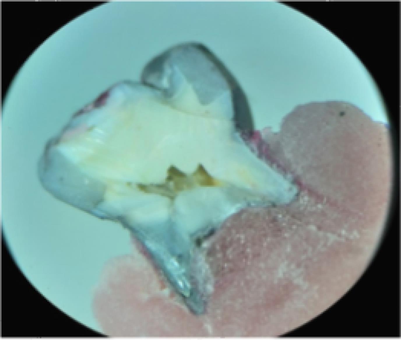
Figure 1.
Sample of an Observed Image by Stereomicroscope
.
Sample of an Observed Image by Stereomicroscope
Microleakage scoring in the gingival wall was as follows:
0 = No dye penetration
1 = Dye penetration to half of the gingival wall
2 = Dye penetration to the entire gingival wall
3 = Dye penetration to gingival and axial walls
After measuring the severity of microleakage in each group, two samples were selected for observation under a scanning electron microscope (SEM; Vega II XMU, Tescan, Czech Republic) (15). First, the samples were immersed in the 6N hydrochloric acid solution (Merk, Germany) for 30 seconds and then rinsed for 5 minutes in water. Then, they were immersed in the 2.5% sodium hypochlorite solution and rinsed again with water for 5 minutes. The samples were dried and covered with a gold foil, and some micrographs were taken from the restoration–tooth interfaces at different magnifications. The data were analyzed with the Mann-Whitney test using SPSS 25.
Results
Fifty deciduous molars with at least one intact proximal wall in the distal or mesial side underwent evaluation. After excluding the improper samples due to the sectioning errors, the data from 22 deciduous teeth in the Cention N group and 23 deciduous teeth in the bulk-fill Tetric N-Ceram composite resin group were statistically analyzed to evaluate the microleakage in both groups. The Kolmogorov-Smirnov test was used to examine the normality of the data. The nonparametric Mann-Whitney test was utilized since the data did not exhibit a normal distribution.
Table 1 presents a comparison of the frequency distribution of microleakage between the two groups; approximately 65.2% of samples in the Tetric N-Ceram bulk-fill composite resin group and 59.1% of samples in the Cention N group had no microleakage (score 0). On the other hand, 4.3% of samples in the Tetric N-Ceram bulk-fill composite resin group and 9.1% of samples in the Cention N group had a score of 3. However, the analysis by the Mann-Whitney test revealed no significant difference between the two groups (Table 2, P > 0.05).
Table 1.
Relative Frequency Distribution of Microleakage of Tetric N-Ceram and Cention N Composites
|
Material
|
Microleakage Scoring
|
|
Score 0
No. (%)
|
Score 1
No. (%)
|
Score 2
No. (%)
|
Score 3
No. (%)
|
Total
No. (%)
|
| Cention N |
13 (59.1) |
3 (13.6) |
4 (18.2) |
2 (9.1) |
22 (100) |
| Tetric N-Ceram |
15 (65.2) |
3 (13.1) |
4 (17.4) |
1 (4.3) |
23 (100) |
Table 2.
Comparison of Microleakage Level Between Tetric N-Ceram and Cention N Composites
|
Group
|
Minimum
|
Maximum
|
Mean Rank
|
P
Value
|
| Cention N |
0 |
3 |
47.91 |
0.44 |
| Tetric N-Ceram |
0 |
3 |
44.21 |
Figures 2 and 3 display the morphology of the teeth interfaces with different types of composite resins under the SEM.
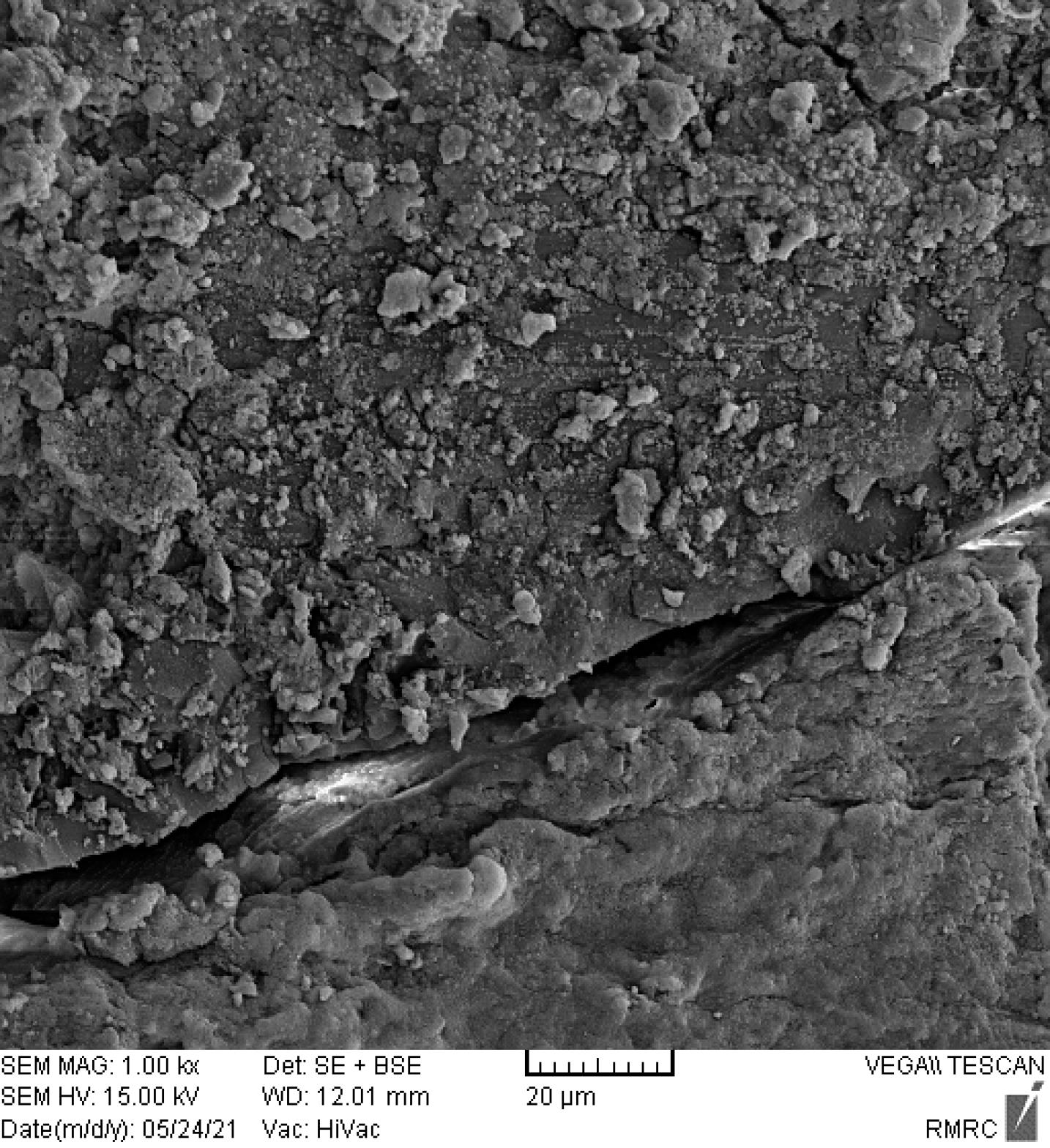
Figure 2.
Tooth Interface With Cention N Under the Electron Microscope With a × 1000 Magnification
.
Tooth Interface With Cention N Under the Electron Microscope With a × 1000 Magnification
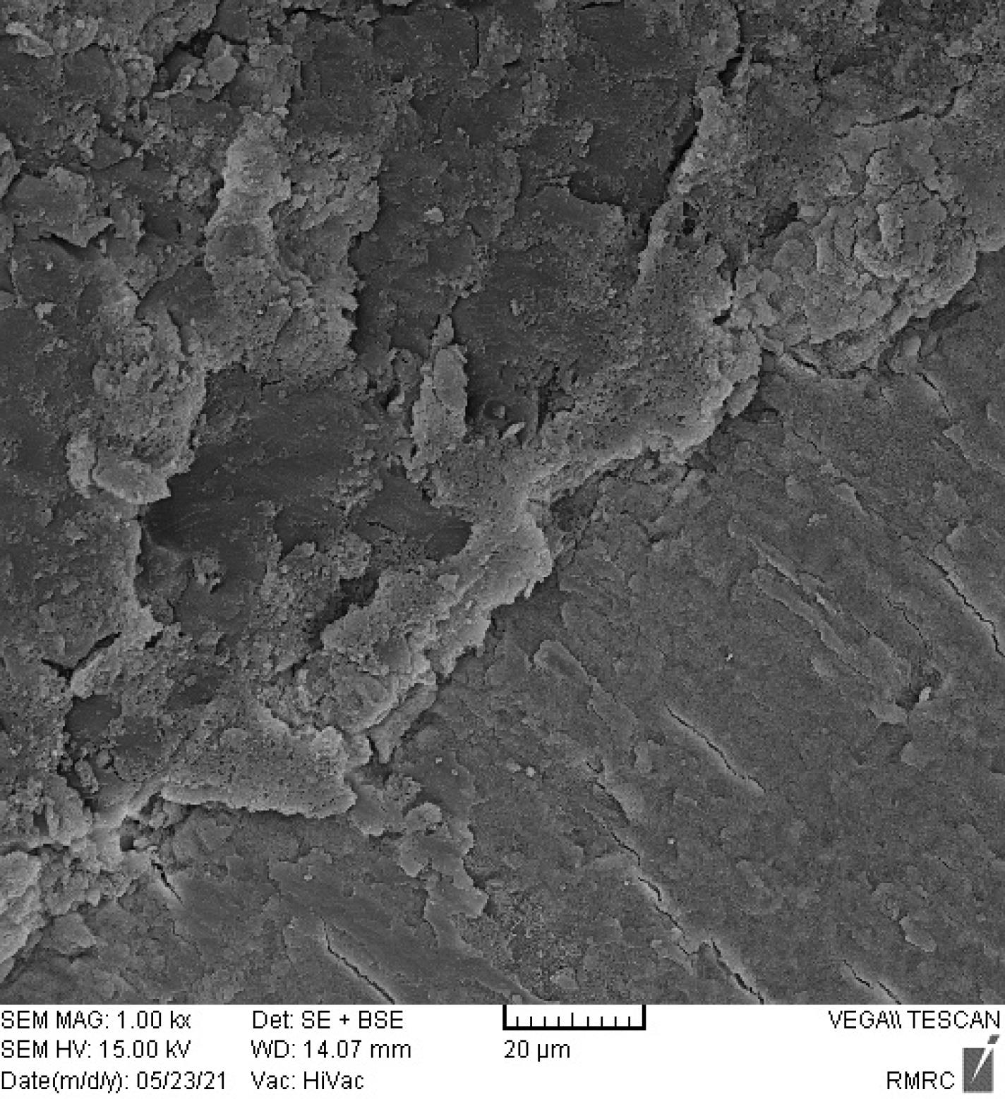
Figure 3.
Tooth Interface With the Tetric N-Ceram Composite Under the Electron Microscope With a × 1000 Magnification
.
Tooth Interface With the Tetric N-Ceram Composite Under the Electron Microscope With a × 1000 Magnification
Discussion
The current study was designed to compare the microleakage of Cention N composites with Tetric N-Ceram bulk-fill composite resins in the class II cavities (box only) of deciduous molars.
The results of the dye penetration by stereomicroscope showed that the means of the microleakage scores in the Cention N and Tetric N-Ceram bulk-fill composite resin groups were not significantly different (P > 0.05), consistent with the results of previous studies. (5,15,16). Punathil et al compared the microleakage of three restorative materials, including nano-filled resin-modified glass ionomer, nanocomposite resin, and Cention N, reporting significant differences in the microleakage of these three materials. The nanocomposite resin had the greatest microleakage. In addition, nano-filled resin-modified glass ionomer exhibited the least microleakage, and Cention N had a moderate level of microleakage (12). One of the reasons for the differences between the current study results and those of the study by Punathil et al is the different types of applied composite resins. The Z350 composite resin was used in their study. This composite resin is a conventional composite and cannot be utilized in bulk. On the other hand, this composite resin does not have the isofillers that Cention N and Tetric N-Ceram bulk-fill composite resins have. Cention N and Tetric N-Ceram bulk-fill composite resins have a special and patented filler named isofiller, which serves as a reliever for polymerization stresses and minimizes shrinkage stresses during the polymerization process. Considering the low elastic modulus of this filler (10 GPa), this isofiller acts like a spring that expands slowly during the polymerization process (11). This isofiller is responsible for the lower polymerization shrinkage, leading to a lower microleakage of the assessed bulk-fill composite resins in the current study compared to the Z350 composite resin used in the study by Punathil et al. In addition, in the mentioned study, the Cention N composite resin was applied without an adhesive, and this might have affected the results and the comparison of the Cention N and Z350 composite resins as a confounding factor. According to the manufacturer’s instructions, the Tetric N bond was utilized as an adhesive for both restorative materials.
To investigate microleakage, various studies have evaluated class II and V cavities (5,17). Class II cavities in the present study had a classic and conservative structure, and the gingival floor was above the cemento-enamel junction on the enamel because the American Association of Pediatric Dentistry has recommended placing stainless steel crowns in wide and deep two-surface restorations (18).
Considering that restorative materials and bonding agents show similar behavior at the beginning of the bonding process, the factor distinguishing the bonding mechanisms from each other is bond continuity and level of microleakage in the long term (19). Similar to other experimental studies, the current study employed a thermocycling process to simulate the thermal stresses of the oral environment and demonstrated that bond continuity was more important than its primary strength. This study used the dye penetration method and 1-mol silver nitrate solution. Silver nitrate is the most common material utilized to assess microleakage, and it easily penetrates the restoration–tooth interface because of its small diameter of particles (0.059 nm). One of the technical problems of working with organic dyes such as methylene blue is the possibility of dissolution and elimination during the cutting process by diamond burs and water cooling systems. However, this is not a problem with silver nitrate because silver nitrate reacts with a radiographic developer solution and can be fixed. Thus, more penetration or movement does not occur in the subsequent preparation stages of the samples. However, due to the small size of the dye particles compared to the typical size of the bacteria (0.5–1 μm), this technique is considered a strict method. Nevertheless, it can be assumed that any restoration that prevents the penetration of silver ions can inhibit bacterial penetration (20). The current study used a qualitative scale to investigate microleakage. Rigsby et al reported similar results in class V restorations with adhesive systems in quantitative and qualitative measurements and showed a proper correlation between quantitative and qualitative measurements (21). Destruction of samples after dye penetration and difficulty in interpretation are some of the disadvantages of dye penetration tests.
Observing the gaps is more reliable, and it seems that it is the first sign of restoration failure. Despite laboratory tests’ restrictions, evaluating margins by SEM is an accurate and reliable method to evaluate the marginal fit of adhesive restorations. This method is not destructive and allows for the evaluation of all the perimeter of the interface between the tooth and restoration before and after exposure to the thermal and mechanical loads and aging processes. On the other hand, dental cracks and marginal gaps are not distinguishable in the evaluation by dye penetration, and picture clarities are lower than those in SEM. Some SEM studies evaluate the gap width. However, the presence or absence of the marginal gaps is more important than the gap width because gap development, regardless of its width, is similar to a gate for liquids and can destroy the tooth–composite resin interface (21). In the current study, two samples of each group were investigated by SEM, and no obvious marginal gap was observed in the restoration–tooth interface in the micrographs. Alonso et al reported that staining the gaps yielded results similar to SEM analysis results (22).
According to the current study results, there was no statistically significant difference in microleakage between Cention N and Tetric N-Ceram composite resins. Considering the bioactive properties of Cention N, it can be considered a suitable treatment option in the class II restorations of deciduous molars. Considering that this was an experimental study, simulating all the clinical conditions was impossible; therefore, we cannot generalize the results of this study to clinical conditions. It is suggested that long-term clinical studies should be executed to confirm the results of this study.
Conclusion
-
None of the restorative materials in the study were without microleakage.
-
Microleakage of the Cention N in the class II restoration in deciduous molars was comparable to the Tetric N-Ceram bulk-fill composite resin (i.e., no statistically significant difference was observed).
Authors’ Contribution
Conceptualization: Baharan Ranjbar Omidi.
Data curation: Farnaz Ghorbani.
Formal analysis: Sara Maleki Kambakhsh.
Funding acquisition: Farnaz Ghorbani.
Investigation: Farnaz Ghorbani.
Methodology: Baharan Ranjbar Omidi.
Project administration: Baharan Ranjbar Omidi, Sara Maleki Kambakhsh.
Resources: Farnaz Ghorbani.
Software: Farnaz Ghorbani.
Supervision: Baharan Ranjbar Omidi, Sara Maleki Kambakhsh.
Validation: Sara Maleki Kambakhsh.
Visualization: Farnaz Ghorbani.
Writing–original draft: Farnaz Ghorbani, Shahrzad Samadian.
Writing–review & editing: Baharan Ranjbar Omidi.
Competing Interests
There is no conflict of interests.
Ethical Approval
This study was approved by the Ethics Committee of Qazvin University of Medical Sciences under the code IR.QUMS.REC.1399.187. There is no conflict with ethical considerations.
Funding
None.
References
- Selwitz RH, Ismail AI, Pitts NB. Dental caries. Lancet 2007; 369(9555):51-9. doi: 10.1016/s0140-6736(07)60031-2 [Crossref] [ Google Scholar]
- Feiz A, Amrollahi N, Ziayi F. Comparative evaluation of microtensile bond strength of four glass-containing materials with primary teeth dentin. Iran J Pediatr 2019; 29(4):e88774. doi: 10.5812/ijp.88774 [Crossref] [ Google Scholar]
- Ranjbar Omidi B, Ferdowsizadeh Naeini F, Dehghan H, Tamiz P, Mohammadi Savadroodbari M, Jabbarian R. Microleakage of an enhanced resin-modified glass ionomer restorative material in primary molars. J Dent (Tehran) 2018; 15(4):205-13. [ Google Scholar]
- Amaireh AI, Al-Jundi SH, Alshraideh HA. In vitro evaluation of microleakage in primary teeth restored with three adhesive materials: ACTIVATM, composite resin, and resin-modified glass ionomer. Eur Arch Paediatr Dent 2019; 20(4):359-67. doi: 10.1007/s40368-019-00428-6 [Crossref] [ Google Scholar]
- Sahadev CK, Bharath MJ, Sandeep R, Remya M, Santhosh PS. An-invitro comparative evaluation of marginal microleakage of Cention-N with bulk-fil SDR and Zirconomer: a confocal microscopic study. Int J Sci Res 2018; 7(7):635-8. doi: 10.21275/art20182483 [Crossref] [ Google Scholar]
- Tiskaya M, Al-Eesa NA, Wong FSL, Hill RG. Characterization of the bioactivity of two commercial composites. Dent Mater 2019; 35(12):1757-68. doi: 10.1016/j.dental.2019.10.004 [Crossref] [ Google Scholar]
- Sunyaruri E, Nainggolan TR, Angelia P, Sumantadireja YH, Gartika M. PRR using Cention N® in children’s teeth. J Appl Dent Med Sci 2019; 5(2):52-8. [ Google Scholar]
- Todd JC. Scientific Documentation: Cention N. Schaan, Liechtenstein: Ivoclar-Vivadent Press; 2016. p. 1-58.
- Assunção CM, Dos Santos NM, Essvein TE, Silva MG, Erhardt MC, Rodrigues JA. Microshear bond strength of adhesive systems on eroded primary enamel and dentin. Pediatr Dent 2020; 42(1):47-52. [ Google Scholar]
- Sumikawa DA, Marshall GW, Gee L, Marshall SJ. Microstructure of primary tooth dentin. Pediatr Dent 1999; 21(7):439-44. [ Google Scholar]
- George P, Bhandary S. A comparative microleakage analysis of a newer restorative material–an ex vivo study. IOSR J Dent Med Sci 2018; 17(12):56-60. [ Google Scholar]
- Punathil S, Almalki SA, AlJameel AH, Gowdar IM, Mc VA, Chinnari K. Assessment of microleakage using dye penetration method in primary teeth restored with tooth-colored materials: an in vitro study. J Contemp Dent Pract 2019; 20(7):778-82. doi: 10.5005/jp-journals-10024-2596 [Crossref] [ Google Scholar]
- Patel P, Shah M, Agrawal N, Desai P, Tailor K, Patel K. Comparative evaluation of microleakage of class II cavities restored with different bulk fill composite restorative systems: an in vitro study. J Res Adv Dent 2016; 5(2):52-62. [ Google Scholar]
- Sawani S, Arora V, Jaiswal S, Nikhil V. Comparative evaluation of microleakage in class II restorations using open vs closed centripetal build-up techniques with different lining materials. J Conserv Dent 2014; 17(4):344-8. doi: 10.4103/0972-0707.136450 [Crossref] [ Google Scholar]
- Dodiya PV, Parekh V, Gupta MS, Patel N, Shah M, Tatu S. Clinical evaluation of Cention-N and nano hybrid composite resin as a restoration of noncarious cervical lesion. J Dent Spec 2019; 7(1):3-5. doi: 10.18231/j.jds.2019.001 [Crossref] [ Google Scholar]
- Sardana A, Kumar M, Taneja S. Comparative evaluation of microleakage and hardness of newer posterior restorative materials. J Oral Biol Craniofac Res 2022; 12(5):733-6. doi: 10.1016/j.jobcr.2022.08.023 [Crossref] [ Google Scholar]
- Baghalian A, Baradaran Nakhjavani Y, Hooshmand T, Motahhary P, Bahramian H. Microleakage of Er:YAG laser and dental bur prepared cavities in primary teeth restored with different adhesive restorative materials. Lasers Med Sci 2013; 28(6):1453-60. doi: 10.1007/s10103-012-1222-0 [Crossref] [ Google Scholar]
- Lopez-Cazaux S, Aiem E, Velly AM, Muller-Bolla M. Preformed pediatric zirconia crown versus preformed pediatric metal crown: study protocol for a randomized clinical trial. Trials 2019; 20(1):530. doi: 10.1186/s13063-019-3559-1 [Crossref] [ Google Scholar]
- de Oliveira JC, Aiello G, Mendes B, Urban VM, Campanha NH, Jorge JH. Effect of storage in water and thermocycling on hardness and roughness of resin materials for temporary restorations. Mater Res 2010; 13(3):355-9. doi: 10.1590/s1516-14392010000300013 [Crossref] [ Google Scholar]
- Baig MM, Mustafa M, Al Jeaidi ZA, Al-Muhaiza M. Microleakage evaluation in restorations using different resin composite insertion techniques and liners in preparations with high c-factor – an in vitro study. King Saud Univ J Dent Sci 2013; 4(2):57-64. doi: 10.1016/j.ksujds.2013.03.002 [Crossref] [ Google Scholar]
- Rigsby DF, Retief DH, Russell CM, Denys FR. Marginal leakage and marginal gap dimensions of three dentinal bonding systems. Am J Dent 1990; 3(6):289-94. [ Google Scholar]
- Caroline Bruschi Alonso R, Maria Correr G, Gonçalves Cunha L, Flávia Sanches Borges A, Maria Puppin-Rontani R, Alexandre Coelho Sinhoreti M. Dye staining gap test: an alternative method for assessing marginal gap formation in composite restorations. Acta Odontol Scand 2006; 64(3):141-5. doi: 10.1080/00016350500474565 [Crossref] [ Google Scholar]