Avicenna J Dent Res. 15(4):163-166.
doi: 10.34172/ajdr.1745
Original Article
Assessment of Periapical Status in Posterior Root Canal-Treated Teeth Using Cone Beam Computed Tomography in the Iranian Population
Maryam Foroozandeh 1  , Azita Ehsani 2, * , Salman Khazaei 3
Author information:
1Department of Oral and Maxillofacial Radiology, Dental School, Hamadan University of Medical Sciences, Hamadan, Iran
2Dental and Maxillofacial Radiology, Department of Oral and Maxillofacial Radiology, Dental School, Hamadan University of Medical Sciences, Hamadan, Iran
3Department of Epidemiology, School of Public Health, Hamadan University of Medical Sciences, Hamadan, Iran
Abstract
Background: Periapical radiolucency occurs due to root canal infections. In this respect, effective root canal treatment and appropriate coronal restoration can aid in preventing these lesions. Thus, the present study aimed to assess the periapical status in posterior root canal-treated teeth by cone-beam computed tomography (CBCT) in the Iranian population.
Methods: The CBCT images of 210 patients (475 endodontically treated molars and premolars) referred to the Hamadan Dental School (from 2020 to 2022) were evaluated in this observational study. The images were obtained by a NewTom 3G CBCT device (NewTom, Verona, Italy) with an FOV of 6×6 inches at 110 kVp and varying milliampers and exposure times according to the patient’s age and body size. They were observed by two maxillofacial radiologists who recorded the periapical status, coronal seal, and endodontic treatment quality of root canal-treated teeth. The Chi-square test was used for data analysis (P≤0.05). Finally, the periapical lesion was determined, along with its association with gender, type of tooth, endodontic treatment quality, and appropriate restoration.
Results: The results revealed no significant association between periapical radiolucency and gender. The prevalence of periapical lesions was higher in males aged 25–50 years, but no significant correlation was found in this regard. There was no significant relationship between tooth type and the presence of periapical lesions, while it had the highest prevalence in maxillary molars. Endodontic treatment quality and coronal restoration showed significant correlations with the presence of periapical radiolucency.
Conclusion: There was no significant association between the presence of periapical radiolucency and gender and age. The prevalence of periapical lesions was higher in males. Cases with poor endodontic treatment and poor restoration demonstrated the highest prevalence of periapical lesions.
Keywords: Cone beam computed tomography, Periapical radiolucency, Restoration, Endodontic treatment
Copyright and License Information
© 2023 The Author(s); Published by Hamadan University of Medical Sciences.
This is an open-access article distributed under the terms of the Creative Commons Attribution License (
http://creativecommons.org/licenses/by/4.0), which permits unrestricted use, distribution, and reproduction in any medium provided the original work is properly cited.
Please cite this article as follows: Foroozandeh M, Ehsani A, Khazaei S. Assessment of periapical status in posterior root canal-treated teeth using cone beam computed tomography in the Iranian population. Avicenna J Dent Res. 2023; 15(4):163-166. doi:10.34172/ajdr.1745
Background
Periapical radiolucency is a complication of root canal infection that may be caused by primary infection and pulp necrosis, persistent infection after root canal treatment, or root canal reinfection due to coronal leakage (1). These infections lead to bone destruction in the periapical region, observed as radiolucency in radiographic images (2).
The quality of endodontic treatment and coronal restoration can affect the periapical radiolucency status (3,4).
To prevent apical periodontitis, root canal treatment should be clean and completely disinfected, with sufficient coronal and apical seals (5). Endodontic treatment quality is primarily evaluated by periapical images and clinical examinations. Nonetheless, cone-beam computed tomography (CBCT) has recently overcome the limitations of two-dimensional imaging and increased the diagnostic accuracy in detecting periapical lesions (2,6). Clinical and histological studies indicated that CBCT can show apical periodontitis before it is observed in conventional images, and its results are closer to the histological gold standard. In other words, CBCT reduces false negative diagnoses and is more sensitive than conventional radiography (2,7). The other applications of CBCT images are evaluating root canal morphology, identifying endodontic errors, and assessing post-treatment results (8).
CBCT imaging provides high-quality, three-dimensional images that eliminate overlapping structures, although it has a higher radiation dose and cost. Moreover, CBCT has an excellent spatial resolution ( < 1 mm) and low dose in comparison with conventional CT scans (9).
The prevalence of periapical radiolucency in panoramic and periapical images in different populations was investigated in several studies (10). However, rare studies have assessed the prevalence of periapical radiolucency by CBCT (11). Evaluating the prevalence of periapical lesions in the population can contribute to predicting the need for additional dental treatments in the future and monitoring the performance of dentists.
The prevalence of periapical lesions in different populations has been investigated worldwide in various studies, and most of them reported a significant relationship between periradicular tissue health and endodontic treatment quality (12–15).
This issue has not yet been studied in the population of Hamadan. Considering the more complex structure of the root canal morphology in premolars and molars that affects the quality of root canal treatment, this study was conducted to assess periapical radiolucency in posterior root canal-treated teeth using CBCT in the population of Hamadan.
Methods
CBCT images of patients visiting Hamadan Dental School during 2020–2022 for implant treatment or surgery were evaluated in this research. Overall, 210 CBCT images were observed by considering the inclusion and exclusion criteria. The images were obtained by a NewTom 3G device (NewTom, Verona, Italy) with field of view (FOV) 6 × 6 inches, 110 KVP, and variable mA and exposure times based on age and body size. They had been saved in the NNT Viewer software. A total of 475 endodontic molar and premolar teeth were examined in this study. The images with at least one endodontically treated premolar or molar were included in the study. Teeth with endo-perio lesions, radiolucent lines similar to fractures, and porcelain crowns and posts were excluded from the study. Before evaluating the images, two maxillofacial radiologists were trained based on the study criteria by viewing 40 slides involving images of root canal-treated teeth (Microsoft, Redmond, WA). Then, CBCT images were observed in a room with reduced light with 0.5 mm slice thickness, 30 mm width, and a 1 mm interval on a 20-inch Samsung monitor (Seoul, South Korea) with a resolution of 1024 × 1200 pixels and a color depth of 32 bytes. The observers were allowed to change the brightness and contrast of the images arbitrarily. In addition, they could view the images in all three orthogonal planes (axial-coronal and sagittal). Disagreements between the observers were solved by counseling. The inter-observer agreement was calculated by reviewing 30% of the images after 2 weeks. The intra- and inter-observer agreements were calculated by the kappa test based on the scoring system (16) as poor (< 0.40), moderate (0.40–0.59), good (0.60–0.74), and excellent (0.75–1.00). Endodontic treatment quality and coronal restoration were evaluated by considering some criteria, including good (no missed canal, homogeneous filling, and filling the length of 0–2 mm from the apex) and poor (there were not one or more items of good endodontic treatment) endodontic treatment. The other criteria were good (sufficient coronal seal, no signs of secondary caries) and poor (open margin restoration, teeth without restoration or partially lost restoration, and the presence of secondary caries) restoration (17-19). The periapical lesion was recorded in the presence of a well-defined periapical radiolucency observed in three-dimensional reconstructions or a periodontal ligament (PDL) space equal to or wider than 0.5 mm (20). Data were analyzed by the chi-square test (P ≤ 0.05) using SPSS 2022 software to determine the periapical lesion and its association between gender, type of tooth, endodontic treatment quality, and appropriate restoration.
Results
Inter- and intra-observer agreement rates were 0.89 and 0.85, indicating excellent agreement. According to Table 1, no significant relationship was found between the incidence of periapical lesions and gender. Of course, the prevalence of periapical lesions was higher in men (P = 0.124). The highest prevalence of these lesions was between the ages of 25 and 50 years, but no significant relationship was observed between age and the occurrence of periapical lesions (P = 0.115). Based on the data in Table 2, there was no significant relationship between the type of tooth and the incidence of periapical lesions, and the highest prevalence was related to maxillary molars.
Table 1.
Periapical Radiolucency Based on Age and Gender
|
|
n
|
With PR, n (%)
|
Without PR, n (%)
|
P
value
|
| Gender |
|
|
|
0.124 |
| Male |
265 |
138 (52.07%) |
127 (47.92%) |
|
| Female |
210 |
57 (27.14%) |
153 (72.85%) |
|
| Age |
|
|
|
0.115 |
| 25 ≥ x |
36 |
16 (44.44%) |
20 (55.55%) |
|
| 25 < x > 50 |
237 |
123 (51.89%) |
114 (48.10%) |
|
| 50 < x > 65 |
202 |
56 (27.72%) |
146 (72.27%) |
|
Note. PR: Periapical radiolucency.
Table 2.
Periapical Radiolucency Based on the Dental Group
|
|
n
|
With PR, n (%)
|
Without PR n (%)
|
P
Value
|
| Dental group |
|
|
|
|
| Premolar maxilla |
125 |
50 (40%) |
75 (60%) |
0.826 |
| Premolar mandible |
95 |
40 (42.10%) |
55 (57.9%) |
| Molar maxilla |
139 |
56 (40.28%) |
83 (59.72%) |
| Molar mandible |
116 |
49 (42.24%) |
67 (57.76%) |
| Total |
475 |
195 |
280 |
|
Note. PR: Periapical radiolucency.
The high prevalence of periapical lesions was found in the presence of poor root canal treatment and poor coronal restoration (Table 3). Periapical status and endodontic errors are shown in Figure 1.
Table 3.
Periapical Radiolucency Based on Endodontic Treatment and Coronal Restoration
|
|
n
|
With PR, n (%)
|
Without PR, n (%)
|
P
Value
|
| Endodontic treatment |
| Good |
317 |
130 (41.01%) |
187 (58.99%) |
< 0.001 |
| Poor |
158 |
65 (41.14%) |
93 (58.86%) |
| Coronal restoration |
|
| Good |
193 |
43 (22.28%) |
150 (77.72%) |
< 0.001 |
| Poor |
282 |
152 (53.90%) |
130 (46.09%) |
Note. PR: Periapical radiolucency.
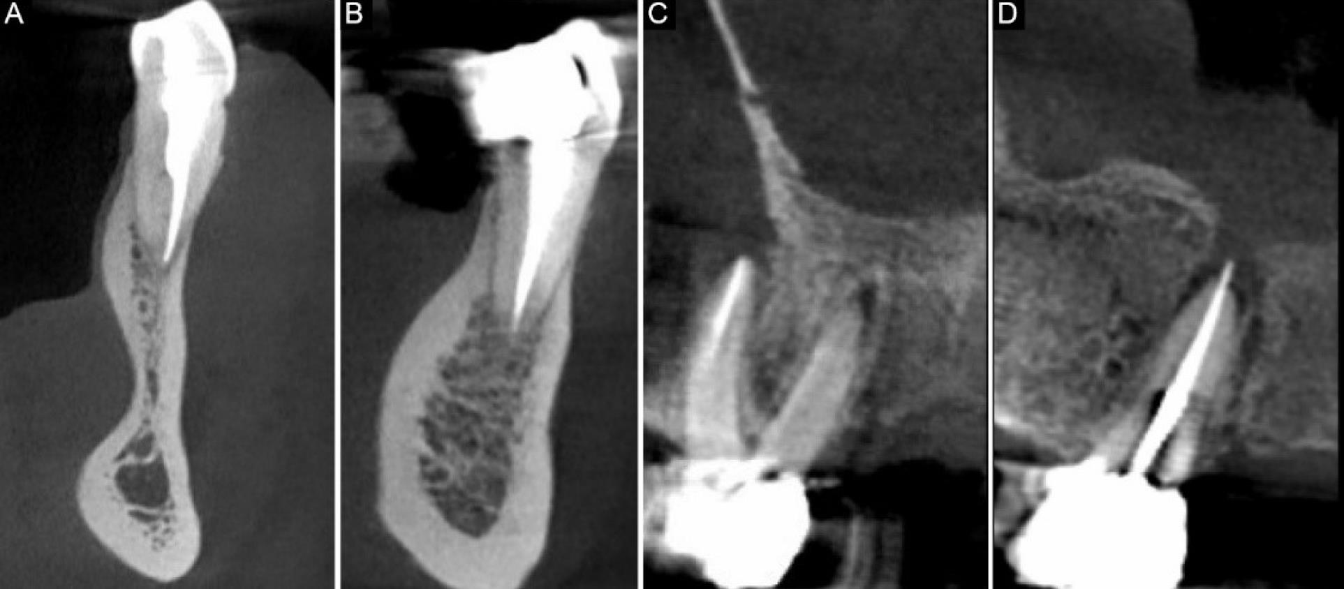
Figure 1.
(A & B) Mandibular Premolars With Good Endodontic Treatment and Good Coronal Restoration, (C) Maxillary Molar With Missing Canal and PA Radiolucency, and (D) Overfilling and PA Radiolucency in Maxillary Premolar
.
(A & B) Mandibular Premolars With Good Endodontic Treatment and Good Coronal Restoration, (C) Maxillary Molar With Missing Canal and PA Radiolucency, and (D) Overfilling and PA Radiolucency in Maxillary Premolar
Discussion
This cross-sectional study evaluated the relationship between periapical radiolucency with endodontic treatment quality and coronal restoration using CBCT. Periapical lesions were visible in plain radiographic images 15–30 days after development, while they could be found in CBCT images only 7 days after development. The assessment of the periapical condition of endodontically treated teeth is an important factor affecting endodontic treatment results (20). CBCT generates 3D images and removes the overlapping of adjacent structures, thereby providing more detailed information that can lead to changes in diagnosis and treatment plans (21,22). However, CBCT is not routinely used to evaluate endodontic treatment quality, except in cases where a proper diagnosis cannot be made by plain radiography. According to the present results, coronal restoration and endodontic treatment quality were significantly related to periapical lesion development, which is in line with the findings of a study by Nascimento et al (17). Although the evaluation of coronal restoration quality is difficult due to various image artifacts in CBCT, clinical examination and intraoral radiography are more reliable methods to examine coronal restoration. Due to the severe artifacts resulting from porcelain crowns and canal posts, these images were excluded from the study. Considering that the severity of these artifacts is different in various imaging protocols and CBCT systems, some studies did not evaluate coronal restoration, which is useful for the assessment of endodontic treatment results (8). Torabinejad et al (23) reported that root canal infection with microorganisms and their products, which were the result of poor coronal restoration, could facilitate reinfection within 3 weeks, suggesting the importance of proper coronal restoration. Further, our results confirmed a statistically significant relationship between proper coronal restoration and periapical radiolucency. Similar to some studies, at different ages, no significant correlation was found between the prevalence of periapical radiolucency and root canal treatment quality (15,24). However, some studies have reported significant relationships between periapical radiolucency and patients over 50 years old (25,26). In the present study, the highest prevalence of lesions was recorded in the age group of 25–50 years (63%), followed by the group over 50 years of age (28%). These results are in accordance with Aysal et al (8). Periapical lesions were more prevalent in men, and the difference was attributed to the biopsychosocial mechanism in some studies (27).
Given that only the presence of periapical radiolucency does not indicate root canal treatment failure, more information is necessary about the patient’s treatment history. In our cross-sectional study, the observed periapical radiolucency could be a healing lesion or bone scar. Hence, it is recommended that prospective longitudinal studies be conducted to find out periapical lesion formation after endodontic treatment, according to the parameters evaluated in this study. Furthermore, it is suggested that more factors, such as the presence of a correlation between coronal and apical seals and which one is more important, undergo investigation.
Conclusion
Overall, no significant association was found between the occurrence of periapical radiolucency and gender and age. The most periapical lesions were observed in males who were in the age range of 25–50 years. The occurrence of periapical radiolucency demonstrated a significant correlation between root canal treatment and coronal restoration status.
Acknowledgements
The authors appreciate the Oral and Maxillofacial Radiology, Dental School, Hamadan University of Medical Science, Hamadan, Iran, for financial and spiritual help.
Authors’ Contribution
Conceptualization: Azita Ehsani.
Data curation: Azita Ehsani.
Formal analysis: Azita Ehsani, Salman Khazaei.
Investigation: Azita Ehsani.
Methodology: Azita Ehsani, Salman Khazaei.
Project administration: Azita Ehsani, Maryam Foroozandeh.
Resources: Azita Ehsani.
Supervision: Maryam Foroozandeh.
Validation: Azita Ehsani.
Visualization: Maryam Foroozandeh.
Writing–original draft: Azita Ehsani.
Writing–review & editing: Azita Ehsani.
Competing Interests
The authors declare that they have no competing interests.
Ethical Approval
This study was confirmed by the Ethics Committee of Hamadan Dental School (Ethics code: IR.UMSHA.REC.1400.821).
Funding
This study did not receive any specific grant from funding agencies in the public, commercial, or not-for-profit sectors.
References
- Nair PN. On the causes of persistent apical periodontitis: a review. Int Endod J 2006; 39(4):249-81. doi: 10.1111/j.1365-2591.2006.01099.x [Crossref] [ Google Scholar]
- Estrela C, Bueno MR, Leles CR, Azevedo B, Azevedo JR. Accuracy of cone beam computed tomography and panoramic and periapical radiography for detection of apical periodontitis. J Endod 2008; 34(3):273-9. doi: 10.1016/j.joen.2007.11.023 [Crossref] [ Google Scholar]
- Gomes AC, Nejaim Y, Silva AI, Haiter-Neto F, Cohenca N, Zaia AA. Influence of endodontic treatment and coronal restoration on status of periapical tissues: a cone-beam computed tomographic study. J Endod 2015; 41(10):1614-8. doi: 10.1016/j.joen.2015.07.008 [Crossref] [ Google Scholar]
- Song M, Park M, Lee CY, Kim E. Periapical status related to the quality of coronal restorations and root fillings in a Korean population. J Endod 2014; 40(2):182-6. doi: 10.1016/j.joen.2013.10.017 [Crossref] [ Google Scholar]
- Ørstavik D. Apical periodontitis: microbial infection and host responses. In: Ørstavik D, ed. Essential Endodontology: Prevention and Treatment of Apical Periodontitis. Wiley; 2019. p. 1-10. 10.1002/9781119272014.ch1.
- Alves Dos Santos GN, Faria-E-Silva AL, Ribeiro VL, Pelozo LL, Candemil AP, Oliveira ML. Is the quality of root canal filling obtained by cone-beam computed tomography associated with periapical lesions? A systematic review and meta-analysis. Clin Oral Investig 2022; 26(8):5105-16. doi: 10.1007/s00784-022-04558-y [Crossref] [ Google Scholar]
- de Paula-Silva FW, Wu MK, Leonardo MR, da Silva LA, Wesselink PR. Accuracy of periapical radiography and cone-beam computed tomography scans in diagnosing apical periodontitis using histopathological findings as a gold standard. J Endod 2009; 35(7):1009-12. doi: 10.1016/j.joen.2009.04.006 [Crossref] [ Google Scholar]
- Aysal Z, Demirturk Kocasarac H, Orhan K, Helvacioglu-Yigit D. Radiological assessment of prevalance and quality of periapical status of endodontic treatments. Med Sci Monit 2022; 28:e936569. doi: 10.12659/msm.936569 [Crossref] [ Google Scholar]
- Saati S, Shokri A, Foroozandeh M, Poorolajal J, Mosleh N. Root morphology and number of canals in mandibular central and lateral incisors using cone beam computed tomography. Braz Dent J 2018; 29(3):239-44. doi: 10.1590/0103-6440201801925 [Crossref] [ Google Scholar]
- Pak JG, Fayazi S, White SN. Prevalence of periapical radiolucency and root canal treatment: a systematic review of cross-sectional studies. J Endod 2012; 38(9):1170-6. doi: 10.1016/j.joen.2012.05.023 [Crossref] [ Google Scholar]
- Dutta A, Smith-Jack F, Saunders WP. Prevalence of periradicular periodontitis in a Scottish subpopulation found on CBCT images. Int Endod J 2014; 47(9):854-63. doi: 10.1111/iej.12228 [Crossref] [ Google Scholar]
- Bürklein S, Schäfer E, Jöhren HP, Donnermeyer D. Quality of root canal fillings and prevalence of apical radiolucencies in a German population: a CBCT analysis. Clin Oral Investig 2020; 24(3):1217-27. doi: 10.1007/s00784-019-02985-y [Crossref] [ Google Scholar]
- Siqueira JF Jr, Rôças IN, Alves FR, Campos LC. Periradicular status related to the quality of coronal restorations and root canal fillings in a Brazilian population. Oral Surg Oral Med Oral Pathol Oral Radiol Endod 2005; 100(3):369-74. doi: 10.1016/j.tripleo.2005.03.029 [Crossref] [ Google Scholar]
- Van der Veken D, Curvers F, Fieuws S, Lambrechts P. Prevalence of apical periodontitis and root filled teeth in a Belgian subpopulation found on CBCT images. Int Endod J 2017; 50(4):317-29. doi: 10.1111/iej.12631 [Crossref] [ Google Scholar]
- Meirinhos J, Martins JNR, Pereira B, Baruwa A, Gouveia J, Quaresma SA. Prevalence of apical periodontitis and its association with previous root canal treatment, root canal filling length and type of coronal restoration - a cross-sectional study. Int Endod J 2020; 53(4):573-84. doi: 10.1111/iej.13256 [Crossref] [ Google Scholar]
- Cicchetti DV. Guidelines, criteria, and rules of thumb for evaluating normed and standardized assessment instruments in psychology. Psychol Assess 1994; 6(4):284-90. doi: 10.1037/1040-3590.6.4.284 [Crossref] [ Google Scholar]
- Nascimento EHL, Gaêta-Araujo H, Andrade MFS, Freitas DQ. Prevalence of technical errors and periapical lesions in a sample of endodontically treated teeth: a CBCT analysis. Clin Oral Investig 2018; 22(7):2495-503. doi: 10.1007/s00784-018-2344-y [Crossref] [ Google Scholar]
- Gambarini G, Piasecki L, Miccoli G, Gaimari G, Nardo DD, Testarelli L. Cone-beam computed tomography in the assessment of periapical lesions in endodontically treated teeth. Eur J Dent 2018; 12(1):136-43. doi: 10.4103/ejd.ejd_320_17 [Crossref] [ Google Scholar]
- Alkis HT, Kustarci A. Radiographic assessment of the relationship between root canal treatment quality, coronal restoration quality, and periapical status. Niger J Clin Pract 2019; 22(8):1126-31. doi: 10.4103/njcp.njcp_129_19 [Crossref] [ Google Scholar]
- Venskutonis T, Plotino G, Tocci L, Gambarini G, Maminskas J, Juodzbalys G. Periapical and endodontic status scale based on periapical bone lesions and endodontic treatment quality evaluation using cone-beam computed tomography. J Endod 2015; 41(2):190-6. doi: 10.1016/j.joen.2014.10.017 [Crossref] [ Google Scholar]
- Pérez-Heredia M, Ferrer-Luque CM, Bravo M, Castelo-Baz P, Ruíz-Piñón M, Baca P. Cone-beam computed tomographic study of root anatomy and canal configuration of molars in a Spanish population. J Endod 2017; 43(9):1511-6. doi: 10.1016/j.joen.2017.03.026 [Crossref] [ Google Scholar]
- Gaêta-Araujo H, Fontenele RC, Nascimento EHL, Nascimento M, Freitas DQ, de Oliveira-Santos C. Association between the root canal configuration, endodontic treatment technical errors, and periapical hypodensities in molar teeth: a cone-beam computed tomographic study. J Endod 2019; 45(12):1465-71. doi: 10.1016/j.joen.2019.08.007 [Crossref] [ Google Scholar]
- Torabinejad M, Ung B, Kettering JD. In vitro bacterial penetration of coronally unsealed endodontically treated teeth. J Endod 1990; 16(12):566-9. doi: 10.1016/s0099-2399(07)80198-1 [Crossref] [ Google Scholar]
- Ureyen Kaya B, Kececi AD, Guldas HE, Orhan H. A retrospective radiographic study of coronal-periapical status and root canal filling quality in a selected adult Turkish population. Med Princ Pract 2013; 22(4):334-9. doi: 10.1159/000346940 [Crossref] [ Google Scholar]
- Alrahabi M, Younes HB. A cross-sectional study of the quality of root canal treatment in Al-Madinah Al-Munawwarah. Saudi Endod J 2016; 6(1):31-5. doi: 10.4103/1658-5984.172005 [Crossref] [ Google Scholar]
- Kielbassa AM, Frank W, Madaus T. Radiologic assessment of quality of root canal fillings and periapical status in an Austrian subpopulation - an observational study. PLoS One 2017; 12(5):e0176724. doi: 10.1371/journal.pone.0176724 [Crossref] [ Google Scholar]
- Shokri A, Ehsani A, Yousefi A. Prevalence of bifid variations of the mandibular canal in an Iranian population using cone-beam computed tomography. Oral Radiol 2023; 39(4):779-83. doi: 10.1007/s11282-023-00698-3 [Crossref] [ Google Scholar]