Avicenna J Dent Res. 17(1):27-34.
doi: 10.34172/ajdr.1655
Review Article
Application of Stem Cells to Treat Xerostomia: A Systematic Review
Parya Atapour Conceptualization, Data curation, Formal analysis, Funding acquisition, Investigation, Methodology, Project administration, Resources, Software, Supervision, Validation, Visualization, Writing – original draft, Writing – review & editing, 1 
Hamidreza Abdolsamadi Conceptualization, Data curation, Formal analysis, Funding acquisition, Investigation, Methodology, Project administration, Resources, Software, Supervision, Validation, Visualization, Writing – original draft, Writing – review & editing, 2
Salman Khazaei Conceptualization, Data curation, Formal analysis, Funding acquisition, Investigation, Methodology, Project administration, Resources, Software, Supervision, Writing – original draft, Writing – review & editing, 3
Mina Jazayeri Conceptualization, Data curation, Formal analysis, Funding acquisition, Investigation, Methodology, Project administration, Resources, Software, Supervision, Validation, Visualization, Writing – original draft, Writing – review & editing, 2, *
Author information:
1Department of Oral and Maxillofacial, Faculty of Dentistry, Tabriz University of Medical Sciences, tabriz, Iran
2Department of Oral and Maxillofacial, Faculty of Dentistry, Hamedan University of Medical Sciences, Hamedan, Iran
3Department of Epidemiology, Health Faculty, Hamedan University of Medical Sciences, Hamedan, Iran
Abstract
Background: This comprehensive systematic review investigated the mil-to long-term efficacy and safety of stem cell therapy in the treatment of xerostomia. The present study aimed to systematically explore available literature on the current state of stem cell research in dentistry and its impacts on the treatment of xerostomia.
Methods: A comprehensive electronic search was conducted in the PubMed, Web of Science, and Scopus databases for human and animal studies published until July 2022. Two independent researchers reviewed the studies based on specific eligibility criteria.
Results: Overall, 439 articles were selected after a comprehensive search. After removing unrelated items, 14 studies were selected for a systematic review. Finally, 14 studies were included in the current work, of which 6 were human clinical trial studies and 8 were animal studies. The articles were on radiation-induced salivary gland (SG) dysfunction, Sjogren syndrome (SS), SG dysfunction, ovariectomy (hypoestrogenic condition), and xerostomia. In all cases, the intervention impression was assessed by the salivary flow rate (SFR) measure, either before/after research or compared to a placebo. The first outcome was to investigate the impact of the intervention on SFR. However, other related variant variables were also extracted, including research design, tissue origin of stem cells, disease model, and participants. In addition, the available cell tracking information was recorded to evaluate the outcome of the transplanted cells. Data meta-analysis was not applied regarding the heterogeneous nature and the small number of included articles encompassing human and murine studies.
Conclusion: Stem cell therapy can be suggested as an adjunctive clinical method to treat dry mouth caused by radiation-induced xerostomia in the neck and head area, SS, hypoestrogenic conditions, and SG dysfunction.
Keywords: Hyposalivation, Salivary hypofunction, Dry mouth, Xerostomia, Stem cell transplantation, Stem cell therapy
Copyright and License Information
© 2025 The Author(s); Published by Hamadan University of Medical Sciences.
This is an open-access article distributed under the terms of the Creative Commons Attribution License (
https://creativecommons.org/licenses/by/4.0), which permits unrestricted use, distribution, and reproduction in any medium provided the original work is properly cited.
Please cite this article as follows: Atapour P, Abdolsamadi H, Khazaei S, Jazayeri M. Application of stem cells to treat xerostomia: a systematic review. Avicenna J Dent Res. 2025;17(1):27-34. doi:10.34172/ajdr.1655
Background
Salivary glands (SGs) have different physiological roles and are important oral cavity structures with critical roles in maintaining oral cavity homeostasis. The SGs protect the mouth’s soft tissues and teeth with antibacterial compounds, enzymes, and electrolytes. The lubrication of the oral cavity provided by saliva is necessary for food taste perception and speech (1,2). The average daily saliva secretion is 1–1.5 L, which plays a significant role in maintaining the functioning of the oral cavity and gastrointestinal tract (3).
SG problems, such as autoimmune diseases, physical traumas, cancer, and infections, affect the patient’s quality of life by affecting SG function (1).
Xerostomia is an irreversible disorder of the SGs. This subjective feeling of dryness exists in systemic disorders, including thyroid disease, liver disease, granulomatous diseases, uncontrolled diabetes, cystic fibrosis, transplant disease, acquired immunodeficiency syndrome, and Sjogren’s syndrome (SS). Genetic abnormalities, surgical injuries, aging, and radiotherapy of neck and head cancers can lead to xerostomia (4,5).
Several treatment options are available for SG dysfunction, such as artificial saliva substitutes, sialogogues, and systemic parasympathomimetics. However, these treatments are temporary and do not repair SGs functionally (6). Recently, stem cell-based therapies have been introduced and extensively studied to treat SG dysfunction and damage to the SGs caused by external radiation (7-10). The results have shown that using stem cells may be an effective treatment alternative for increasing the saliva flow rate, thereby improving dry mouth (11-14). Stem cells are undifferentiated cells that can regenerate themselves and, concurrently, distinguish into more specialized cells. They can be transformed into many different cell lines through differentiation. There are various stem cell types in terms of the type of cells they produce and their location (15).
After birth, different sources of stem cells are recognized in dental tissues, including tooth follicles (16), dental pulp (17), exfoliated deciduous teeth (18), periodontal ligament (19), and apical root papilla (20). Stem cells derived from exfoliated deciduous teeth are an interesting source compared to other human stem cells and are obtained by non-invasive approaches (18). Today, these cells are employed for treating such disorders as nervous system diseases (21), dentin and pulp regeneration (22), jaw regeneration (23), diabetes (24), corneal damage (25), immune system diseases (26), liver damage (27), kidney damage (28), and lung damage (29).
The present study sought to provide a systematic review of the current state of research on stem cells in dentistry and their impact on the treatment of xerostomia by using a systematic search in a valid international database.
Materials and Methods
Data Sources
The current research was conducted to determine the application of stem cells for the treatment of xerostomia.
To this end, the guidelines of the Preferred Reporting Items for Systematic Reviews and Meta-Analyses were followed. A systematic search was conducted on relevant documents published in major databases, including PubMed, Web of Science, and Scopus. The search was performed with maximum sensitivity using a combination of free text and subject headings, such as MESH, without any restriction in language or time until July 2022.
Search Strategy
The keyword combinations used for the search strategy were stem cell OR stem cell transplantation OR stem cell therapy, “AND” hyposalivation OR salivary hypofunction OR dry mouth OR xerostomia.
In addition, more resources were assessed by reviewing the reference list of selected articles for further studies. To ensure the proper selection of papers relevant to the inclusion criteria, two independent researchers (L.G. and P.R.) performed an electronic search on the mentioned websites and evaluated the articles. In cases of disagreement, decisions were made in consultation with other team members through negotiation.
Eligibility Criteria
The present study reviewed randomized controlled trials, non-random intervention studies, quasi-experimental studies, and animal studies. All the mentioned studies were reviewed without considering their publication date or language. Keywords and titles were also searched manually to ensure no research was missed.
The keywords used for the search strategy included hyposalivation, salivary hypofunction, dry mouth-xerostomia, stem cells, stem cell transplantation, and stem cell therapy.
Data Extraction
First, the initial search results of the mentioned databases were entered into Endnote software. Retrieved studies were then listed, and their relevance to the exclusion and inclusion criteria of the study were separately examined by the two researchers. Disagreements between the authors were resolved through negotiations. Two extraction forms were electronically designed to extract the needed information from the entered studies. The first included study information such as the first author, publication year, country, research design and type, study population, stem cell donor, tissue origin of stem cells, disease model, and effect.
Results
Selection of Articles
In our initial systematic search of the mentioned databases, 439 studies were obtained until July 2022 (Figure 1).
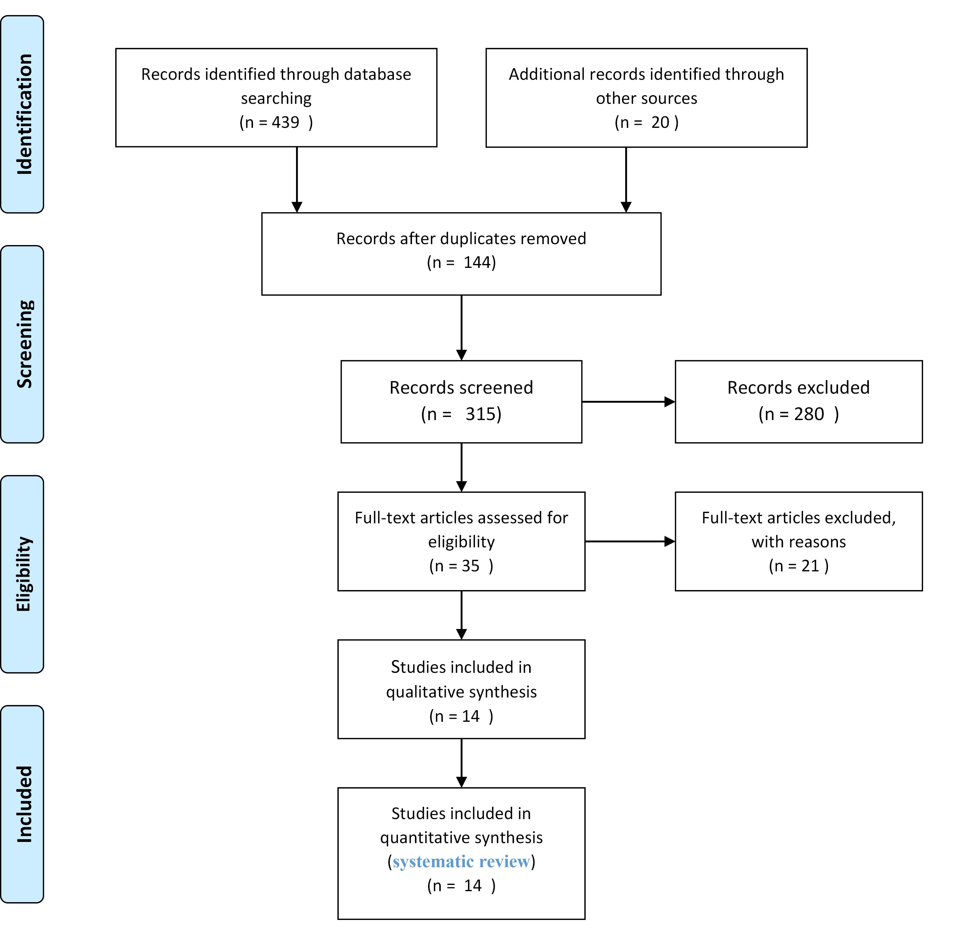
Figure 1.
Diagram of the Search Strategy
.
Diagram of the Search Strategy
Duplicate records were removed, and articles irrelevant to the subject of the present study were excluded by screening the abstracts and titles. Lastly, 14 studies were included in the current work, of which 6 were human clinical trial studies and 8 were animal studies. The articles were about radiation-induced xerostomia (RIX)-related SG dysfunction, SS, SG dysfunction, ovariectomy (hypoestrogenic condition), and xerostomia. In all cases, the intervention impression was assessed using a salivary flow rate (SFR) measure, either before/after research or compared to a placebo.
The first outcome was to investigate the impact of the intervention on SFR. However, other related variant variables were also extracted, such as research design, tissue origin of stem cells, disease model, and participants.
The available cell tracking information was recorded to evaluate the outcome of the transplanted cells. Data meta-analysis was not applied concerning the heterogeneous nature and the small number of included articles consisting of human and murine studies.
Human Studies
Two studies measured the SFR in adult recipients of mesenchymal stem cells with radiation-induced xerostomia and SS. In one of the human studies, the subjects experienced mini-liposuction, from which adipose stem cells were ex vivo extended in a good manufacturing practice-approved clean room facility. All subjects underwent a core-needle sample of one of the submandibular glands and abdominal liposuction (about 60 mL of adipose tissue) (30). In another study, the Stem Cell Center of Jiangsu Province was used to prepare umbilical cord mesenchymal stem cells (UCMSCs). UCMSCs (1 × 10⁶ per kg of body weight) were utilized for treatment by intravenous infusion without premedication, such as antihistamines and steroids. The primary outcome of using adipose stem cells was an alteration in the whole unstimulated SFR. The secondary outcomes included efficacy, safety, changed quality of life, and quantitative and qualitative saliva assessments. The data were assessed at baseline (one month before treatment) and one and four months after the intervention. The studies that employed UCMSCs demonstrated that the unstimulated and stimulated SFR of the group with primary Sjogren syndrome (SS) increased significantly one month after mesenchymal stem cell therapy. This index continuously rose on later follow-up visits (31) (Table 1).
Table 1.
Characteristics of Human Studies
|
Effect
|
Cause of
Dry Mouth
|
Type of Cells
|
Study Participates
|
Donor
|
Participants
|
Study Design
|
Country
|
References
|
| The primary outcome was alterations in the whole unstimulated salivary flow rate. |
Radiation-induced
xerostomia |
Adipose-derived MSCs |
30 cases |
Human |
Human |
Randomized controlled trial |
Denmark |
(30) |
Note. MSC, mesenchymal stem cell.
Human-Animal Studies
Four animal studies used human MSCs to treat ionizing radiation (IR)-injured SGs. The results revealed that the stem cell transplanted group increased saliva secretion compared with saline or phosphate-buffered saline (PBS)-treated mice groups 8 and 12 weeks after IR. However, SFR in the control group (not IR) was higher than in other groups (32-34). Su et al investigated the cell transplantation of labial gland stem cell-derived exosomes (LGSCE) into mice injured by 13-Gy IR SGs. The results showed that 16 weeks after IR, the SFR difference between saline-treated mice and LSCE was not statistically different (P > 0.05) (35). Dong et al used an irradiated-submandibular gland mouse model to study the therapeutic effects of human dental pulp stem cell-derived small extracellular vesicle (SEV) against cellular senescence. These authors assessed saliva production 18 days after irradiation. The SFR, or size, did not represent a remarkable improvement in the 25 Gy þ sEV group compared to the 25 Gy þ PBS group (36), the results of which are provided in Table 2.
Table 2.
Characteristics of Human-Animal Studies
|
Effect
|
Cause of Dry Mouth
|
Type of Cell
|
Study Participates
|
Donor
|
Papulation
|
Study Design
|
Country
|
References
|
| The SFR or size was not remarkably improved in the 25 Gy þ sEV group than in the 25 Gy þ PBS group. |
Irradiation with 25 G |
Human dental pulp stem cell |
Control group
(only PBS, no IR)
25 Gy þ PBS
25 Gy þ sEV |
Human |
Female mice
(7 weeks old) |
Experimental |
Japan |
Dong et al (36) |
LSCE-treated mice showed a 50-60% greater SFR level than saline-treated mice at 8 and 12 weeks after IR (P < 0.05).
At 16 weeks after IR, the SFR difference between saline- and LSCE-treated mice was decreased to 32% (P > 0.05, no statistical difference). |
Irradiation with 13G |
Labial stem cell extract |
6 in the saline group (these mice took 13-Gy IR, being injected with saline)
6 in the LSCE group (IR-injured mice injected with the experimental treatment) |
Human |
C3H female mice
(8 weeks old) |
Experimental |
Canada |
SU et al (35) |
After 60 days, elevated secretion of saliva was observed in the hSGSC transplanted group compared with the PBS-treated group
(P < 0.05). |
Irradiation 25Gy |
Mesenchymal stem cell |
IR + PBS group
IR + hSGSC group
Control group |
Human |
Male Wistar rats
(6 weeks old) |
Experimental |
South Korea |
Jeong et al (32) |
| A significant increase was observed in the saliva flow rate in mice transplanted with 100,000 human salisphere cells after irradiation (P < 0.001). |
Irradiated with a single X-ray dose of 5 Gy |
Salivary gland stem cell
(salisphere) |
9 in the control group,
9 in the irradiated, non-transplanted group,
18 in the irradiated + transplanted group |
Human |
NOD.Cg-Prkdcscid Il2rgtm1Wjl/SzJ |
Experimental |
Netherland |
Pringle
et al (33) |
| hADSCs group demonstrated a 1.51-fold greater output than the irradiation group (P < 0.05). |
Gy 18
irradiation |
Adipose tissue-derived stem cell |
30 in the group,
30 in the 18 Gy irradiation group,
30 in the 18 Gy + hADSC group |
Human |
Prague-Dawley male (SD) rat |
Experimental |
China |
Xiong et al (34) |
PBS: Phosphate-buffered saline; sEV: small extracellular vesicle is correct; LSCE: Labial stem cell extract; IR: Irradiation; SFR: Salivaryflow rate; hSGSC: Human salivary gland stem cells.
Animal Studies
Among the animal studies reviewed in this paper, 3 were performed on the irradiation-induced salivary hypofunction mouse model. They administered a single dose of 15-18 Gy to the submandibular glands of the mice. In these studies, MSCs were transplanted to improve post-irradiation SG injury. The results showed that SFR was significantly higher in stem cell-treated mice compared to the PBS-treated mice at 8/12 weeks post-IR; however, it was higher in the control group (not IR) than in other groups (10,37,38). The other 3 animal studies evaluated the effects of MSCs obtained from non-dental and dental sources of the mice donors on the hyposalivation derived by SS. These studies were conducted by injecting stem cells into the mice (i.e., the animal model of SS). The results revealed a considerable increase in the SFR of mice with stem cell transplantation compared with the PBS group. The SFR of the untreated mice (control) group declined rapidly (31,39,40). Abd El-Haleem et al investigated the impact of a hypoestrogenic condition induced by ovariectomy (OVX) on the parotid gland of albino rats. They also evaluated the potential therapeutic effect of MSCs. Bone marrow was removed from the tibiae and femurs of 10 six-week old male rats. The results demonstrated that the mean SFR was significantly lower in the OVX group than in the control group. Moreover, a significant elevation was noted in the OVX + BM-MSCs group compared to the OVX group (41). Najafi et al examined the possible capacity of MSCs (separated from the submandibular SG of healthy rats) for restoring function and regenerating the necrotic SG in the rat animal model. Based on the obtained results, the mean secretions of mucin and serous in the treatment group indicated a significant increase compared with the positive control groups (SG atrophic/necrotic without treatment (42), the details of which are summarized in Table 3.
Table 3.
Characteristics of Animal to Animal Studies
|
Effect
|
Cause of Dry Mouth
|
Type of Cell
|
Study Participates
|
Stem Cell Donor
|
Papulation
|
Study Design
|
Country
|
References
|
| 8 weeks after IR, the salivary flow rate of animals showed a lower level in the PBS/DPEC/MAEC/BMDC groups compared to the non-irradiated group. The SFR rate demonstrated a significant increase in mice treated with MAECs and DPECs than in PBS-treated mice (P = 0.0263, P = 0.0452). |
Irradiation 15 Gy |
DPECs |
7 in dental pulp endothelial cells
7 in the aortic vascular endothelial cell line
7 in bone marrow-derived cells
7 in fetal bovine serum |
Female and male C57BL/6J green fluorescent protein mice (6 weeks old) |
Male C57BL/6J mice
(6 weeks old) |
Experimental |
Japan |
Yamamura et al (37) |
| The mean salivary flow rate was significantly reduced (P < 0.05) in the OVX group in comparison to the control group, while it was elevated (P < 0.05) in the OVX + MSCs group in comparison to the OVX group. |
Ovariectomy |
MSC |
18 in the control group
18 in the OVX group
18 in the group treated with OVX + MSC |
Tibiae and femurs of 10 male albino rats (6 weeks old) |
Nulliparous female albino rats
(8–10 weeks old) |
Experimental |
Egypt |
Abd El-Haleem et al (41) |
| A constant reduction of SFR was noted in the control group, while MSCs-/MSCE-treated groups indicated a significantly higher SFR compared to the control group (P ≤ 0.01) |
SS |
MSC |
8 in the group treated with MSC
12 in the group treated with MSCE
11 in the control group |
Male C57BL/6 (8 weeks old) |
Female NOD mice
(8 weeks old) |
Experimental |
Canada |
Abughanam et al (39) |
The mice treated with BM-cMSC presented a significant enhancement in the production of saliva in comparison with the mice treated with a PBS injection (P < 0.05).
A significant reduction was observed in SFR in PBS-injected mice in comparison with normal mice (P < 0.001). |
Irradiation 15 Gy |
BM-cMSC |
8 in no radiation (normal)
8 in irradiated + PBS (control)
8 in the group treated with BM-cMSCs following irradiation |
C57BL/6 mice
(8-9 weeks old) |
C57BL/6 mice |
Experimental |
South Korea |
Lim et al (10) |
| Significantly higher SFRs were observed in the group treated with ADSC 8 weeks following radiation compared to the irradiated group (P < 0.05) up to approximately 80% of the normal SFR. |
Irradiation
18 Gy |
ADSC |
10 in the radiation + ADSC group
10 in the radiation + PBS group
10 in the no-radiation and no-cell transplant group |
C57BL/6 mice |
C57BL/6 female mic
(8 weeks old) |
Experimental |
China |
Li et al (38) |
| A significant elevation was noted in the SFR of 21-week-old mice with SHED transplantation in comparison to the PBS and untreated groups (P < 0.01). |
SS |
SHED |
SHED diluted in PBS (200 μL) was instilled into the tail veins of 7 weeks old mice. PBS (200 μL) was injected as a control. |
Female non-obese diabetic mice |
Mice |
Experimental |
China |
Du et al (40) |
In the prevention group, a sustained saliva flow rate was observed in mice receiving BM-MSC infusion (P < .977).
The saliva flow rate of untreated NOD/LtJ mice declined rapidly (P < .008).
In addition, a significant improvement was found in the saliva flow rate in the treatment group following the infusion of BM-MSC (P < .045). |
SS |
BM-MSC |
6 in the prevention group (early stage of SS)
6 in the treatment group (developed stage of SS)
6 in the untreated group |
Male BALB/c mice |
Female NOD/LtJ mice |
Experimental |
China |
Xu et al (31) |
A significant increase was observed in the mean secretions of mucin and serous in the treatment group in comparison with the positive control groups
(P < 0.001). |
Necrotic submandibular SG |
MSC |
7 in the negative control group (healthy rats) 7 in the positive control group (SG atrophic/necrotic without treatment)
7 in the SG necrotic/atrophy with MSC transplantation group |
Submandibular SG of healthy rats |
Sprague–Dawley rats (200–220 g) |
Experimental |
Iran |
Najafi et al (42) |
Note. MSC: Mesenchymal stem cells; DPECs: Dental pulp endothelial cells; PBS: Phosphate-buffered saline; DPEC: Dental pulp endothelial cells; MAEC: Mouse aortic vascular endothelial cell; BMDC: Bone marrow-derived cells; SFR: Salivary flow rate; OVX: Ovariectomy; MSCE: Mesenchymal stem cell extract; BM-cMSC: Marrow-derived clonal mesenchymal stem cells; ADSC: Adipose stem cell; SHED: Stem cells from exfoliated deciduous teeth; BM-cMSC: Bone marrow mesenchymal stem cell extract; SG: Salivary gland; SS, Sjogren syndrome.
Discussion
This systematic review evaluated current scientific knowledge on the effect of stem cell therapy on treating dry mouth caused by RIX in the neck and head area, SS, hypoestrogenic conditions, and SG dysfunction.
All articles included in this review showed a significant elevation in SFR following stem cell therapy, except for the study by Dong et al. They reported no significant enhancement in the SFR in the 25 Gy þ sEV group compared to the 25 Gy þ PBS group 18 days post-irradiation. In addition, Su et al found that the SFR difference between saline-treated mice and LSCE was not statistically different 16 weeks after the cell transplantation of LSCE into mice injured by 13-Gy IR (35,36).
The quality of life declines in cases with irreversible damage to SG function. Despite progress made in recent decades, no definitive cure has been found so far. In this respect, creating alternative therapies is needed to restore the function of damaged acinar cells (43,44).
Treatment with undifferentiated stem cells is a component of regenerative or alternative medicine. Stem cell therapy is believed to change the face of the disease, thereby relieving the pain (4,44).
MSCs are pluripotent stem cells that can differentiate into various types of cells, including ductal and epithelial cells. As a result of their anti-inflammatory effects, the potential for repair of damaged tissues, and the low immunization of these cells, they are a great option for in vitro and in vivo empirical surveys and clinical disease treatment (4,43-45).
MSCs are found in miscellaneous tissue, including the umbilical cord blood placenta (46) and fat (47), and hADSCs are human adipose tissue-derived MSCs. These cells are available sources for stem cell therapy and have been used in numerous animal and clinical studies (48). Inflammation is suppressed by UCMSCs and bone marrow MSCs, and SG functioning is improved in cases with SS and in the rat model (31).
Researchers isolated mesenchymal-like and tissue-specific stem cells from human SGs and transferred them into radiation-damaged rat SGs. Based on duct and acinar structure, they reported that these stem cells could improve decreased secretion, recover acinar structure, and reduce the number of apoptotic cells. They further indicated that oral stem cells could have a therapeutic impact on hyposalivation (32,33).
After birth, different stem cell sources are recognized in dental tissues, including tooth follicles (16), periodontal ligaments (19), dental pulp (17), apical root papilla (20), and exfoliated deciduous teeth. Stem cells isolated from exfoliated deciduous teeth are the cells from human exfoliated teeth. Stem cells derived from exfoliated deciduous teeth provide an interesting source compared to other human stem cells gained using non-invasive approaches (18).
Following the bone marrow stem cells (BMSCs) injection for treating NOD mice, the cells can migrate to the site of inflammation (the submandibular gland), the liver, spleen, lung, and kidney, as well as the tissues of NOD mice. Then, the number of BMSCs distributed in the lung, kidney, and spleen declined one week after infusion. However, large numbers of BMSCs remained in the SGs. Other studies have shown that BMSCs placed in the submandibular gland have immune-regulating roles and ultimately exert therapeutic impacts (31).
Despite studies conducted in this field, it is still unclear whether stem cells from human exfoliated deciduous teeth (SHED) can move to affected areas and perform their function. Du et al showed no migration of the injected SHED to the submandibular gland. This injection affected the spleen and liver from the first hour, lasting up to one week. The release of SHED into the liver influenced the blood supply, and SHED attracted the attention of the spleen, the largest immune organ. The results also revealed that SFR was significantly higher in 21-week-old SHED-grafted mice than in the PBS and untreated groups. Du et al reported that SHED has a protective effect on SG secretory function. SHED reduces SS-induced hyposalivation due to reduced apoptosis, autophagy in the submandibular glands, decreased peripheral inflammatory cytokines, and a localized inflammatory microenvironment (4,31,41-48).
Some studies have used small available salivary tissue to provide sufficient MSCs for treatment because the methods utilized to extract stem cells from the human parotid and submandibular glands are invasive (32). In this respect, Su et al extracted and employed human minor SGs, which required less invasive surgery. They reported that the duration of LSCE treatment lasted up to 9 weeks but did not last up to 16 weeks (35).
Similarly, Xu et al found that the allogeneic injection of MSC is a satisfactory therapy for SS and has considerable implications for a further survey of MSCs. These cells can be considered a therapeutic solution for cases with SS and patients with other autoimmune problems (31).
Another noteworthy point in applying the results of clinical studies of RIX mice is that radiotherapy treatments to treat neck and head cancer are fragmented and often take up to several weeks. Nevertheless, diets in mouse studies are single-dose. Furthermore, the duration from the end of radiotherapy to the beginning of MSC therapy can be crucial.
MSCs can lower the inflammatory response, thereby decreasing the immune-induced parenchyma destruction that occurs in the acute phase of radiotherapy (49). Given the overall rise in SFR following the treatment of MCS and the moderate degree of differentiation of MSCs into acinar cells noted in mouse surveys, using MSC therapy for RIX can be considered a “window of opportunity”.
To evaluate the efficacy of stem cells, Lane et al started treatment with mesenchymal cells 24 h following irradiation (50). Likewise, Lim et al started therapy 11 days following irradiation. However, using this method in clinical settings was impossible due to various factors, including the length of the radiation therapy process in cases with neck and head cancer. Nevertheless, it was necessary to obtain a more appropriate model of MSCs for RIX, given that the expected lifespan of the mice used in this regard was about 2 years (10). Accordingly, Kojima et al started MSC therapy 10 weeks after SG irradiation and observed a 51% increase in SFR (51).
Some laboratory studies have demonstrated the potential risk of stimulating cancer growth when using MSCs to treat RIX. However, studies of MSCs in cases with previous cancer and in many clinical settings reported no increase in cancer incidence (52).
Various sources of relevant studies and databases were thoroughly searched in the present work. The selection of studies was not limited to controlled and randomized trials. Objective decisions were made for selecting or excluding studies. However, the present study’s findings should be interpreted with caution regarding animal surveys’ external and internal validity since it is not straightforward to interpret them in clinical surveys.
Conclusion
We did complete research from various databases and resources and did not limit the studies to randomized and centralized trials. The decision to choose or exclude the studies was clearly objective; however, our findings should be interpreted with usual caution about the credibility of internal and external animal studies.
Competing Interests
None declared.
Ethical Approval
Not applicable.
Funding
None.
References
- Porcheri C, Mitsiadis TA. Physiology, pathology and regeneration of salivary glands. Cells 2019; 8(9):976. doi: 10.3390/cells8090976 [Crossref] [ Google Scholar]
- de Paula F, Teshima TH, Hsieh R, Souza MM, Nico MM, Lourenco SV. Overview of human salivary glands: highlights of morphology and developing processes. Anat Rec (Hoboken) 2017; 300(7):1180-8. doi: 10.1002/ar.23569 [Crossref] [ Google Scholar]
- Mattingly A, Finley JK, Knox SM. Salivary gland development and disease. Wiley Interdiscip Rev Dev Biol 2015; 4(6):573-90. doi: 10.1002/wdev.194 [Crossref] [ Google Scholar]
- Lombaert I, Movahednia MM, Adine C, Ferreira JN. Concise review: salivary gland regeneration: therapeutic approaches from stem cells to tissue organoids. Stem Cells 2017; 35(1):97-105. doi: 10.1002/stem.2455 [Crossref] [ Google Scholar]
- Millsop JW, Wang EA, Fazel N. Etiology, evaluation, and management of xerostomia. Clin Dermatol 2017; 35(5):468-76. doi: 10.1016/j.clindermatol.2017.06.010 [Crossref] [ Google Scholar]
- See L, Mohammadi M, Han PP, Mulligan R, Enciso R. Efficacy of saliva substitutes and stimulants in the treatment of dry mouth. Spec Care Dentist 2019; 39(3):287-97. doi: 10.1111/scd.12370 [Crossref] [ Google Scholar]
- Sui Y, Zhang S, Li Y, Zhang X, Hu W, Feng Y. Generation of functional salivary gland tissue from human submandibular gland stem/progenitor cells. Stem Cell Res Ther 2020; 11(1):127. doi: 10.1186/s13287-020-01628-4 [Crossref] [ Google Scholar]
- Choi JS, An HY, Shin HS, Kim YM, Lim JY. Enhanced tissue remodelling efficacy of adipose-derived mesenchymal stem cells using injectable matrices in radiation-damaged salivary gland model. J Tissue Eng Regen Med 2018; 12(2):e695-706. doi: 10.1002/term.2352 [Crossref] [ Google Scholar]
- Nicolay NH, Lopez Perez R, Debus J, Huber PE. Mesenchymal stem cells – a new hope for radiotherapy-induced tissue damage?. Cancer Lett 2015; 366(2):133-40. doi: 10.1016/j.canlet.2015.06.012 [Crossref] [ Google Scholar]
- Lim JY, Yi T, Choi JS, Jang YH, Lee S, Kim HJ. Intraglandular transplantation of bone marrow-derived clonal mesenchymal stem cells for amelioration of post-irradiation salivary gland damage. Oral Oncol 2013; 49(2):136-43. doi: 10.1016/j.oraloncology.2012.08.010 [Crossref] [ Google Scholar]
- Eisbruch A, Ten Haken RK, Kim HM, Marsh LH, Ship JA. Dose, volume, and function relationships in parotid salivary glands following conformal and intensity-modulated irradiation of head and neck cancer. Int J Radiat Oncol Biol Phys 1999; 45(3):577-87. doi: 10.1016/s0360-3016(99)00247-3 [Crossref] [ Google Scholar]
- Murdoch-Kinch CA, Kim HM, Vineberg KA, Ship JA, Eisbruch A. Dose-effect relationships for the submandibular salivary glands and implications for their sparing by intensity modulated radiotherapy. Int J Radiat Oncol Biol Phys 2008; 72(2):373-82. doi: 10.1016/j.ijrobp.2007.12.033 [Crossref] [ Google Scholar]
- Ortholan C, Chamorey E, Benezery K, Thariat J, Dassonville O, Poissonnet G. Modeling of salivary production recovery after radiotherapy using mixed models: determination of optimal dose constraint for IMRT planning and construction of convenient tools to predict salivary function. Int J Radiat Oncol Biol Phys 2009; 73(1):178-86. doi: 10.1016/j.ijrobp.2008.03.068 [Crossref] [ Google Scholar]
- Roesink JM, Moerland MA, Battermann JJ, Hordijk GJ, Terhaard CH. Quantitative dose-volume response analysis of changes in parotid gland function after radiotherapy in the head-and-neck region. Int J Radiat Oncol Biol Phys 2001; 51(4):938-46. doi: 10.1016/s0360-3016(01)01717-5 [Crossref] [ Google Scholar]
- Oliveri RS. Epigenetic dedifferentiation of somatic cells into pluripotency: cellular alchemy in the age of regenerative medicine?. Regen Med 2007; 2(5):795-816. doi: 10.2217/17460751.2.5.795 [Crossref] [ Google Scholar]
- Yao S, Pan F, Prpic V, Wise GE. Differentiation of stem cells in the dental follicle. J Dent Res 2008; 87(8):767-71. doi: 10.1177/154405910808700801 [Crossref] [ Google Scholar]
- Gronthos S, Mankani M, Brahim J, Robey PG, Shi S. Postnatal human dental pulp stem cells (DPSCs) in vitro and in vivo. Proc Natl Acad Sci U S A 2000; 97(25):13625-30. doi: 10.1073/pnas.240309797 [Crossref] [ Google Scholar]
- Miura M, Gronthos S, Zhao M, Lu B, Fisher LW, Robey PG. SHED: stem cells from human exfoliated deciduous teeth. Proc Natl Acad Sci U S A 2003; 100(10):5807-12. doi: 10.1073/pnas.0937635100 [Crossref] [ Google Scholar]
- Seo BM, Miura M, Gronthos S, Bartold PM, Batouli S, Brahim J. Investigation of multipotent postnatal stem cells from human periodontal ligament. Lancet 2004; 364(9429):149-55. doi: 10.1016/s0140-6736(04)16627-0 [Crossref] [ Google Scholar]
- Sonoyama W, Liu Y, Yamaza T, Tuan RS, Wang S, Shi S. Characterization of the apical papilla and its residing stem cells from human immature permanent teeth: a pilot study. J Endod 2008; 34(2):166-71. doi: 10.1016/j.joen.2007.11.021 [Crossref] [ Google Scholar]
- Taghipour Z, Karbalaie K, Kiani A, Niapour A, Bahramian H, Nasr-Esfahani MH. Transplantation of undifferentiated and induced human exfoliated deciduous teeth-derived stem cells promote functional recovery of rat spinal cord contusion injury model. Stem Cells Dev 2012; 21(10):1794-802. doi: 10.1089/scd.2011.0408 [Crossref] [ Google Scholar]
- Rosa V, Zhang Z, Grande RH, Nör JE. Dental pulp tissue engineering in full-length human root canals. J Dent Res 2013; 92(11):970-5. doi: 10.1177/0022034513505772 [Crossref] [ Google Scholar]
- Jahanbin A, Rashed R, Hamidi Alamdari D, Koohestanian N, Ezzati A, Kazemian M, et al. Success of maxillary alveolar defect repair in rats using osteoblast-differentiated human deciduous dental pulp stem cells. J Oral Maxillofac Surg 2016;74(4):829.e1-829.e9. doi: 10.1016/j.joms.2015.11.033.
- Kanafi MM, Rajeshwari YB, Gupta S, Dadheech N, Nair PD, Gupta PK. Transplantation of islet-like cell clusters derived from human dental pulp stem cells restores normoglycemia in diabetic mice. Cytotherapy 2013; 15(10):1228-36. doi: 10.1016/j.jcyt.2013.05.008 [Crossref] [ Google Scholar]
- Gomes JA, Geraldes Monteiro B, Melo GB, Smith RL, da Silva MC, Lizier NF. Corneal reconstruction with tissue-engineered cell sheets composed of human immature dental pulp stem cells. Invest Ophthalmol Vis Sci 2010; 51(3):1408-14. doi: 10.1167/iovs.09-4029 [Crossref] [ Google Scholar]
- Ma L, Makino Y, Yamaza H, Akiyama K, Hoshino Y, Song G. Cryopreserved dental pulp tissues of exfoliated deciduous teeth is a feasible stem cell resource for regenerative medicine. PLoS One 2012; 7(12):e51777. doi: 10.1371/journal.pone.0051777 [Crossref] [ Google Scholar]
- Yamaza T, Alatas FS, Yuniartha R, Yamaza H, Fujiyoshi JK, Yanagi Y. In vivo hepatogenic capacity and therapeutic potential of stem cells from human exfoliated deciduous teeth in liver fibrosis in mice. Stem Cell Res Ther 2015; 6(1):171. doi: 10.1186/s13287-015-0154-6 [Crossref] [ Google Scholar]
- Hattori Y, Kim H, Tsuboi N, Yamamoto A, Akiyama S, Shi Y. Therapeutic potential of stem cells from human exfoliated deciduous teeth in models of acute kidney injury. PLoS One 2015; 10(10):e0140121. doi: 10.1371/journal.pone.0140121 [Crossref] [ Google Scholar]
- Wakayama H, Hashimoto N, Matsushita Y, Matsubara K, Yamamoto N, Hasegawa Y. Factors secreted from dental pulp stem cells show multifaceted benefits for treating acute lung injury in mice. Cytotherapy 2015; 17(8):1119-29. doi: 10.1016/j.jcyt.2015.04.009 [Crossref] [ Google Scholar]
- Grønhøj C, Jensen DH, Glovinski PV, Jensen SB, Bardow A, Oliveri RS. First-in-man mesenchymal stem cells for radiation-induced xerostomia (MESRIX): study protocol for a randomized controlled trial. Trials 2017; 18(1):108. doi: 10.1186/s13063-017-1856-0 [Crossref] [ Google Scholar]
- Xu J, Wang D, Liu D, Fan Z, Zhang H, Liu O. Allogeneic mesenchymal stem cell treatment alleviates experimental and clinical Sjögren syndrome. Blood 2012; 120(15):3142-51. doi: 10.1182/blood-2011-11-391144 [Crossref] [ Google Scholar]
- Jeong J, Baek H, Kim YJ, Choi Y, Lee H, Lee E. Human salivary gland stem cells ameliorate hyposalivation of radiation-damaged rat salivary glands. Exp Mol Med 2013; 45(11):e58. doi: 10.1038/emm.2013.121 [Crossref] [ Google Scholar]
- Pringle S, Wang X, Verstappen G, Terpstra JH, Zhang CK, He A. Salivary gland stem cells age prematurely in primary Sjögren’s syndrome. Arthritis Rheumatol 2019; 71(1):133-42. doi: 10.1002/art.40659 [Crossref] [ Google Scholar]
- Xiong X, Shi X, Chen F. Human adipose tissue-derived stem cells alleviate radiation-induced xerostomia. Int J Mol Med 2014; 34(3):749-55. doi: 10.3892/ijmm.2014.1837 [Crossref] [ Google Scholar]
- Su X, Liu Y, Bakkar M, ElKashty O, El-Hakim M, Seuntjens J. Labial stem cell extract mitigates injury to irradiated salivary glands. J Dent Res 2020; 99(3):293-301. doi: 10.1177/0022034519898138 [Crossref] [ Google Scholar]
- Dong J, Sakai K, Koma Y, Watanabe J, Liu K, Maruyama H. Dental pulp stem cell-derived small extracellular vesicle in irradiation-induced senescence. Biochem Biophys Res Commun 2021; 575:28-35. doi: 10.1016/j.bbrc.2021.08.046 [Crossref] [ Google Scholar]
- Yamamura Y, Yamada H, Sakurai T, Ide F, Inoue H, Muramatsu T. Treatment of salivary gland hypofunction by transplantation with dental pulp cells. Arch Oral Biol 2013; 58(8):935-42. doi: 10.1016/j.archoralbio.2013.02.015 [Crossref] [ Google Scholar]
- Li Z, Wang Y, Xing H, Wang Z, Hu H, An R. Protective efficacy of intravenous transplantation of adipose-derived stem cells for the prevention of radiation-induced salivary gland damage. Arch Oral Biol 2015; 60(10):1488-96. doi: 10.1016/j.archoralbio.2015.07.016 [Crossref] [ Google Scholar]
- Abughanam G, Elkashty OA, Liu Y, Bakkar MO, Tran SD. Mesenchymal stem cells extract (MSCsE)-based therapy alleviates xerostomia and keratoconjunctivitis sicca in Sjogren’s syndrome-like disease. Int J Mol Sci 2019; 20(19):4750. doi: 10.3390/ijms20194750 [Crossref] [ Google Scholar]
- Du ZH, Ding C, Zhang Q, Zhang Y, Ge XY, Li SL. Stem cells from exfoliated deciduous teeth alleviate hyposalivation caused by Sjögren syndrome. Oral Dis 2019; 25(6):1530-44. doi: 10.1111/odi.13113 [Crossref] [ Google Scholar]
- Abd El-Haleem MR, Selim AO, Attia GM. Bone marrow-derived mesenchymal stem cells ameliorate parotid injury in ovariectomized rats. Cytotherapy 2018; 20(2):204-17. doi: 10.1016/j.jcyt.2017.10.003 [Crossref] [ Google Scholar]
- Najafi S, Nosrati H, Faraji Z, Mohamadnia A, Shirian S, Mortazavi SM. Reconstruction of necrotic submandibular salivary gland using mesenchymal stem cells. Heliyon 2020; 6(10):e05162. doi: 10.1016/j.heliyon.2020.e05162 [Crossref] [ Google Scholar]
- Khalili S, Liu Y, Kornete M, Roescher N, Kodama S, Peterson A. Mesenchymal stromal cells improve salivary function and reduce lymphocytic infiltrates in mice with Sjögren’s-like disease. PLoS One 2012; 7(6):e38615. doi: 10.1371/journal.pone.0038615 [Crossref] [ Google Scholar]
- Sumita Y, Liu Y, Khalili S, Maria OM, Xia D, Key S. Bone marrow-derived cells rescue salivary gland function in mice with head and neck irradiation. Int J Biochem Cell Biol 2011; 43(1):80-7. doi: 10.1016/j.biocel.2010.09.023 [Crossref] [ Google Scholar]
- Yoo C, Vines JB, Alexander G, Murdock K, Hwang P, Jun HW. Adult stem cells and tissue engineering strategies for salivary gland regeneration: a review. Biomater Res 2014; 18:9. doi: 10.1186/2055-7124-18-9 [Crossref] [ Google Scholar]
- Erices A, Conget P, Minguell JJ. Mesenchymal progenitor cells in human umbilical cord blood. Br J Haematol 2000; 109(1):235-42. doi: 10.1046/j.1365-2141.2000.01986.x [Crossref] [ Google Scholar]
- Zuk PA, Zhu M, Ashjian P, De Ugarte DA, Huang JI, Mizuno H. Human adipose tissue is a source of multipotent stem cells. Mol Biol Cell 2002; 13(12):4279-95. doi: 10.1091/mbc.e02-02-0105 [Crossref] [ Google Scholar]
- Schäffler A, Büchler C. Concise review: adipose tissue-derived stromal cells--basic and clinical implications for novel cell-based therapies. Stem Cells 2007; 25(4):818-27. doi: 10.1634/stemcells.2006-0589 [Crossref] [ Google Scholar]
- Uccelli A, Moretta L, Pistoia V. Mesenchymal stem cells in health and disease. Nat Rev Immunol 2008; 8(9):726-36. doi: 10.1038/nri2395 [Crossref] [ Google Scholar]
- Lin CY, Chang FH, Chen CY, Huang CY, Hu FC, Huang WK. Cell therapy for salivary gland regeneration. J Dent Res 2011; 90(3):341-6. doi: 10.1177/0022034510386374 [Crossref] [ Google Scholar]
- Kojima T, Kanemaru S, Hirano S, Tateya I, Ohno S, Nakamura T. Regeneration of radiation damaged salivary glands with adipose-derived stromal cells. Laryngoscope 2011; 121(9):1864-9. doi: 10.1002/lary.22080 [Crossref] [ Google Scholar]
- Fu KK, Clery M, Ang KK, Byhardt RW, Maor MH, Beitler JJ. Randomized phase I/II trial of two variants of accelerated fractionated radiotherapy regimens for advanced head and neck cancer: results of RTOG 88-09. Int J Radiat Oncol Biol Phys 1995; 32(3):589-97. doi: 10.1016/0360-3016(95)00078-d [Crossref] [ Google Scholar]