Avicenna J Dent Res. 15(4):167-172.
doi: 10.34172/ajdr.1637
Original Article
Evaluation of Dentin Cracks by Stereomicroscope after Preparation of Mesiobuccal Canal of Maxillary First Molars Using Edge Taper Platinum and ProTaper Gold Rotary Files: A Laboratory Study
Narjes Hoshyari 1  , Seyedali Seyedmajidi 2 , Anahita Lotfizadeh 3 , Eghlima Malakan 3 , Abolfazl Hosseinnataj 4 , Azam Haddadi Kohsar 1, *
Author information:
1Department of Endodontics, Dental Research Center, Mazandaran University of Medical Sciences, Sari, Iran
2Dental Materials Research Center, Health Research Institute, Babol University of Medical Sciences, Babol, Iran
3Dentist, Mazandaran, Iran
4Department of Biostatistics, Faculty of Health, Mazandaran University of Medical Sciences, Sari, Iran
Abstract
Background: Successful root canal treatment depends on correct cleaning, widening, and shaping of the root canal system. Root canal preparation using rotary files can cause dentin cracks. This study aimed to evaluate the number of dentin cracks created after the preparation of the mesiobuccal root canal of the first maxillary molars by ProTaper Gold and Edge taper platinum rotary files using a stereomicroscope.
Methods: In this laboratory study, 72 maxillary first molars were collected and randomly divided into three groups (n=24): control group, ProTaper Gold group, and Edge taper platinum group. Mesiobuccal canal preparation was performed in Edge taper platinum and ProTaper Gold groups up to file size F2. All roots were cut horizontally at distances of 3, 6, and 9 mm from the apex with a disc. The sections were examined under a stereomicroscope at 40x magnification. Fisher’s exact test and Chi-square test were used in SPSS version 22.0 for analyzing data. A significant level of 5% was considered.
Results: In the control group, no cracks were observed in any of the sections. At 3 and 9 mm from the apex, the ProTaper Gold group had the highest frequency of cracks and at 6 mm from the apex, the Edge taper platinum group had the highest frequency of cracks. There was no significant difference in the frequency of dentin cracks between ProTaper Gold and Edge taper platinum groups in any of the sections.
Conclusion: Both ProTaper Gold and Edge taper platinum systems caused the formation of dentin cracks. Preparation of root canals by Edge taper platinum and ProTaper Gold rotary files causes the formation of dentin cracks to the same extent.
Keywords: Dentin cracks, First maxillary molar, Mesiobuccal root canal, Stereomicroscope, Rotary files
Copyright and License Information
© 2023 The Author(s); Published by Hamadan University of Medical Sciences.
This is an open-access article distributed under the terms of the Creative Commons Attribution License (
http://creativecommons.org/licenses/by/4.0), which permits unrestricted use, distribution, and reproduction in any medium provided the original work is properly cited.
Please cite this article as follows: Hoshyari N, Seyedmajidi S, Lotfizadeh A, Malakan E, Hosseinnataj A, Haddadi Kohsar A. Evaluation of dentin cracks by stereomicroscope after preparation of mesiobuccal canal of maxillary first molars using edge taper platinum and protaper gold rotary files: a laboratory study. Avicenna J Dent Res. 2023; 15(4):167-172. doi:10.34172/ajdr.1637
Background
Successful root canal treatment depends on correct cleaning, widening, and shaping of the root canal (1). Mechanical instrumentation includes cleaning and shaping and by this method, the effectiveness of detergents and antibacterial agents in eradicating bacteria and eliminating bacterial by-products is achieved. Therefore, enough space for three-dimensional obturation is prepared (2,3). The anatomy and morphology of the root canal system pose other challenges to disinfection. Isthmuses, inter-canal and intra-canal connections, and round and oval canals make root canal disinfection challenging (4-8).
Inaccurate root canal instrumentation may have inevitable consequences. During the biomechanical preparation of root canals, the contact between the instrument and the canal walls puts pressure on the dentin and may produce craze lines (9-11). Cracks may spread over a long period and cause vertical root fracture (VRF) (12). Root fracture in 10.9%-31% of cases causes tooth extraction (13). Root fracture may occur after the extension of microcracks or craze lines due to occlusal stress (14).
Rotary nickel-titanium (Ni-Ti) files were developed in the 1980s. Compared to manual instruments, they have shorter instrumentation time and better performance (2). Conversely, Ni-Ti rotary files can cause more friction and stress than manual files (6). In addition, canals with more curvature may increase the pressure on the rotary files, resulting in perforation, canal transportation, lodging, or instrument fracture (15-17). Instruments and instrumentation techniques should be selected based on their ability to form (especially in canals with curvature) and the possibility of achieving faster and non-deflection preparations (6,17).
Various Ni-Ti file systems are commercially available, based on their cross-sectional shape, groove angle, tapper, depth of flutes, and number of spirals or flutes per unit length. They all have different characteristics, which may affect file performance (17-19).
The ProTaper instruments include Ni-Ti files with a conical design. ProTaper Next has an eccentric rectangular cross-section that gives the file a snake-like motion as it progresses through the root canal. ProTaper Gold has a triangular cross-section made by proprietary metallurgy that increases flexibility and resistance to cyclic fatigue (15,20-23).
Heat-treated Ni-Ti rotary files are available with the same shape, structure, and function as the ProTaper Gold file, including edge taper platinum, flex gold, and Pro-T. These files are recently known as replica-like systems. Among them, the edge taper platinum file has more flexibility and lower rotational resistance than the ProTaper Gold file. Although replica-like systems are sold and used daily around the world, not enough studies have been done in this field (24).
Commonly used methods for assessing dentin defects are radiography, dental sections, and plastic blocks (25). The stereomicroscope has been used as a tool to study the roots in the evaluation of apical anatomy and to evaluate the sealing capability using different filling materials and techniques. It can also be used as a valuable tool for students in the field of endodontics to evaluate debridement and obturation techniques (26).
In this study, the number of dentin cracks created after the preparation of the mesiobuccal canal of the first maxillary molars by ProTaper Gold and edge taper platinum rotary files was investigated using a stereomicroscope.
Materials and Methods
This Ex-vivo study was carried out after receiving ethical approval (IR.MAZUMS.REC.1400.396). The samples were chosen from the extracted first upper molars which were collected from dental clinics in Sari (Mazandaran, Iran). Based on the results of previous similar studies and taking into account the value of C = 7.85 (a test power of 80% and a significance level of 5%), the total sample size was calculated to be 72 teeth, which were divided into three groups (24 teeth in each group) (27).
Collected teeth were cleaned by a periodontal scaler and kept in distilled water to prevent dehydration during the study. Additionally, the teeth were disinfected with sodium hypochlorite 2.5% and investigated for initial evaluation of microcracks by stereomicroscope at a magnification of 25x (Dewinter, Milano, Italy).
The inclusion criteria of the study were maxillary first molars with mature roots having apex with moderate canal curves (20°-40°( measured according to Schneider’s method (28), roots without caries and external resorption, and the maximum width of the end of the canal which matches the file size number 15.
Teeth having reduced pulp space, pulp stones, calcified canals, hypercementosis, root caries, internal or external root resorption, previous root canal therapy, open apex, and fully curved canals were excluded from the study (27).
For the preparation of the samples, the access cavity was prepared with a fissure diamond bur, and the orifice of the mesiobuccal canal was identified by an endodontic catheter.
Canals with a K file size 10 or maximum 15 (Dentsply Maillefer, Ballaigues, Switzerland) which had a snag in apical constriction were selected. The second mesiobuccal canals were not considered. Then, the crowns were cut with a fissure bur (Brasseler USA, Savannah, GA, USA). The working length of all samples was 13 mm. The working length was determined visually using file number 10. The rate of penetration into the canal and the working length, 1 mm shorter than the anatomic apex, were recorded using ISO #10 K-file and #15 K-file, respectively. Afterward, the degree of root curvatures was assessed.
First, periapical radiographs were conducted. Then, root curvatures were measured by Schneider’s method.
For tooth preparation, collected teeth were randomly divided into three groups of 24.
-
Group 1: control group, in which teeth remained without preparation.
-
Group 2: canals were prepared by ProTaper Gold file (Dentsply Maillefer, Ballaigues, Switzerland) and electromotor at 300 rpm and 3N torque. According to the manufacturer’s instructions, an apical canal preparation was done up to file size F2 (25#0.08)
-
Group 3: canals were prepared by Edge taper platinum file (EdgeEndo, Albuquerque, NM, USA) and electromotor at 300 rpm and 4N torque according to the manufacturer’s instruction, and apical canal preparation was done up to file size F2 (25#0.06). Each canal was prepared with 1 file package and the rotation time of each canal was 5-10 seconds.
After preparation, the samples were mounted in resin. Then, all the roots were cut at distances of 3, 6, and 9 mm from the apex with a diamond disc (a thickness of 0.3 mm under water cooling conditions) and a three-axis cutting machine (NemoFanavaran Pars, Mashhad, Iran).
The sections were then examined under a stereomicroscope (Dewinter Technologies, Milano, Italy) at 40x magnification. Digital images were recorded with a digital camera attached to a stereomicroscope at 25x and 40x magnifications and examined by two endodontists for dentin cracks. The obtained data were divided into two groups (presence and absence of cracks). The data were presented using descriptive statistics (frequency and percentage). To compare the frequency of dentin cracks between groups, Fisher’s exact test and chi-square test were used in SPSS version 22.0 at the significance level of 5%.
Results
The frequency of teeth with dentin cracks in each of the studied groups was evaluated at different cross-sections. At 3 mm from the apex (apical), dentin cracks were observed only in the ProTaper Gold group (3 cases). The number of cracks in this section was not significantly different between the three groups (Table 1).
Table 1.
The Frequency of Dentin Cracks at 3 mm From the Apex in the Study Groups
|
Group
|
Frequency
|
Percent
|
Chi-square Test
|
P
Value
|
| ProTaper Gold |
3 |
12.5 |
4.27 |
0.102 |
| Edge taper platinum |
0 |
0.0 |
| Control |
0 |
0.0 |
At 6 mm from the apex (middle), 8 cases of dentin cracks were observed in the ProTaper Gold group, and 10 cases of dentin cracks were observed in the Edge taper platinum group. Additionally, no cases were observed in the control group. The number of cracks was significantly different between the 3 groups (Table 2).
Table 2.
The Frequency of Dentin Cracks at 6 mm From the Apex in the Study Groups
|
Groups
|
Frequency
|
Percent
|
Chi-square Test
|
P
Value
|
| ProTaper Gold |
8 |
33.3 |
12.44 |
0.002 |
| Edge taper platinum |
10 |
41.7 |
| Control |
0 |
0.0 |
At 9 mm from the apex (coronal), 15 cases of dentin cracks were observed in the ProTaper Gold group, and 10 cases of dentin cracks were observed in the Edge taper platinum group. Besides, no cases were observed in the control group. In this section, the number of dentin cracks was significantly different between the 3 groups (Table 3).
Table 3.
The Frequency of Dentin Cracks at 9 mm From the Apex in the Study Groups
|
Groups
|
Frequency
|
Percent
|
Chi-square Test
|
P
value
|
| ProTaper Gold |
15 |
62.5 |
21.45 |
<0.001 |
| Edge taper platinum |
10 |
41.7 |
| Control |
0 |
0.0 |
No cracks were observed in the control group in any of the sections. At 3 and 9 mm from the apex, the ProTaper Gold group had the highest frequency and at 6 mm from the apex, the Edge taper platinum group had the highest frequency of the cracked teeth.
Comparing the frequency of cracked teeth between ProTaper Gold and Edge taper platinum groups in different sections, no significant difference was observed between all three sections (Table 4).
Table 4.
Frequency of Dentin Cracks in ProTaper and Edge Taper Groups in Different Sections
|
Section
|
Groups
|
Frequency of Cracks
|
Chi-square Test
|
P
value
|
| 3 mm |
ProTaper Gold |
3 |
3.20 |
0.234 |
| Edge taper platinum |
0 |
| 6 mm |
ProTaper Gold |
8 |
0.36 |
0.551 |
| Edge taper platinum |
10 |
| 9 mm |
ProTaper Gold |
15 |
2.09 |
0.149 |
| Edge taper platinum |
10 |
The images related to the examination of cracks by stereomicroscope in the ProTaper Gold, Edge taper Platinum, and control groups are shown in Figures 1 to 3.
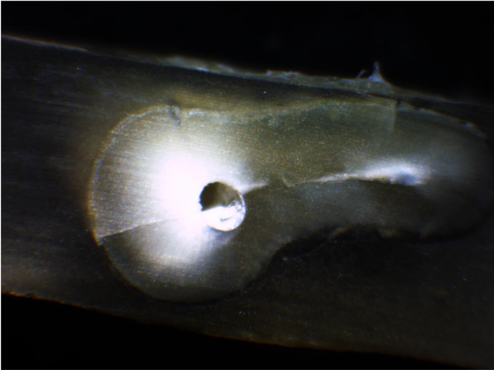
Figure 1.
Dentin Cracks Created by ProTaper Gold Files
.
Dentin Cracks Created by ProTaper Gold Files
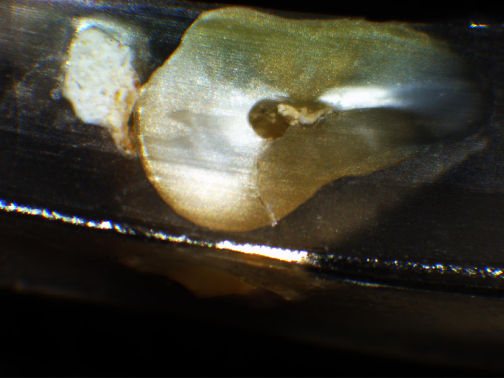
Figure 2.
Dentin cracks Created by Edge Taper Platinum Files
.
Dentin cracks Created by Edge Taper Platinum Files
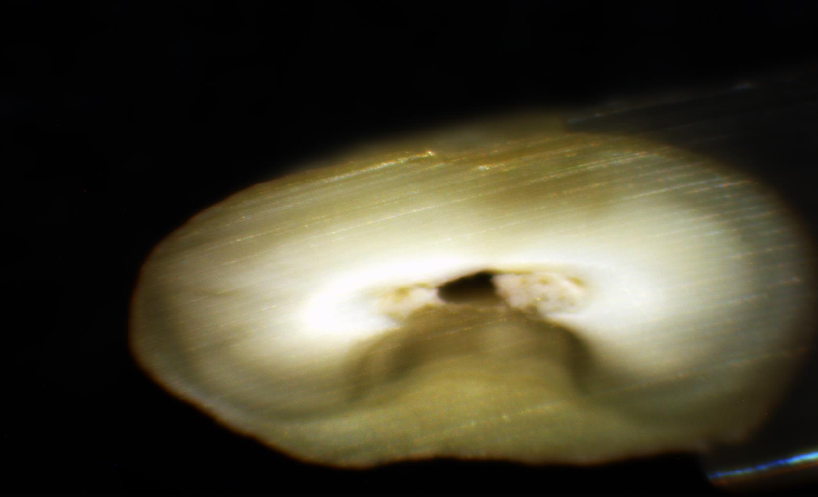
Figure 3.
Control Group (Without Dentin Cracks)
.
Control Group (Without Dentin Cracks)
Discussion
There are many different Ni-Ti file systems on the market, and dentists are trying to find studies that compare these systems clinically. Considering that the maxillary first molar teeth are very prone to fracture due to their narrow mesiodistal dimensions, they were used in the present study (29). The stereomicroscope has been used as a tool to study the roots in the examination of the apex anatomy and evaluate sealing capability using different filling materials and techniques (30). It can also be used as a useful teaching tool. Therefore, we examined the number of dentinal cracks created by the preparation of the mesiobuccal canal of the first maxillary molars with ProTaper Gold and Edge taper platinum rotary files. The examination was done utilizing a stereomicroscope.
By comparing the number of cracked teeth at different root levels between the ProTaper Gold and Edge taper platinum groups, no significant difference was observed between the studied systems at 3, 6, or 9 mm from the apex. This finding was consistent with the findings of studies by Fráter et al (30) and Karataş et al (31). In these studies, there was no significant difference between 3, 6, or 9 mm sections. In a study conducted by Fráter et al, there was no difference between ProTaper Gold files and other studied file systems in producing dentinal cracks.
In the present study, there was no significant difference in the number of microcracks at 3 mm (apical one-third) between the 3 groups (ProTaper, Edge taper, and control), and only in the ProTaper group, cracks were observed at 3 mm (3 cases). However, at 6 mm (middle third) and 9 mm (cervical third), the number of cracks was significantly different between the 3 groups. In the control group, no cracks were observed in any of the sections.
In an in vitro study, Chole et al compared the formation of dentin cracks after root canal preparation with PtoTaper Universal, ProTaper Next, and ProTaper Gold rotary files. At 3 and 9 mm, ProTaper Universal, ProTaper Next, and ProTaper Gold had the highest number of dentin cracks, respectively. At 6 mm from the apex, the number of dentin cracks created by ProTaper Next and ProTaper Gold was equal. In their study, it was reported that generally, ProTaper Next and ProTaper Gold rotary files produced significantly fewer dentin cracks than ProTaper Universal (32).
In the present study, at 3 and 9 mm from the apex, the ProTaper Gold group had the highest number of cracks and at 6 mm from the apex, the Edge taper platinum produced the highest number of cracks. In general, we did not find a significant difference between the two groups of files. The differences could generally indicate that the number of dentin cracks formed by the Edge taper platinum file, especially in apical and cervical sections, may be lower than that of the cracks formed by ProTaper Next and ProTaper Gold files.
The results of two studies conducted by Chole et al (32) and Nishad et al (33), who examined apical dentin cracks due to root canal preparation with ProTaper Universal, ProTaper Next, and ProTaper Gold rotary files, are contradictory.
In the study done by Chole et al, the roots were only cut at 3 mm from the apex for examination with a stereomicroscope. The control group remained intact and the other groups were prepared with files. The results of this study showed that the number of dentin defects was significantly lower in ProTaper Gold and ProTaper Universal groups (32). In the study conducted by Nishad et al, no significant difference was observed between ProTaper Universal and ProTaper Next and between ProTaper Gold and ProTaper Next (33). However, this study also introduced ProTaper Gold as one of the groups which had the least number of dentin cracks. Overall, in line with both previous studies, the superiority of this file over the other two groups can be noticed.
In an in vitro study, Hussien et al compared the rate of dentin crack formation between the RECIPROC blue, ProTaper Gold, ProTaper Next, and RECIPROC nickel-titanium rotary file systems. The results showed that dentin defects were seen in all groups (except for the control group), as in the present study, the ProTaper Gold file created dentin cracks in all 3, 6, and 9 mm sections. However, in the present study, the Edge Taper Platinum file was also examined and it was indicated that it did not cause any dentin cracks in the apical sections, which could show the superiority of this file over the ProTaper Gold file in apical sections. In the study of Hussien et al, no significant difference was observed in any of the intra-group and inter-group comparisons (34). In the present study, a two-by-two comparison of ProTaper Gold and Edge taper platinum files did not demonstrate any significant difference. In their study, the root samples were sectioned horizontally at 2, 4.5, and 7 mm from the apex. Based on the results, nickel-titanium rotary systems were not significantly different from others.
Although the number of cracks in different sections was not significantly different, the number of cracks in both Edge taper platinum and ProTaper Gold rotary files at 9 mm from the apex was higher compared to other sections. This finding was consistent with the results of the study by Harandi et al who stated that microcracks occurred mainly in the coronal part (9 mm) and no microcracks occurred in the control group (27). The systems used in the study conducted by Harandi et al included ProTaper Universal (rotary, multi-file system), Neolix (rotary, single-file system), and SafeSider (reciprocation movement, multi-file system). Besides, a stereomicroscope was used to assess cracks. In summary, it can be assumed that crack formation cannot be attributed to a single factor. Instead, crack formation is the result of several factors, possibly incremental and/or synergistic, such as tip design, cross-section design, taper, Ni-Ti alloy production process, and so on (11). Given the limitations of this study, it should be considered that the in vitro approach can produce a false positive effect due to the extraction forces to which the teeth are exposed during extraction, the storage conditions, and the sectioning method (35,36).
The sectioning method is not reliable because if it is not accompanied by a CT scan, cracks in the dentin cannot be ruled out. The sectioning method itself may also help to form dentin cracks; however, it is unlikely to significantly affect the results. Untreated (but sectioned) control specimens showed no cracks at all. Several other studies also concluded the same (31,37-39).
Problems with the use of extracted teeth have been highlighted by De-Deus et al (40); however, the present findings seem to contradict the possible role of tooth extraction in the formation of dentinal cracks within the root canal (41). Overall, while it is theoretically impossible to rule out the influence of these factors, the complete absence of cracks in the control samples is a strong argument against the role of the factors mentioned above in this study.
As there is still no clear agreement on the interpretation of the collected results from microscopic evaluations after sectioning and CT evaluation, future research should be performed with both evaluation methods in similar samples. The need for future research in this field has been clearly emphasized by Zaslansky et al (42).
Conclusion
Both ProTaper Gold and Edge taper platinum systems caused the formation of dentin cracks. Preparation of root canals by Edge taper platinum and ProTaper Gold rotary files causes the formation of dentin cracks to the same extent.
Acknowledgments
We would like to thank the Research Center and Research Vice-Chancellor of Mazandaran University of Medical Sciences for helping us in the implementation of this research.
Authors’ Contribution
Conceptualization: Narjes Hoshyari, Azam Haddadi Kohsar.
Data curation: Eghlima Malakan, Seyedali Seyedmajidi.
Formal analysis: Seyedali Seyedmajidi.
Funding acquisition: Azam Haddadi.
Investigation: Anahita Lotfizadeh, Eghlima Malakan.
Methodology: Azam Haddadi, Seyedali Seyedmajidi.
Project administration: Narjes Hoshyari.
Resources: Abolfazl Hosseinnataj.
Software: Abolfazl Hosseinnataj.
Supervision: Azam Haddadi.
Validation: Narjes Hoshyari.
Visualization: Seyedali Seyedmajidi.
Writing–original draft: Eghlima Malakan, Anahita Lotfizadeh.
Writing–review & editing: Anahita Lotfizadeh, Azam Haddadi.
Competing Interests
The authors have no conflict of interests to declare.
Ethical Approval
This study was approved by ethical committee of Mazandaran University of Medical Sciences (ethical approval No. IR.MAZUMS.REC.1400.396).
Funding
This research was funded by the research center of Mazandaran University of Medical Sciences.
References
- Vallaeys K, Chevalier V, Arbab-Chirani R. Comparative analysis of canal transportation and centring ability of three Ni-Ti rotary endodontic systems: ProTaper®, MTwo® and Revo-STM, assessed by micro-computed tomography. Odontology 2016; 104(1):83-8. doi: 10.1007/s10266-014-0176-z [Crossref] [ Google Scholar]
- do Amaral RO, Leonardi DP, Gabardo MC, Coelho BS, de Oliveira KV, Baratto Filho F. Influence of cervical and apical enlargement associated with the WaveOne system on the transportation and centralization of endodontic preparations. J Endod 2016; 42(4):626-31. doi: 10.1016/j.joen.2015.12.005 [Crossref] [ Google Scholar]
- Liu W, Wu B. Root canal surface strain and canal center transportation induced by 3 different nickel-titanium rotary instrument systems. J Endod 2016; 42(2):299-303. doi: 10.1016/j.joen.2015.10.023 [Crossref] [ Google Scholar]
- Adcock W, Trout NA, Vercoe D, Taylor DK, Shiner VJ Jr, Sorensen TS. Solvolysis of (Z)-5-trimethylstannyl 2-adamantyl p-bromobenzenesulfonate: mechanistic implications of a record-breaking secondary alpha-deuterium kinetic isotope effect for an SN1 substrate. J Org Chem 2003; 68(13):5399-402. doi: 10.1021/jo0300892 [Crossref] [ Google Scholar]
- De-Deus G, Souza EM, Barino B, Maia J, Zamolyi RQ, Reis C. The self-adjusting file optimizes debridement quality in oval-shaped root canals. J Endod 2011; 37(5):701-5. doi: 10.1016/j.joen.2011.02.001 [Crossref] [ Google Scholar]
- Peters OA. Current challenges and concepts in the preparation of root canal systems: a review. J Endod 2004; 30(8):559-67. doi: 10.1097/01.don.0000129039.59003.9d [Crossref] [ Google Scholar]
- Peters OA, Paqué F. Root canal preparation of maxillary molars with the self-adjusting file: a micro-computed tomography study. J Endod 2011; 37(1):53-7. doi: 10.1016/j.joen.2010.08.047 [Crossref] [ Google Scholar]
- Schilder H. Cleaning and shaping the root canal. Dent Clin North Am 1974; 18(2):269-96. [ Google Scholar]
- Hartmann MS, Barletta FB, Camargo Fontanella VR, Vanni JR. Canal transportation after root canal instrumentation: a comparative study with computed tomography. J Endod 2007; 33(8):962-5. doi: 10.1016/j.joen.2007.03.019 [Crossref] [ Google Scholar]
- Estrela C, de Almeida Decurcio D, Rossi-Fedele G, Silva JA, Guedes OA, Borges ÁH. Root perforations: a review of diagnosis, prognosis and materials. Braz Oral Res 2018; 32(Suppl 1):e73. doi: 10.1590/1807-3107bor-2018.vol32.0073 [Crossref] [ Google Scholar]
- Yoldas O, Yilmaz S, Atakan G, Kuden C, Kasan Z. Dentinal microcrack formation during root canal preparations by different NiTi rotary instruments and the self-adjusting file. J Endod 2012; 38(2):232-5. doi: 10.1016/j.joen.2011.10.011 [Crossref] [ Google Scholar]
- Bier CA, Shemesh H, Tanomaru-Filho M, Wesselink PR, Wu MK. The ability of different nickel-titanium rotary instruments to induce dentinal damage during canal preparation. J Endod 2009; 35(2):236-8. doi: 10.1016/j.joen.2008.10.021 [Crossref] [ Google Scholar]
- Fuss Z, Lustig J, Katz A, Tamse A. An evaluation of endodontically treated vertical root fractured teeth: impact of operative procedures. J Endod 2001; 27(1):46-8. doi: 10.1097/00004770-200101000-00017 [Crossref] [ Google Scholar]
- Shantiaee Y, Dianat O, Mosayebi G, Namdari M, Tordik P. Effect of root canal preparation techniques on crack formation in root dentin. J Endod 2019; 45(4):447-52. doi: 10.1016/j.joen.2018.12.018 [Crossref] [ Google Scholar]
- Capar ID, Ertas H, Ok E, Arslan H, Ertas ET. Comparative study of different novel nickel-titanium rotary systems for root canal preparation in severely curved root canals. J Endod 2014; 40(6):852-6. doi: 10.1016/j.joen.2013.10.010 [Crossref] [ Google Scholar]
- Gergi R, Rjeily JA, Sader J, Naaman A. Comparison of canal transportation and centering ability of twisted files, PathFile-ProTaper system, and stainless steel hand K-files by using computed tomography. J Endod 2010; 36(5):904-7. doi: 10.1016/j.joen.2009.12.038 [Crossref] [ Google Scholar]
- Lopes HP, Gambarra-Soares T, Elias CN, Siqueira JF Jr, Inojosa IF, Lopes WS. Comparison of the mechanical properties of rotary instruments made of conventional nickel-titanium wire, M-wire, or nickel-titanium alloy in R-phase. J Endod 2013; 39(4):516-20. doi: 10.1016/j.joen.2012.12.006 [Crossref] [ Google Scholar]
- Al-Manei KK, Al-Hadlaq SM. Evaluation of the root canal shaping ability of two rotary nickel-titanium systems. Int Endod J 2014; 47(10):974-9. doi: 10.1111/iej.12243 [Crossref] [ Google Scholar]
- García M, Duran-Sindreu F, Mercadé M, Bueno R, Roig M. A comparison of apical transportation between ProFile and RaCe rotary instruments. J Endod 2012; 38(7):990-2. doi: 10.1016/j.joen.2012.03.022 [Crossref] [ Google Scholar]
- Ceyhanli KT, Erdilek N, Tatar I, Cetintav B. Comparative micro-computed tomography evaluation of apical root canal transportation with the use of ProTaper, RaCe and Safesider systems in human teeth. Aust Endod J 2014; 40(1):12-6. doi: 10.1111/aej.12014 [Crossref] [ Google Scholar]
- Bernardes RA, Rocha EA, Duarte MA, Vivan RR, de Moraes IG, Bramante AS. Root canal area increase promoted by the EndoSequence and ProTaper systems: comparison by computed tomography. J Endod 2010; 36(7):1179-82. doi: 10.1016/j.joen.2009.12.033 [Crossref] [ Google Scholar]
- Gagliardi J, Versiani MA, de Sousa-Neto MD, Plazas-Garzon A, Basrani B. Evaluation of the shaping characteristics of ProTaper Gold, ProTaper NEXT, and ProTaper Universal in curved canals. J Endod 2015; 41(10):1718-24. doi: 10.1016/j.joen.2015.07.009 [Crossref] [ Google Scholar]
- Zhao D, Shen Y, Peng B, Haapasalo M. Root canal preparation of mandibular molars with 3 nickel-titanium rotary instruments: a micro-computed tomographic study. J Endod 2014; 40(11):1860-4. doi: 10.1016/j.joen.2014.06.023 [Crossref] [ Google Scholar]
- Alcalde M, Duarte MAH, Amoroso Silva PA, Souza Calefi PH, Silva E, Duque J. Mechanical properties of ProTaper Gold, EdgeTaper Platinum, flex gold and pro-T rotary systems. Eur Endod J 2020; 5(3):205-11. doi: 10.14744/eej.2020.48658 [Crossref] [ Google Scholar]
- Elsherief SM, Zayet MK, Hamouda IM. Cone-beam computed tomography analysis of curved root canals after mechanical preparation with three nickel-titanium rotary instruments. J Biomed Res 2013; 27(4):326-35. doi: 10.7555/jbr.27.20130008 [Crossref] [ Google Scholar]
- Schindler WG. The stereo microscope: an aid to evaluate root canal debridement and obturation. J Endod 1986; 12(8):359-62. doi: 10.1016/s0099-2399(86)80039-5 [Crossref] [ Google Scholar]
- Harandi A, Mirzaeerad S, Mehrabani M, Mahmoudi E, Bijani A. Incidence of dentinal crack after root canal preparation by ProTaper Universal, Neolix and SafeSider systems. Iran Endod J 2017; 12(4):432-8. doi: 10.22037/iej.v12i4.17597 [Crossref] [ Google Scholar]
- Zhang R, Hu T. Root canal curvature. Int Endod J 2010; 43(7):616-8. doi: 10.1111/j.1365-2591.2010.01727.x [Crossref] [ Google Scholar]
- Bhushan J, Gupta G, Gupta A. The ability of different nickel-titanium rotary instruments to induce dentinal micro-cracks during root canal preparation. Dent J Adv Stud 2018; 6(2-3):71-5. doi: 10.1055/s-0038-1676917 [Crossref] [ Google Scholar]
- Fráter M, Jakab A, Braunitzer G, Tóth Z, Nagy K. The potential effect of instrumentation with different nickel titanium rotary systems on dentinal crack formation-an in vitro study. PLoS One 2020; 15(9):e0238790. doi: 10.1371/journal.pone.0238790 [Crossref] [ Google Scholar]
- Karataş E, Gündüz HA, Kırıcı DÖ, Arslan H, Topçu MÇ, Yeter KY. Dentinal crack formation during root canal preparations by the twisted file adaptive, ProTaper Next, ProTaper Universal, and WaveOne instruments. J Endod 2015; 41(2):261-4. doi: 10.1016/j.joen.2014.10.019 [Crossref] [ Google Scholar]
- Chole D, Kamble S, Bakle S, Gandhi N, Hatte N, Bawa P. Effects of 3 single-file system (ProTaper Universal, ProTaper Next, ProTaper Gold) on crack formation in dentin after root canal preparation-an in vitro study. IOSR J Dent Med Sci 2019; 18(11):47-50. doi: 10.9790/0853-1811124750 [Crossref] [ Google Scholar]
- Nishad SV, Shivamurthy GB. Comparative analysis of apical root crack propagation after root canal preparation at different instrumentation lengths using ProTaper Universal, ProTaper Next and ProTaper Gold rotary files: an in vitro study. Contemp Clin Dent 2018; 9(Suppl 1):S34-S8. doi: 10.4103/ccd.ccd_830_17 [Crossref] [ Google Scholar]
- Hussien SW, Al-Gharrawi HA. Incidence of dentinal root defects caused by RECIPROC blue, ProTaper Gold, ProTaper NEXT and RECIPROC nickel titanium rotary instruments. J Contemp Dent Pract 2019; 20(3):291-7. [ Google Scholar]
- Coelho MS, Card SJ, Tawil PZ. Visualization enhancement of dentinal defects by using light-emitting diode transillumination. J Endod 2016; 42(7):1110-3. doi: 10.1016/j.joen.2016.04.006 [Crossref] [ Google Scholar]
- Shemesh H, Lindtner T, Portoles CA, Zaslansky P. Dehydration induces cracking in root dentin irrespective of instrumentation: a two-dimensional and three-dimensional study. J Endod 2018; 44(1):120-5. doi: 10.1016/j.joen.2017.07.025 [Crossref] [ Google Scholar]
- Pawar AM, Thakur B, Kfir A, Kim HC. Dentinal defects induced by 6 different endodontic files when used for oval root canals: an in vitro comparative study. Restor Dent Endod 2019; 44(3):e31. doi: 10.5395/rde.2019.44.e31 [Crossref] [ Google Scholar]
- Li SH, Lu Y, Song D, Zhou X, Zheng QH, Gao Y. Occurrence of dentinal microcracks in severely curved root canals with ProTaper Universal, WaveOne, and ProTaper Next file systems. J Endod 2015; 41(11):1875-9. doi: 10.1016/j.joen.2015.08.005 [Crossref] [ Google Scholar]
- Pedullà E, Genovesi F, Rapisarda S, La Rosa GR, Grande NM, Plotino G. Effects of 6 single-file systems on dentinal crack formation. J Endod 2017; 43(3):456-61. doi: 10.1016/j.joen.2016.10.038 [Crossref] [ Google Scholar]
- De-Deus G, Cavalcante DM, Belladonna FG, Carvalhal J, Souza EM, Lopes RT. Root dentinal microcracks: a post-extraction experimental phenomenon?. Int Endod J 2019; 52(6):857-65. doi: 10.1111/iej.13058 [Crossref] [ Google Scholar]
- Arashiro FN, De-Deus G, Belladonna FG, Cavalcante DM, Coelho MS, Silva E. Dentinal microcracks on freshly extracted teeth: the impact of the extraction technique. Int Endod J 2020; 53(4):440-6. doi: 10.1111/iej.13239 [Crossref] [ Google Scholar]
- Zaslansky P, Prates Soares A, Shemesh H. Root dentinal microcracks - absence of evidence is not evidence of absence. Int Endod J 2020; 53(1):135-6. doi: 10.1111/iej.13207 [Crossref] [ Google Scholar]