Avicenna J Dent Res. 15(2):63-69.
doi: 10.34172/ajdr.2023.1618
Original Article
An Investigation of the Clinico-histopathologic Trend of Salivary Gland Tumors in Amir Alam Hospital During 2010- 2019: A Retrospective Study
Amirhosein Ghaemi 1  , Noushin Jalayer Naderi 2, * , Farzad Yazdani Biucki 3
Author information:
1Faculty of Dentistry, Shahed University, Tehran, Iran
2Department of Oral and Maxillofacial Pathology, Faculty of Dentistry, Shahed University, Tehran, Iran
3Department of Pathology, Amir Alam Hospital, Tehran University of Medical Sciences, Tehran, Iran
Abstract
Background: The incidence and mortality rates of salivary gland tumors have increased according to previous evidence. No study has so far focused on the trend of clinical and histopathologic patterns of salivary gland tumors in Iran. Therefore, the aim was to investigate the incidence and clinico-histopathologic trend of salivary gland tumors in a retrospective, cross-sectional, institutional study from 2010-2019 in Amir Alam hospital.
Methods: The archived medical records were collected from patients with the histopathologic diagnosis of benign and malignant salivary gland tumors from Amir Alam hospital, Tehran during (April-April) 2010- 2019. Demographic data and histopathologic features, including tumor size, lymph node involvement, vascular invasion, perineural involvement, and histopathologic differentiation were retrieved, and the samples were categorized and reviewed based on the new classification of head and neck tumors. Finally, the frequencies of characteristics were determined and expressed as numbers (percentage values).
Results: Of 1203 salivary gland tumors, 77.6% and 22.4% were benign and malignant, respectively. The incidence of benign tumors was increased from 37 (22.2%) in 2010 to 178 (364.9%) in 2019. In the collection of the total samples, the incidence of malignant tumors was relatively steady from 23 (13.8%) samples in 2010 to 27 (55.35%) in 2019. However, an increase in the incidence of tumors with low-grade differentiation was found from 12.5% in 2010 to 80% in 2019.
Conclusions: The incidence of benign and malignant salivary tumors with a higher degree of malignancy had an increasing trend in Amir Alam hospital during 2010-2019.
Keywords: Diseases, Epidemiology, Salivary glands, Trends
Copyright and License Information
© 2023 The Author(s); Published by Hamadan University of Medical Sciences.
This is an open-access article distributed under the terms of the Creative Commons Attribution License (
http://creativecommons.org/licenses/by/4.0), which permits unrestricted use, distribution, and reproduction in any medium provided the original work is properly cited.
Please cite this article as follows: Ghaemi A, Jalayer Naderi N, Yazdani Biucki F. An investigation of the clinico-histopathologic trend of salivary gland tumors in amir alam hospital during 2010-2019: A retrospective study. Avicenna J Dent Res. 2023; 15(2):63-69. doi:10.34172/ ajdr.2023.1618
Background
Salivary gland neoplasms account for 3%-10% of all neck and jaw neoplasms (1). The prevalence of salivary gland tumors varies in different geographical areas, but in total, an annual prevalence of 1-6.5 per 100 000 people has been reported in this regard. This rate is 4.7 per year for benign and 0.9 per year for malignant tumors (2). Based on reports, the incidence of benign and malignant salivary tumors in Iran is similar to the global configuration (3-7). Although the reports show similarities in the incidence of salivary gland tumors, factors such as the improvement of the salivary gland classification system and racial/ethnic differences can lead to differences in outcomes. Despite the available global information on the incidence of salivary gland tumors, the trend of benign and malignant salivary lesions has not been studied yet. The incidence (8-11) and mortality rate (9) of salivary gland tumors have increased according to previous studies. A population-based historical cohort study in the United States demonstrated that the incidence of malignant tumors of major salivary glands increased from 10.4 per 1 000 000 to 16 per 1 000 000 in 1973-2009. The study represented an increased rate ofparotid tumors measuring 0-2.0 cm (8).
Previous studies mainly focused on the incidence of salivary gland tumors based on demographic patterns. These studies evaluated limited characteristics rather than incidence trends. No study has so far been performed on the trend of clinical and histopathologic patterns of salivary gland tumors in Iran. Accordingly, the aim was to investigate theclinico-histopathologic trend of salivary gland tumors in Amir Alam hospital during 2010-2019 in a retrospective study. Considering that the Amir Alam hospital is the center of head and neck diseases, the findings may reflect the current state of salivary gland tumors in Iran.
Materials and Methods
The current retrospective cross-sectional study was approved by the Ethics Committee of Shahed University under registration number IR.SHAHED.REC.1400.080.
The archived medical records of patients with the histopathologic diagnosis of benign and malignant salivary gland tumors from Amir Alam hospital, Tehran, Iran from April 2010 to April 2019 were statistical society. The inclusion criterion was including the records with complete demographic and histopathologic information. On the other hand, the exclusion criteria were incomplete data on lymph node involvement and vascular, perineural invasion, metastatic tumors and tumors of stroma, including lymphomas, hemangioma, and lipoma, and incisional biopsies.
The included records were reviewed, and the samples were categorized based on the new classification of head and neck tumors (1). To maintain confidentiality, the samples were recorded only based on the number of records while removing the name of patients. The demographic data comprising age, gender, and anatomical location of tumors were recorded, as well as registering the histopathologic features, including the tumor size, lymph node involvement, vascular invasion, perineural involvement, and histopathologic differentiation of malignant tumors.
Overall, 1203 salivary gland tumors were recorded from 2010-2019. The frequencies of demographic characteristics and histopathologic features were determined and presented as numbers (percentage values). The SPSS software for windows (version 21, SPSS Inc., IBM Co., Chicago, Illinois, USA) was used to determine percentages and mean values. Eventually, statistical findings were interpreted descriptively.
Results
Of the 1203 salivary gland tumors, 933 (77.6%) and 270 (22.4%) cases were benign and malignant, respectively. In addition, there were 616 (51.2%) and 587 (48.8%) salivary tumors in males and females, respectively. The male-to-female ratio was 1.04:1. Of all samples, 479 (51.3%) and 454 (48.7%) benign salivary tumors were increased in males and females, respectively. The male-to-female ratio was 1.05:1. Of the 270 malignant salivary gland tumors, 137 (50.7%) and 133 (49.3%) were in males and females with the male-to-female ratio of 1.03:1, respectively.
The age ranged from 2 to 91 years with a mean of 43.86 ± 15.79 years and a median age of 43 years. In benign and malignant cases, the age ranged from 10 to 82 (mean of 42.2 ± 14. 9 years) and 2 to 91 (mean of 49.57 ± 17. 39 years) years, respectively. There were 1091 (90.7%) and 112 (9.3%) salivary gland tumors in major and minor salivary glands, respectively. In benign tumors, 869 (93.1%) and 64 (6.9%) cases occurred in major and minor salivary glands, respectively. In malignant tumors, 222 (82.2%) cases occurred in major salivary glands, while 48 (17.8%) cases belonged to minor salivary glands. The frequency of the demographic features of benign and malignant salivary gland tumors during 2010-2019 is summarized in Tables 1 and 2, respectively.
Table 1.
Frequency of the Demographic Features of Benign Salivary Gland Tumors During 2010-2019 in Amir Alam Hospital
|
Variables
|
Years
|
|
2010
|
2011
|
2012
|
2013
|
2014
|
2015
|
2016
|
2017
|
2018
|
2019
|
| Age |
|
|
|
|
|
|
|
|
|
|
| 1-10 |
0 |
0 |
0 |
0 |
0 |
2 |
0 |
0 |
0 |
0 |
| 11-20 |
3 |
1 |
6 |
3 |
11 |
3 |
6 |
15 |
3 |
10 |
| 21-30 |
6 |
9 |
8 |
14 |
21 |
22 |
19 |
18 |
23 |
27 |
| 31-40 |
10 |
8 |
5 |
14 |
19 |
28 |
24 |
32 |
41 |
37 |
| 41-50 |
7 |
6 |
8 |
14 |
26 |
11 |
29 |
15 |
33 |
40 |
| 51-60 |
6 |
4 |
11 |
13 |
16 |
20 |
20 |
19 |
29 |
42 |
| 61-70 |
4 |
2 |
3 |
11 |
7 |
11 |
14 |
9 |
9 |
19 |
| 71-80 |
1 |
1 |
0 |
7 |
3 |
1 |
2 |
1 |
7 |
2 |
| > 81 |
0 |
0 |
0 |
0 |
0 |
0 |
1 |
0 |
0 |
1 |
| Gender |
|
|
|
|
|
|
|
|
|
|
| Males |
20 |
16 |
15 |
38 |
56 |
44 |
65 |
50 |
75 |
100 |
| Females |
17 |
15 |
26 |
38 |
47 |
54 |
50 |
59 |
70 |
78 |
| Location |
|
|
|
|
|
|
|
|
|
|
| Buccal mucosa |
0 |
0 |
0 |
0 |
1 |
0 |
0 |
0 |
0 |
0 |
| Palate |
2 |
1 |
3 |
2 |
4 |
4 |
2 |
3 |
4 |
5 |
| Tongue |
0 |
0 |
0 |
0 |
0 |
0 |
0 |
0 |
0 |
0 |
| Retro molar |
2 |
1 |
2 |
3 |
3 |
6 |
6 |
1 |
2 |
7 |
| Maxillary sinus |
0 |
0 |
0 |
0 |
0 |
0 |
0 |
0 |
0 |
0 |
| Parotid |
30 |
25 |
27 |
62 |
84 |
81 |
97 |
96 |
131 |
156 |
| Sub mandibular gland |
3 |
4 |
8 |
9 |
11 |
7 |
10 |
9 |
8 |
10 |
| Sub lingual gland |
0 |
0 |
1 |
0 |
0 |
0 |
0 |
0 |
0 |
0 |
Note. Values stated in number.
Table 2.
Frequency of the Demographic Features of Malignant Salivary Gland Tumors During 2010-2019 in Amir Alam Hospital
|
Variables
|
Years
|
|
2010
|
2011
|
2012
|
2013
|
2014
|
2015
|
2016
|
2017
|
2018
|
2019
|
| Age |
|
|
|
|
|
|
|
|
|
|
| 1-10 |
1 |
1 |
1 |
0 |
0 |
1 |
0 |
0 |
0 |
0 |
| 11-20 |
2 |
0 |
2 |
1 |
2 |
0 |
1 |
1 |
1 |
1 |
| 21-30 |
4 |
0 |
2 |
1 |
3 |
3 |
2 |
4 |
3 |
2 |
| 31-40 |
4 |
2 |
1 |
4 |
2 |
6 |
9 |
5 |
6 |
2 |
| 41-50 |
3 |
1 |
4 |
7 |
7 |
6 |
5 |
7 |
7 |
7 |
| 51-60 |
4 |
1 |
9 |
7 |
10 |
5 |
4 |
11 |
6 |
5 |
| 61-70 |
1 |
3 |
1 |
6 |
4 |
8 |
5 |
8 |
4 |
5 |
| 71-80 |
4 |
0 |
1 |
2 |
2 |
2 |
3 |
1 |
4 |
3 |
| > 81 |
0 |
0 |
0 |
0 |
0 |
2 |
2 |
0 |
1 |
2 |
| Gender |
|
|
|
|
|
|
|
|
|
|
| Males |
14 |
4 |
10 |
17 |
15 |
14 |
18 |
13 |
15 |
17 |
| Females |
9 |
4 |
11 |
11 |
15 |
19 |
13 |
24 |
17 |
10 |
| Location |
|
|
|
|
|
|
|
|
|
|
| Buccal mucosa |
0 |
0 |
0 |
0 |
1 |
0 |
0 |
2 |
0 |
0 |
| Palate |
0 |
2 |
1 |
3 |
4 |
3 |
3 |
0 |
0 |
3 |
| Tongue |
1 |
0 |
1 |
1 |
0 |
2 |
1 |
1 |
1 |
1 |
| Retro molar |
1 |
0 |
1 |
0 |
1 |
0 |
0 |
0 |
0 |
2 |
| Maxillary sinus |
1 |
0 |
2 |
3 |
3 |
0 |
1 |
1 |
0 |
1 |
| Parotid |
20 |
5 |
14 |
18 |
19 |
23 |
21 |
28 |
27 |
17 |
| Sub mandibular gland |
0 |
1 |
2 |
2 |
2 |
5 |
5 |
2 |
3 |
1 |
| Sub lingual gland |
0 |
0 |
0 |
1 |
0 |
0 |
0 |
3 |
1 |
2 |
Note. Values stated in number.
Pleomorphic adenoma (n = 808, 67.2%) and papillary cystadenoma lymphomatosum (Warthin tumor, n = 125, 10.4%) were the most frequent benign tumors. Further, the most prevalent malignant tumors were adenoid cystic carcinoma and mucoepidermoid carcinoma (n = 92, 7.6%, and n = 75, 6.2%, respectively). The histopathologic features of malignant tumors are presented in Table 3.
Table 3.
Frequency of the Histopathologic Findings of Malignant Salivary Gland Tumors During 2010-2019 in Amir Alam Hospital
|
Features
|
Number
|
Percent
|
| Grade |
|
|
| Well |
37 |
43 |
| Moderate |
18 |
20.9 |
| Poor |
31 |
36 |
| Size (cm) |
|
|
| 0-2 |
91 |
33.7 |
| 2.1-4 |
124 |
45.9 |
| > 4.1 |
55 |
20.4 |
| Perineural invasion |
|
|
| With invasion |
105 |
38.9 |
| Without invasion |
165 |
61.1 |
| Vascular invasion |
|
|
| With invasion |
29 |
10.7 |
| Without invasion |
241 |
89.3 |
| Lymph node involvement |
|
|
| With involvement |
|
|
| 0-4 |
11 |
4.1 |
| 5-10 |
6 |
2.2 |
| > 10 |
5 |
1.9 |
| Without involvement |
248 |
91 |
There was an increase in the incidence of benign tumors in parotid glands from 2010-2019. The incidence of pleomorphic adenoma and papillary cystadenoma lymphomatosum (Warthin tumor) increased during 2010-2019; however, there was no change in the incidence of malignant salivary gland tumors (Table 4, Figure 1).
Table 4.
Incidence of Benign and Malignant Salivary Gland Tumors During 2010-2019 in Amir Alam Hospital
|
Tumors
|
Years
|
|
2010
|
2011
|
2012
|
2013
|
2014
|
2015
|
2016
|
2017
|
2018
|
2019
|
| Benign tumors |
|
|
|
|
|
|
|
|
|
|
Pleomorphic adenoma
Warthin tumor |
34 |
30 |
36 |
67 |
91 |
84 |
91 |
99 |
131 |
145 |
| 3 |
1 |
5 |
9 |
12 |
14 |
24 |
10 |
14 |
33 |
| Malignant tumors |
|
|
|
|
|
|
|
|
|
|
| Adenoid cystic carcinoma |
2 |
4 |
5 |
13 |
12 |
16 |
10 |
13 |
9 |
8 |
| Mucoepidermoid carcinoma |
8 |
2 |
9 |
7 |
5 |
10 |
9 |
6 |
9 |
10 |
| Acinic cell adenocarcinoma |
4 |
1 |
3 |
3 |
2 |
0 |
4 |
6 |
5 |
2 |
| Basal cell adenocarcinoma |
0 |
0 |
0 |
2 |
1 |
0 |
1 |
2 |
1 |
0 |
| Adenocarcinoma, not otherwise specified |
4 |
0 |
2 |
3 |
4 |
2 |
1 |
4 |
2 |
1 |
| Carcinosarcoma |
1 |
0 |
0 |
0 |
0 |
1 |
0 |
0 |
0 |
0 |
| Clear cell adenocarcinoma |
0 |
0 |
1 |
0 |
0 |
0 |
0 |
0 |
0 |
0 |
| Myoepithelial carcinoma |
1 |
0 |
1 |
0 |
3 |
3 |
2 |
2 |
3 |
0 |
| Carcinoma ex pleomorphic adenoma |
0 |
1 |
0 |
0 |
0 |
0 |
1 |
1 |
3 |
5 |
| Malignant lymphoepithelial lesion |
2 |
0 |
0 |
0 |
0 |
0 |
0 |
0 |
0 |
0 |
| Salivary duct carcinoma |
0 |
0 |
0 |
0 |
3 |
1 |
3 |
3 |
0 |
1 |
| Small cell carcinoma |
1 |
0 |
0 |
0 |
0 |
0 |
0 |
0 |
0 |
0 |
Note. Values stated in number.
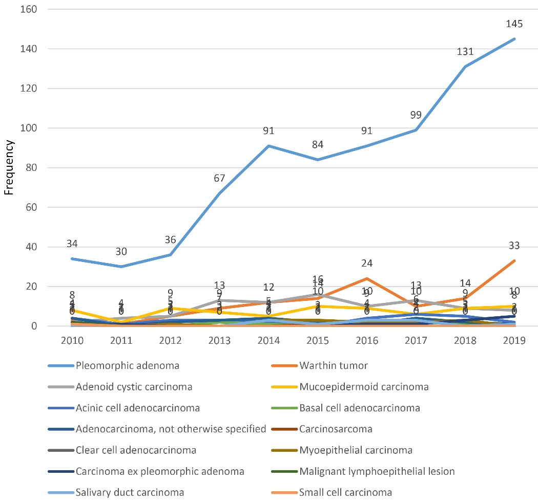
Figure 1.
Incidence Trends of Benign and Malignant Salivary Tumors 2010-2019 in Amir Alam hospital. Note. Values stated in number
.
Incidence Trends of Benign and Malignant Salivary Tumors 2010-2019 in Amir Alam hospital. Note. Values stated in number
In malignant salivary gland tumors, there was an increase in the incidence of high-grade tumors. Tumor size measuring 2.1-4 cm had an increasing trend during 2010-2019. An increased trend was observed in cases with the involvement of 0-4 lymph nodes. Invasion to vascular and perineural tissues showed increased trends. The trend of histopathologic features in malignant salivary gland tumors during 2010-2019 is depicted in Figure 2.
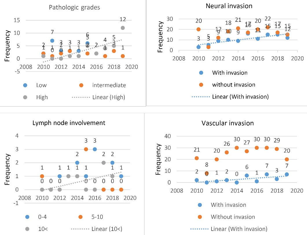
Figure 2.
The Trend of Histopathologic Parameters of Malignant Salivary Gland Tumors During 2010-2019 in Amir Alam Hospital. Note. Values mentioned in number
.
The Trend of Histopathologic Parameters of Malignant Salivary Gland Tumors During 2010-2019 in Amir Alam Hospital. Note. Values mentioned in number
Discussion
The findings of the study revealed that the frequency of benign salivary gland tumors has increased in Amir Alam hospital during 2010-2019. An increase in the incidence of high-grade malignant tumors was noted as well.
Benign and malignant salivary tumors were most prevalent in the fourth and sixth decades of life, respectively, which is consistent with the findings of previous studies (4,12,13). However, the present results contradict those of Ansari et al, Jaafari-Ashkavandi et al, and Rahrotaban et al, representing the most prevalence of malignant tumors in the mean age of 47, 50.6, and 45 years old, respectively (14-16).
In the current study, the prevalence of benign and malignant salivary gland tumors was higher in males, which corroborates the results of some studies (12,13,17). However, it is not consistent with the findings of some other studies, showing a higher prevalence of salivary tumors in females (4,5,18). The different prevalence between ages and genders in different studies needs further investigation because it may be related to a genetic base in some ethnic/races. Pleomorphic adenoma was the most common benign tumor with 86% of cases. Although this finding is in line with those of previous studies (3,5,15,17), the prevalence of malignant salivary gland tumors did not match in different studies (3,14,16,17). Consistent with our study findings, some previous studies reported that adenoid cystic carcinoma was the most common malignant tumor of the salivary glands (3,15,19,20). A number of studies have shown that mucoepidermoid carcinoma was the most prevalent malignant tumor of salivary glands (21,22). These discrepancies can be due to the type of sampling or the genetic-related setting of different races.
The results of the present study showed that the rate of salivary gland tumors has slightly increased in recent years. In a population-based historical cohort study from 5 states of the United States, Del Signore and Megwalu found that the incidence of the malignant tumors of major salivary gland increased from 10.4 per 1 000 000 to 16 per 1 000 000 during in 1973-2009 (8). Accordingly, studies in Poland and China reported a growing incidence of salivary gland tumors (9,10), which conforms to the findings of the present study. Although this increase may be due to lifestyle changes in human societies such as the prevalence of smoking (23), increasing public awareness of the symptoms of cancer and the level of public dental services can also lead to early detection of the disease. Contrary, Shu et al reported no increase in the incidence of malignant parotid gland tumors in Swedish and Nordic adults during 1970-2009 (24). Although this may be due to the level of health care in Northern Europe and the consequent early detection of tumors, genetic differences between races may play a role in this regard. This is a topic that needs further research.
In spite of the stable incidence of malignant tumors, an increasing incidence of high-grade tumors was recorded in the current study. The results of the present study showed that in the past decade, the trend of well and moderate differentiated salivary tumors had a decreasing rate, while tumors with poor differentiation have increased, which is consistent with the findings of Gupta et al, demonstrating that poorly differentiated tumors of parotid malignancies had a highest upward trend from 1973 to 2015 (25).
In the present study, the malignant tumors of salivary glands with 2-4 cm diameter had the highest frequency among the samples, which contradicts the results of Del Signore and Megwalu, indicating that the incidence of salivary tumors smaller than 2 cm had an increasing rate (8). This finding may be due to the early detection of salivary tumors with small sizes.
Based on the findings of this study, concurrent with an increase in the tumor size, the rate of vascular involvement and lymph node metastasis has also increased in the recent decade. Nonetheless, there was no study to compare the trend of these variables. This finding needs further biologic studies since the vascular invasion and lymph node metastases indicate more aggressive behavior of a malignant tumor.
In this study, the outcome of the malignant tumors was not detected because of operational limitations on following the patient. Despite this limitation, the study was completed in a referral head and neck center that admits patients from all over Iran, thus it may reflect the current situation of salivary gland tumors in Iran. The research society was the most important strength of the present study. Changes in the incidence of salivary gland tumors in relation to the age and gender of patients were not investigated in this study. It is suggested that the trend of salivary tumors should be separately examined for each tumor with regard to ages and genders in future studies.
Conclusions
During 2010-2019, the incidence of benign and malignant salivary tumors with a higher degree of malignancy had an increasing trend in Amir Alam hospital. Our findings marked the importance of further research on the risk factors and even early diagnosis and management of salivary gland cancer.
Acknowledgments
The authors thank Mr. S. Parisa Kiani for kindly assistance in archive retrieval. This article is the result of thesis No. 892 of Amirhosein Ghaemi, a graduate student of dentistry from the Faculty of Dentistry, Shahed University, Tehran, Iran.
Authors’ Contribution
Conceptualization: Noushin Jalayer Naderi.
Data curation: Amirhosein Ghaemi, Noushin Jalayer Naderi, Farzad Yazdani Biucki.
Formal analysis: Amirhosein Ghaemi, Noushin Jalayer Naderi.
Investigation: Amirhosein Ghaemi, Noushin Jalayer Naderi, Farzad Yazdani Biucki.
Methodology: Noushin Jalayer Naderi, Farzad Yazdani Biucki.
Project administration: Noushin Jalayer Naderi.
Supervision: Noushin Jalayer Naderi.
Validation: Noushin Jalayer Naderi, Farzad Yazdani Biucki.
Visualization: Amirhosein Ghaemi, Noushin Jalayer Naderi, Farzad Yazdani Biucki.
Writing – original draft: Amirhosein Ghaemi, Noushin Jalayer Naderi, Farzad Yazdani Biucki.
Writing – review & editing: Amirhosein Ghaemi, Noushin Jalayer Naderi, Farzad Yazdani Biucki.
Competing Interests
There is no conflict of interests.
References
- El-Naggar AK, Chan JKC, Grandis JR, Takata T, Slootweg PJ. World Health Organization Classification of Tumors. 4th ed. Lyon, France: International Agency for Research on Cancer Press; 2017.
- Neville BW, Dam DD, Allen CM, Bouquoct JE. Oral & Maxillofacial Pathology. 2nd ed. Philadelphia, PA: W.B. Saunders Company; 2016.
- Sentani K, Ogawa I, Ozasa K, Sadakane A, Utada M, Tsuya T. Characteristics of 5015 salivary gland neoplasms registered in the Hiroshima Tumor Tissue Registry over a period of 39 years. J Clin Med 2019; 8(5):566. doi: 10.3390/jcm8050566 [Crossref] [ Google Scholar]
- Galdirs TM, Kappler M, Reich W, Eckert AW. Current aspects of salivary gland tumors–a systematic review of the literature. GMS Interdiscip Plast Reconstr Surg DGPW 2019; 8:Doc12. doi: 10.3205/iprs000138 [Crossref] [ Google Scholar]
- Vasconcelos AC, Nör F, Meurer L, Salvadori G, de Souza LB, Vargas PA. Clinicopathological analysis of salivary gland tumors over a 15-year period. Braz Oral Res 2016; 30:S1806-83242016000100208. doi: 10.1590/1807-3107BOR-2016.vol30.0002 [Crossref] [ Google Scholar]
- Fomete B, Adebayo ET, Ononiwu CN. Management of salivary gland tumors in a Nigerian tertiary institution. Ann Afr Med 2015; 14(3):148-54. doi: 10.4103/1596-3519.152071 [Crossref] [ Google Scholar]
- Al Sarraj Y, Nair SC, Al Siraj A, AlShayeb M. Characteristics of salivary gland tumours in the United Arab Emirates. Ecancermedicalscience 2015; 9:583. doi: 10.3332/ecancer.2015.583 [Crossref] [ Google Scholar]
- Del Signore AG, Megwalu UC. The rising incidence of major salivary gland cancer in the United States. Ear Nose Throat J 2017; 96(3):E13-E6. doi: 10.1177/014556131709600319 [Crossref] [ Google Scholar]
- Kordzińska-Cisek I, Grzybowska-Szatkowska L. Salivary gland cancer—epidemiology. Nowotwory 2018; 68(1):22-7. doi: 10.5603/njo.2018.0005 [Crossref] [ Google Scholar]
- Li LJ, Li Y, Wen YM, Liu H, Zhao HW. Clinical analysis of salivary gland tumor cases in West China in past 50 years. Oral Oncol 2008; 44(2):187-92. doi: 10.1016/j.oraloncology.2007.01.016 [Crossref] [ Google Scholar]
- Bjørndal K, Krogdahl A, Therkildsen MH, Overgaard J, Johansen J, Kristensen CA. Salivary gland carcinoma in Denmark 1990-2005: a national study of incidence, site and histology Results of the Danish Head and Neck Cancer Group (DAHANCA). Oral Oncol 2011; 47(7):677-82. doi: 10.1016/j.oraloncology.2011.04.020 [Crossref] [ Google Scholar]
- Rezaei F, Tavakoli P, Mozaffari HR, Azari M. Prevalence of salivary gland tumors in patients referred-pathology departments of Kermanshah hospitals, Iran, 2007-2012. Res J Med Sci 2016; 10(5):487-91. doi: 10.36478/rjmsci.2016.487.491 [Crossref] [ Google Scholar]
- Khajavi M, Peyvandi A, Naghibzadeh B, Ahmadi Roozbahani N. Salivary gland tumors in patients referring to Loghman Hakim hospital in a 10-Year Period. Pajoohande 2010;15(1):45-8. [Persian].
- Ansari MH. Salivary gland tumors in an Iranian population: a retrospective study of 130 cases. J Oral Maxillofac Surg 2007; 65(11):2187-94. doi: 10.1016/j.joms.2006.11.025 [Crossref] [ Google Scholar]
- Jaafari-Ashkavandi Z, Ashraf MJ, Moshaverinia M. Salivary gland tumors: a clinicopathologic study of 366 cases in southern Iran. Asian Pac J Cancer Prev 2013; 14(1):27-30. doi: 10.7314/apjcp.2013.14.1.27 [Crossref] [ Google Scholar]
- Rahrotaban S, Masoomi P, Moradi M, Sharifpour Vajari S. Frequency of salivary gland tumors in two referral center of Qazvin University of Medical Sciences from 1999 to 2009. J Res Dent Sci 2010;7(2):27-31. [Persian].
- Shishegar M, Ashraf MJ, Azarpira N, Khademi B, Hashemi B, Ashrafi A. Salivary gland tumors in maxillofacial region: a retrospective study of 130 cases in a southern Iranian population. Patholog Res Int 2011; 2011:934350. doi: 10.4061/2011/934350 [Crossref] [ Google Scholar]
- Sando Z, Fokouo JV, Mebada AO, Djomou F, Ndjolo A, Oyono JL. Epidemiological and histopathological patterns of salivary gland tumors in Cameroon. Pan Afr Med J 2016; 23:66. doi: 10.11604/pamj.2016.23.66.5105 [Crossref] [ Google Scholar]
- Shamloo N, Ghanadan A, Hashemiyan FS, Ghorbanpour M. Epidemiologic characteristics of salivary gland tumors in an Iranian population. Shiraz E-Med J 2021; 22(9):e107675. doi: 10.5812/semj.107675 [Crossref] [ Google Scholar]
- Chinipardaz Z, Khalili M, Abouzeid F. Salivary gland tumors: a retrospective study of 339 cases in an Iranian population from 1986 to 2006. Int Dent J Stud Res 2013; 1:19-23. [ Google Scholar]
- Fu JY, Wu CX, Shen SK, Zheng Y, Zhang CP, Zhang ZY. Salivary gland carcinoma in Shanghai (2003-2012): an epidemiological study of incidence, site and pathology. BMC Cancer 2019; 19(1):350. doi: 10.1186/s12885-019-5564-x [Crossref] [ Google Scholar]
- Ghartimagar D, Ghosh A, Shrestha MK, Thapa S, Talwar OP. Histopathologic profile of salivary gland tumors among specimens from a tertiary care hospital: a descriptive cross-sectional study. JNMA J Nepal Med Assoc 2020; 58(230):729-35. doi: 10.31729/jnma.4898 [Crossref] [ Google Scholar]
- Li TI, Chiang MT, Chiu KC, Lai CH, Liu SY, Shieh YS. The association of betel quid, alcohol, and cigarettes with salivary gland tumor-a case-control study. J Dent Sci 2017; 12(2):151-5. doi: 10.1016/j.jds.2016.12.002 [Crossref] [ Google Scholar]
- Shu X, Ahlbom A, Feychting M. Incidence trends of malignant parotid gland tumors in Swedish and Nordic adults 1970 to 2009. Epidemiology 2012; 23(5):766-7. doi: 10.1097/EDE.0b013e31825988fa [Crossref] [ Google Scholar]
- Gupta A, Koochakzadeh S, Neskey DM, Nguyen SA, Lentsch EJ. Incidence and survival trends of parotid malignancies over 42 years. Head Neck 2020; 42(9):2308-15. doi: 10.1002/hed.26172 [Crossref] [ Google Scholar]