Avicenna J Dent Res. 12(3):81-85.
doi: 10.34172/ajdr.2020.17
Original Article
Comparative Study of the Effect of Flavonoids and the Whey Extract on Enamel Microhardness: An In Vitro Study
Shokufeh Nobahar 1, Shahla Mirzaeei 2, Leila Simaei 3, *  , Zohreh Ahmadi 4
Author information:
1Assistant Professor, Department of Pediatric Dentistry, Dental School, Kermanshah University of Medical Sciences, Kermanshah, Iran.
2Associated Professor of Pharmaceutics School of Pharmacy, Nano Drug Delivery Research Center Research Institute for Health Technology, Kermanshah University of Medical Sciences Iran.
3Assistant Professor, Department of Pediatric Dentistry, Dental School, Guilan University of Medical Sciences, Guilan, Iran.
4Dentist, Private Practice, Kermanshah, Iran.
Abstract
Background: Preventing caries or stopping primary caries lesions is one of the most important goals of oral health care. The aim of this study was to compare the effect of flavonoids and the whey extract on tooth enamel by measuring the microhardness of enamel following its demineralization.
Methods: In general, 42 samples of healthy enamel were prepared in this experimental-laboratory study. After measuring the initial microhardness using Vickers hardness tester )Buehler, Lake Bluff, IL, USA(, the samples were immersed in the demineralization solution for 4 days and re-tested for microhardness. Then, the samples were randomly divided into three groups of 14 and treated, including the grape seed extract w/v8%, the whey extract, and the artificial saliva. The samples were in contact with the treatment material for 8 days in an incubator at 37°C and then their microhardness was evaluated again.
Results: The analysis of covariance showed that there was a significant difference in the amount of microhardness changes between the study groups (P < 0.001) so that microhardness changes were the highest in the grape seed extract group while they decreased in the control group. However, the post hoc test showed no statistically significant difference between the whey extract and grape seed extract groups (P = 1.000). Finally, the control group had a statistically significant difference from the whey extract and grape seed extract groups.
Conclusions: Both grape seed extract and whey extract increased remineralization and the microhardness of tooth enamel compared to the control group. Thus, these two substances can be considered as natural and effective substances in the non-invasive treatment of carious lesions.
Keywords: Enamel, Remineralization, Microhardness, Whey exrtact, Grape seed extract, Flavonoids
Copyright and License Information
© 2020 The Author(s); Published by Hamadan University of Medical Sciences.
This is an open-access article distributed under the terms of the Creative Commons Attribution License (
http://creativecommons.org/licenses/by/4.0), which permits unrestricted use, distribution, and reproduction in any medium provided the original work is properly cited.
Citation: Nobahar S, Mirzaeei S, Simaei L, Ahmadi S. Comparative study of the effect of flavonoids and the whey extract on enamel microhardness: an in vitro study. Avicenna J Dent Res. 2020;12(3):81-85. doi: 10.34172/ajdr.2020.16.
Background
Highlights
-
GSE had positive effect in enamel remineralization.
-
whey extract had positive effect in enamel remineralization.
-
GSE and whey extract can be use in preventive dentistry.
The tooth structure in the oral environment is exposed to continuous demineralization and remineralization. There will be no impairment in the tooth structure unless there is a balance between these two processes. However, if demineralization occurs more than remineralization due to many reasons including caries attack, the tooth structure will be destroyed (1).
Dental caries as a destructive lesion is predictable and one of the most important goals of dental care is to prevent or stop this process (2).
Historically, fluoride is the most commonly used compound for promoting enamel remineralization. However, attempts are ongoing to find effective cariostatic compounds without the unfavorable effects of fluoride because of its potential toxicity in high concentrations or incorrect use.
On the way of finding an alternative choice for fluoride, evidence shows that milk and dairy products such as cheese are cariostatic in humans and animal models via its phosphoproteins, casein, and calcium (3).
Casein phosphopeptide amorphous calcium phosphate (CPP-ACP) is one of the most important factors in dairy products that can affect the caries process by inhibiting demineralization alone or combined with the enhancement of remineralization (1,4-7).
The whey extract is a dairy product containing high amounts of CPP-ACP that can naturally inhibit tooth demineralization (8).
On the other hand, using antimicrobial agents is another caries preventive tool and in recent years, the implementation of natural components and the herbal antimicrobial agent is one of the most used subjects in new dental approaches and preventive care studies (9).
Natural flavonoids derived from herbs are among these agents that are highly interesting for researchers because of their anti-inflammatory, antibacterial, and antioxidant properties in combination with their effects on proteins such as collagen, its receptor molecules, or cations such as calcium and iron.
The role of several flavonoids such as hesperidin, the tea extract, genipin, and proanthocyanidin (PA) is proven in the dental field and oral health. PA, which is found in the pine tree skin, elm tree, and the grape seed and in vegetables and fruits by lower concentrations, can inhibit organic acid production and biofilm formation by cariogenic bacteria (10).
Its caries protective mechanism on dentin is increasing collagen synthesis and the mechanical properties of the collagen matrix of the demineralized dentin (11), accelerates the conversion of insoluble collagen to soluble collagen, and decreases the degeneration of the collagen matrix (4). It has protective effects on dentin and enhances enamel remineralization (9) and can inhibit the proliferation of Streptococcus mutans and biofilm formation by this microorganism (2).
Although PA is a herbal substance, its clinical application as a nutritional supplement is safer compared to synthetic compounds and is more easily available and inexpensive (9). Further, it can be added to gels, toothpastes, fluoride varnishes, or chlorhexidine as a cariostatic agent for local/topical uses (2).
The positive effects of this natural extract are determined on dentin although more studies should be done on enamel defect remineralization.
Da Silva et al assessed the surface hardness of 3 groups of bovine teeth (i.e., fluoride group, grape seed extract group, and control group) to evaluate the effects of the grape seed extract on minimizing or inhibiting enamel and dentin demineralization. At the end of the study, the fluoride group showed the maximum surface roughness in the enamel and the dentin tissue, and generally, fluoride and grape extract groups had higher surface hardness compared to the control group (12).
Rezvani et al evaluated the effect of the whey extract and CPP-ACP on enamel microhardness and compared it with artificial saliva. The findings indicated the enhancement of enamel microhardness in all 3 groups. The highest microhardness was found in the whey extract group (30%), followed by artificial saliva and CPP-ACP groups with 17% and 8%, respectively (13).
This study aimed to compare the efficacy of the grape seed extract (rich in PA) and the whey extract for the remineralization of enamel lesions by measuring enamel microhardness following its demineralization by the simulation of the intraoral cariogenic environment.
Materials and Methods
In general, 42 extracted caries-free, unrestored erupted permanent first and second premolars were used in this in-vitro experimental study. The teeth were not hypo-plastic and were collected during the past 5 months. Then, the teeth were disinfected in a 0.5% chloramine solution and placed in distilled water for up to 1 month. After polishing surfaces, each sample was evaluated for the absence of any dentin exposure using a low-speed handpiece and water. The roots were then cut using a diamond disc 1 mm under CEJ, and sample surfaces were polished using 400, 600, and 1200-grit abrasive papers) Silicon carbide, 400, 600, and 1200-grit, Germany). To evaluate the microhardness of samples, teeth were mounted in an auto-polymerizing acrylic resin ) GC-UNIFAST III, GC Company, Japan( in metal molds measuring 2 cm x2 cm such that a piece of the buccal enamel (4 mm ×6 mm) remained exposed and the sample number was carved at the bottom of each frame by a bur (Figure 1). Then, they were placed in distilled water and transferred to the metallography laboratory of Material Engineering School of Hamadan University for measuring microhardness. After drying the samples, microhardness was measured at the center of the polished area in each sample using Vickers hardness tester) Buehler, Lake Bluff, IL, USA(. For this purpose, 50 g load was applied to three points at the center of each sample with 500, 1000, and 1500 µ intervals for 10 seconds by the diamond indenter of the device (Figure 2). Each load application created a diamond shape indentation on the samples. The two diameters of each indentation were measured by a micrometer under a microscope. The mean value indicating the microhardness of each sample was referred to as the ‘Vickers hardness number’ (VHN).
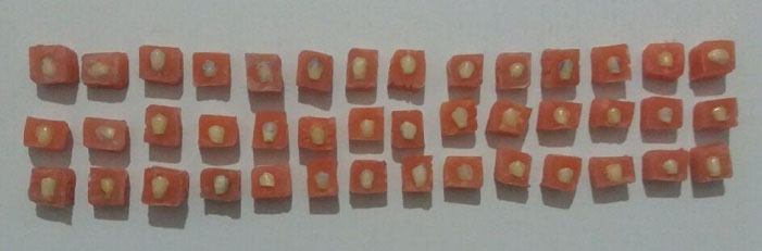
Figure 1.
Mounted Samples.
.
Mounted Samples.
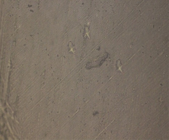
Figure 2.
Measuring Microhardness by Vickers Hardness Taster.
.
Measuring Microhardness by Vickers Hardness Taster.
To induce caries, the samples were immersed in the demineralizing solution, (2.2 mM of CaCl2.2H2O, 2.2 mM of KH2PO2, and 45 mM of acetate, pH = 4.6) which was prepared in the Pharmaceutics Department of School of Pharmacy, Kermanshah University of Medical Sciences and incubated at 37°C for 96 hours. After demineralization and measuring the microhardness of samples for the second time, the samples were randomly assigned to 3 groups of 14 and subjected to the following treatment while they were incubated at 37°C for 8 days (9). In group 1, the samples were immersed in 8w/v% grape seed extract emulsion (ground grape seeds, 100 g were extracted with ethanol to water by the maceration method and then filtered). In group 2, the samples were immersed in the whey extract (The yogurt was centrifuged at 10°C for 10 minutes by centrifugation at 4°C (12). Then, the lower phase (sediment) was examined as the whey extract, and in group 3 (control), the samples were subjected to artificial saliva (CaCl2_2H2O 0.7 mmol/L; MgCl2 0.2 mmol/L; KH2PO4 4.0 mmol/L; HEPES buffer as an acid form of 20.0 mmol/L; KCl 30.0 mmol/L), and the remineralizing solution was refreshed every two days in order to minimize probable errors. After the completion of the treatment, samples in all three groups were immersed in distilled water and sent to a laboratory for the measurement of microhardness for the third time.
To assess the possible difference in the change of microhardness, the percentage of the change was calculated using the analysis of covariance (ANCOVA) test and data were analyzed using SPSS, version 18 (SPSS Inc., IL, the USA) under the confidence level of 95%.
Results
The results of ANOVA showed that the baseline microhardness was significantly different among the groups (P = 0.044, Table 1). Thus, ANCOVA was used to compare microhardness after the treatment and demineralization. The results demonstrated a significant difference in microhardness changes among the study groups (P<0.001) such that the microhardness in the grape seed extract group experienced the greatest increase while it decreased in the control group. Based on the post hoc test results, no significant difference was observed between the whey extract and grape seed extract groups although the control group had significant differences with the whey extract and grape seed extract groups (P < 0.05, Table 2).
Table 1.
Mean, Standard Deviation, Maximum and Minimum Microhardness in the 3 Groups at Different Time Points
|
Group
|
Statistic
|
Baseline Microhardness
|
Microhardness After Demineralization
|
Microhardness After Treatment
|
Difference
|
Percentage of Changes in Microhardness
|
| Control |
Number |
14 |
14 |
14 |
14 |
14 |
| Mean |
413.76c |
184.78 |
172.38 |
-12.40a |
-9.93a |
| Standard deviation |
68.18 |
100.78 |
80.27 |
129.16 |
114.01 |
| Minimum |
313 |
39.23 |
40.95 |
-176.54 |
-206.72 |
| Maximum |
571 |
413 |
290.30 |
240.80 |
282.77 |
| Whey extract |
Number |
14 |
14 |
14 |
14 |
14 |
| Mean |
358.35a |
94.16 |
127.94 |
33.78b |
13.18a |
| Standard deviation |
47.28 |
44.23 |
62.94 |
69.67 |
25.71 |
| Minimum |
268 |
28.80 |
50.35 |
-56.50 |
-19.15 |
| Maximum |
413 |
162.60 |
246.30 |
204.80 |
69 |
| Grape seed extract |
Number |
14 |
14 |
14 |
14 |
14 |
| Mean |
370.51ab |
168.22 |
219.04 |
50.82b |
36.07a |
| Standard deviation |
60.22 |
95.05 |
101.66 |
59.44 |
35.12 |
| Minimum |
279 |
28.40 |
38.10 |
-34.93 |
-9.98 |
| Maximum |
492 |
355 |
364 |
151.55 |
92.71 |
|
P value |
0.044 |
- |
- |
<0.001 |
0.051 |
Table 2.
Pairwise Comparison of the Mean Change in the 3 Groups
|
Group (I)
|
Group (J)
|
Mean Difference (I-J)
|
Standard Error
|
P
Value
|
95% Confidence Interval
|
|
Lower Bound
|
Upper Bound
|
| Control |
Whey extract |
-93.525* |
26.883 |
0.004 |
-161.030 |
-26.021 |
| Grape seed extract |
-114.331* |
25.713 |
0.001 |
-178.897 |
-49.764 |
| Whey extract |
Control |
93.525* |
26.883 |
0.004 |
26.021 |
161.030 |
| Grape seed extract |
-20.805 |
25.678 |
1.000 |
-85.284 |
43.674 |
| Grape seed |
Control |
114.331* |
25.713 |
0.001 |
49.764 |
178.897 |
| Whey extract |
20.805 |
25.678 |
1.000 |
-43.674 |
-85.284 |
Note. *Mean difference is significant at 0.05.
However, the results of ANCOVA revealed no significant difference in the percentage of changes in microhardness among the study groups (P = 0.051). Given that this value was borderline significant, the hypothesis regarding the difference in the percentage of the change of microhardness among the groups could not be rejected definitely. Figures 3-5 show the mean microhardness, mean change, and mean percentage of changes in the microhardness of the three groups.
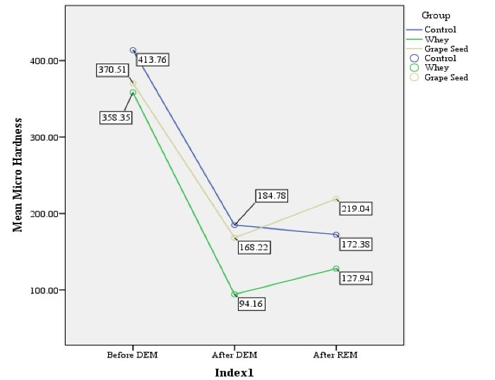
Figure 3.
Mean Microhardness of the Groups at Different Time Points.
.
Mean Microhardness of the Groups at Different Time Points.
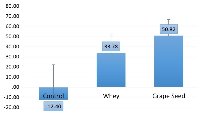
Figure 4.
Mean Change of Microhardness in the Groups.
.
Mean Change of Microhardness in the Groups.
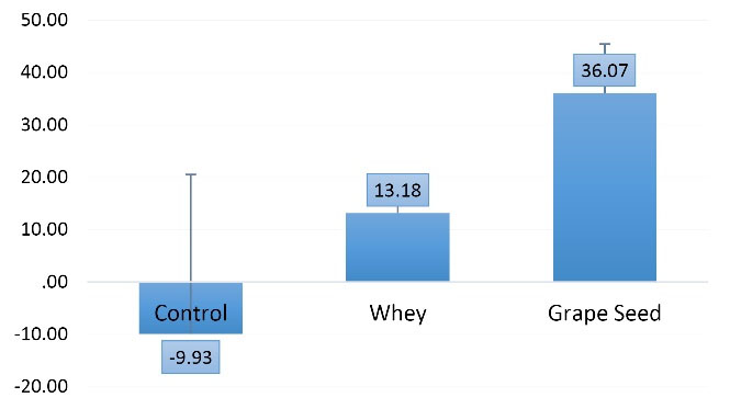
Figure 5.
Mean Percentage of Changes in Microhardness in the Groups.
.
Mean Percentage of Changes in Microhardness in the Groups.
Discussion
This study sought to compare the mean value and changes in enamel microhardness following the use of the grape seed extract, the whey extract, and artificial saliva (as the control group) at baseline, after demineralization, and after treatment. Since enamel microhardness and enamel mineral content are correlated, the comparison of mean percentage and surface microhardness changes in 3 phases (i.e., before demineralization, after demineralization, and after treatment) showed the efficacy level of these substances.
The baseline microhardness of the grape seed extract and whey extract groups was 370.51 VHN and 358.35 VHN, respectively which were close to the values reported by Rezvani et al. However, the baseline microhardness in the control group was 413.76 VHN in our study, which is different from that of the study by Rezvani et al. Our results revealed that both the grape seed extract and whey extract increased the microhardness of teeth. This increase was slightly greater in the grape seed extract group although the difference between the two groups did not reach statistical significance (13).
In another study, Ferrazzano et al concluded that dairy products can decrease enamel demineralization via three mechanisms. Milk proteins are absorbed by the enamel surface and inhibit enamel demineralization. In addition, milk fat is absorbed by the enamel surface and exerts a protective effect, and finally, milk enzymes can efficiently decrease the count of acidogenic bacteria in dental plaque (8).
In the above-mentioned study, the supernatant at the surface of a tube containing yogurt was used as the remineralizing solution, and their finding showed that natural CPP present in probiotic yogurt enhanced the process of enamel remineralization. In the current study, yogurt dregs were used as the whey extract, which contradicts the findings of the above-mentioned study (8).
The results of the current study represented that the grape seed extract increases the microhardness of teeth, which is in agreement with the results of Mirkarimi et al (9). Moreover, Silva et al (12) evaluated the effect of the grape seed extract on bovine enamel and dentin and concluded that the hardness of enamel and dentin in the grape seed extract group was higher compared to the control group. They further reported that the grape seed extract had a greater effect on dentin compared to the enamel due to the higher action potential on collagen matrix crosslinks although its effect on the enamel and dentin was less than that of fluoride.
Da Silva et al (12) believed that the deposition of minerals is the main reason for the effect of the grape seed extract on tooth enamel. In the same way, Tang et al stated that gallic acid as the main constituent of the grape seed extract enhanced the deposition of minerals, especially in the superficial enamel layer (14).
In the current study, the whey extract increased the microhardness of the samples and a significant difference existed between the whey extract and the artificial saliva (control) groups. Likewise, Rezvani et al compared the effect of CPP-ACP and the whey extract (as a natural CPP-ACP) on enamel microhardness and achieved promising findings in terms of the efficacy of the whey extract on enamel microhardness. In their study, an increase in microhardness was 30%, 17%, and 8% in the whey extract, the artificial saliva, and the CPP-ACP groups, respectively (13). In our study, a 9% reduction in microhardness was found in the artificial saliva group, which does not match the findings of Rezvani et al probably due to the difference in the methodology of the two studies and the duration of contact with the materials.
To achieve more reliable findings about the efficacy of these compounds, some recommendations are provided as follows:
-
It is better to compare the effect of the supernatant of centrifuged yogurt with its sediment on enamel microhardness;
-
Considering the limitations of in-vitro studies, clinical studies are required in this respect.
Conclusions
According to the results of this study, the grape seed extract and the whey extract had positive efficacy for the remineralization of enamel lesions in-vitro and may be used as natural, effective substances for the non-invasive treatment of carious lesions in children. Considering the limitations in the use of fluoride, especially in children, the grape seed extract and the whey extract can serve as natural and safe alternatives in conservative and preventive dentistry.
Conflict of Interest Disclosures
The authors declare that they have no conflict of interests.
Ethical Statement
This study was approved by the research ethics commitee of kermanshah university of medical sciences.
Authors’ Contribution
SN and SM developed the concept of the study.ZH and SM performed laboratory steps.LS was primiraly responsible for writing the manuscribe.all authors approved the final.
References
- Kamath P, Nayak R, Kamath SU, Pai D. A comparative evaluation of the remineralization potential of three commercially available remineralizing agents on white spot lesions in primary teeth: an in vitro study. J Indian Soc Pedod Prev Dent 2017; 35(3):229-37. doi: 10.4103/jisppd.jisppd_242_16 [Crossref] [ Google Scholar]
- Zhao W, Xie Q, Bedran-Russo AK, Pan S, Ling J, Wu CD. The preventive effect of grape seed extract on artificial enamel caries progression in a microbial biofilm-induced caries model. J Dent 2014; 42(8):1010-8. doi: 10.1016/j.jdent.2014.05.006 [Crossref] [ Google Scholar]
- Salehzadeh Esfahani K, Mazaheri R, Pishevar L. Effects of treatment with various remineralizing agents on the microhardness of demineralized enamel surface. J Dent Res Dent Clin Dent Prospects 2015; 9(4):239-45. doi: 10.15171/joddd.2015.043 [Crossref] [ Google Scholar]
- Chandak S, Bhondey A, Bhardwaj A, Pimpale J, Chandwani M. Comparative evaluation of the efficacy of fluoride varnish and casein phosphopeptide-amorphous calcium phosphate in reducing Streptococcus mutans counts in dental plaque of children: An in vivo study. J Int Soc Prev Community Dent 2016; 6(5):423-9. doi: 10.4103/2231-0762.192936 [Crossref] [ Google Scholar]
- Maden EA, Acar Ö, Altun C, Polat GG. The effect of casein phosphopeptide-amorf calcium phosphate and acidulated phosphate fluoride gel on dental erosion in primary teeth: an in vitro study. J Clin Pediatr Dent 2017; 41(4):275-9. doi: 10.17796/1053-4628-41.4.275 [Crossref] [ Google Scholar]
- Thierens LAM, Moerman S, Elst CV, Vercruysse C, Maes P, Temmerman L. The in vitro remineralizing effect of CPP-ACP and CPP-ACPF after 6 and 12 weeks on initial caries lesion. J Appl Oral Sci 2019; 27:e20180589. doi: 10.1590/1678-7757-2018-0589 [Crossref] [ Google Scholar]
- Dashper SG, Catmull DV, Liu SW, Myroforidis H, Zalizniak I, Palamara JE. Casein phosphopeptide-amorphous calcium phosphate reduces Streptococcus mutans biofilm development on glass ionomer cement and disrupts established biofilms. PLoS One 2016; 11(9):e0162322. doi: 10.1371/journal.pone.0162322 [Crossref] [ Google Scholar]
- Ferrazzano GF, Cantile T, Quarto M, Ingenito A, Chianese L, Addeo F. Protective effect of yogurt extract on dental enamel demineralization in vitro. Aust Dent J 2008; 53(4):314-9. doi: 10.1111/j.1834-7819.2008.00072.x [Crossref] [ Google Scholar]
- Mirkarimi M, Eskandarion S, Bargrizan M, Delazar A, Kharazifard MJ. Remineralization of artificial caries in primary teeth by grape seed extract: an in vitro study. J Dent Res Dent Clin Dent Prospects 2013; 7(4):206-10. doi: 10.5681/joddd.2013.033 [Crossref] [ Google Scholar]
- Epasinghe DJ. Applications of Proanthocyanidin in Dentistry [dissertation]. Hong Kong: University of Hong Kong; 2014.
- Epasinghe DJ, Yiu CK, Burrow MF, Tsoi JK, Tay FR. Effect of flavonoids on the mechanical properties of demineralised dentine. J Dent 2014; 42(9):1178-84. doi: 10.1016/j.jdent.2014.07.002 [Crossref] [ Google Scholar]
- da Silva AP, Gonçalves RS, Borges AF, Bedran-Russo AK, Shinohara MS. Effectiveness of plant-derived proanthocyanidins on demineralization on enamel and dentin under artificial cariogenic challenge. J Appl Oral Sci 2015; 23(3):302-9. doi: 10.1590/1678-775720140304 [Crossref] [ Google Scholar]
- Rezvani MB, Karimi M, Akhavan Rasoolzade R, Haghgoo R. Comparing the effects of whey extract and casein phosphopeptide-amorphous calcium phosphate (CPP-ACP) on enamel microhardness. J Dent (Shiraz) 2015; 16(1):49-53. [ Google Scholar]
- Tang CF, Fang M, Liu RR, Dou Q, Chai ZG, Xiao YH. The role of grape seed extract in the remineralization of demineralized dentine: micromorphological and physical analyses. Arch Oral Biol 2013; 58(12):1769-76. doi: 10.1016/j.archoralbio.2013.09.007 [Crossref] [ Google Scholar]