Avicenna J Dent Res. 15(3):138-141.
doi: 10.34172/ajdr.2023.1723
Case Report
Eruption of an Uncommon Impacted Mandibular Canine in a Large Inflamed Dentigerous Cyst With the Combined Marsupialization and Orthodontic Treatment: A Case Report
Vahid Mollabashi 1  , Hana Salehisaheb 2, Hamid Naderi 3, * , Nafiseh Shamloo 4, Faezeh Yousefi 5
Author information:
1DDS, Associate Professor of Orthodontics, Department of Orthodontics, School of Dentistry, Hamadan University of Medical Sciences, Hamadan, Iran
2DDS, Assistant Professor of Orthodontics, Department of Orthodontics, School of Dentistry, Kurdistan University of Medical Sciences, Kurdistan, Iran
3DDS, Resident of Oral & Maxillofacial Surgery, Department of Oral & Maxillofacial Surgery, School of Dentistry, Hamadan University of Medical Sciences, Hamadan, Iran
4DDS, Associate Professor of Oral & Maxillofacial Pathology, Department of Oral and Maxillofacial Pathology, School of Dentistry, Shahid Beheshti University of Medical Sciences, Tehran, Iran
5Associate Professor of Oral and Maxillofacial Radiology, Department of Oral and Maxillofacial Radiology, School of Dentistry, Hamadan University of Medical Sciences, Hamadan, Iran
Abstract
A dentigerous cyst is a benign and asymptomatic lesion that may become extremely large and then interfere with tooth eruption. Marsupialization is known as the best treatment option for large cysts involving an unerupted permanent tooth. A 16-year-old female has been referred with the chief complaint of an unerupted tooth, extraoral and intraoral swelling, and pain. Radiographic examination demonstrated a unilocular, well-defined, and large radiolucent lesion extended from the distal of the right mandibular canine approximately to the left first mandibular molar. The mandibular left canine was also embedded within the lesion. The selected treatment plan was marsupialization, followed by performing orthodontic treatment of the unerupted mandibular canine. This clinical report presented the successful eruption of an impacted mandibular canine over a 4-years follow-up. The findings of this study revealed that the combination of marsupialization and orthodontic treatment is an effective treatment for large jaw cysts with impacted teeth.
Keywords: Dentigerous cyst, Marsupialization, Impacted mandibular canine, Orthodontic treatment
Copyright and License Information
© 2023 The Author(s); Published by Hamadan University of Medical Sciences.
This is an open-access article distributed under the terms of the Creative Commons Attribution License (
http://creativecommons.org/licenses/by/4.0), which permits unrestricted use, distribution, and reproduction in any medium provided the original work is properly cited.
Please cite this article as follows: Mollabashi V, Salehisaheb H, Naderi H, Shamloo N, Yousefi F. Eruption of an uncommon impacted mandibular canine in a large inflamed dentigerous cyst with the combined marsupialization and orthodontic treatment: a case report. Avicenna J Dent Res. 2023; 15(3):138-141. doi:10.34172/ajdr.2023.1723
Background
A dentigerous cyst is known as the second most common odontogenic cyst in the oral and maxillofacial region (1). In most cases, dentigerous cysts have no clinical symptoms and are diagnosed on routine radiographic examinations.
The treatment modalities for dentigerous cysts are enucleation, marsupialization, or the decompression of the cyst via fenestration (2). In patients diagnosed with large cysts, orthodontic treatment is often needed after the marsupialization to help the eruption of the impacted tooth (3).
This is a case report of a patient with an unerupted mandibular left canine associated with a large dentigerous cyst treated by marsupialization. After performing the marsupialization technique, the tooth erupted somewhat spontaneously. Ultimately orthodontic treatment was used to align this tooth.
Case Report
A 16-year-old female with class II division I malocclusion has been referred to the orthodontic department of the dental school with the chief complaint of an unerupted tooth, extraoral and intraoral swelling, and pain. Panoramic radiography displayed a unilocular, well-defined, and large radiolucent lesion which was extended from the distal of the right mandibular canine approximately to the left first mandibular molar. In addition, the mandibular left canine was embedded within the lesion, as it was placed horizontally on the lower border of the mandible (Figure 1).
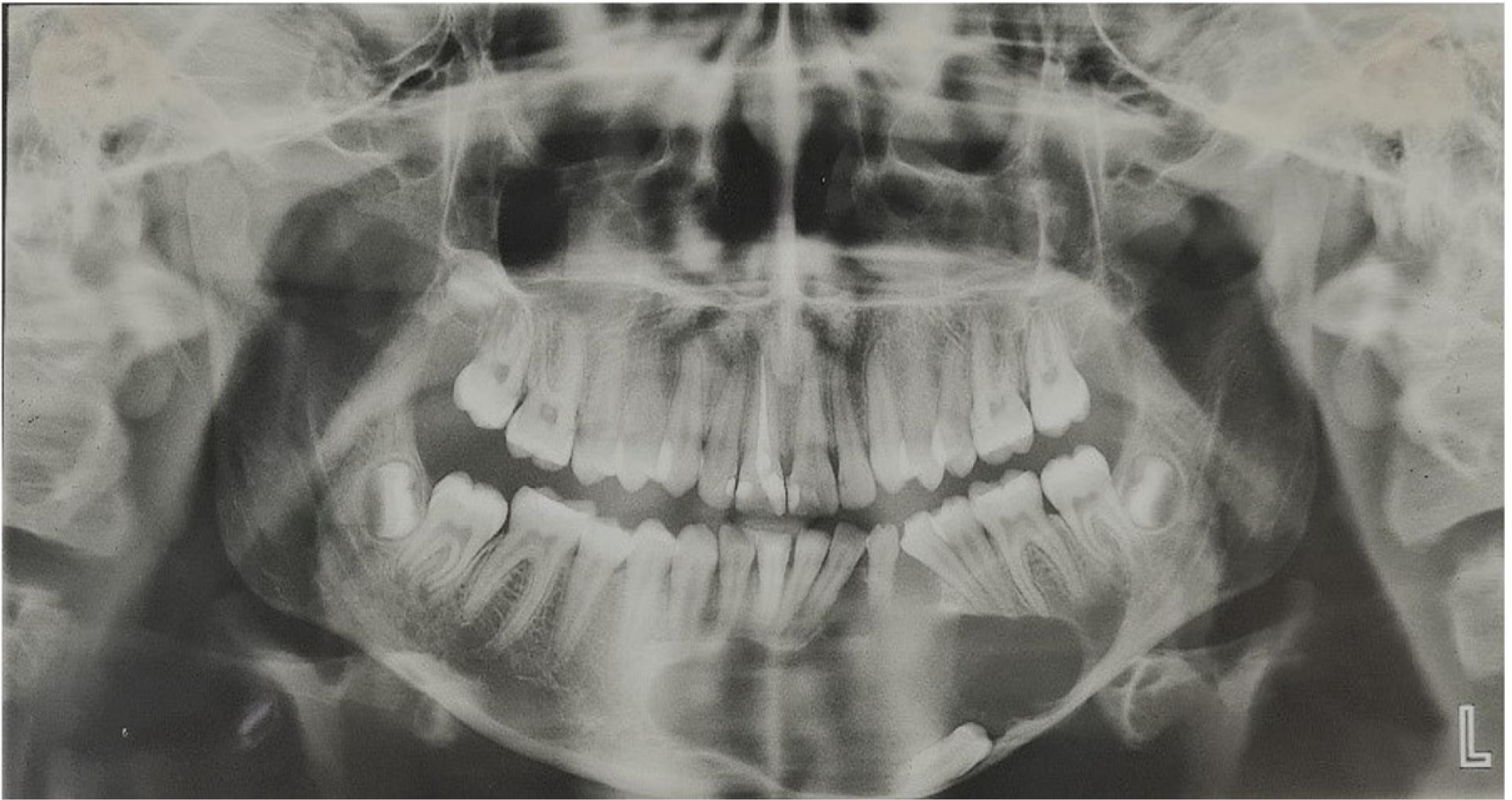
Figure 1.
Panoramic View Before Starting the Treatment. Note. A unilocular, well-defined, and large radiolucent lesion was extended from the distal of the right mandibular canine approximately to the left first mandibular molar. The mandibular left canine was also embedded within the lesion
.
Panoramic View Before Starting the Treatment. Note. A unilocular, well-defined, and large radiolucent lesion was extended from the distal of the right mandibular canine approximately to the left first mandibular molar. The mandibular left canine was also embedded within the lesion
The patient was referred to an oral and maxillofacial surgeon. In the cone-beam computed tomography view, lingual cortex was intact. The perforation of the buccal cortex was evident in some areas. There was no tooth mobility. The vitality test of the mandibular teeth was normal. A dentigerous cyst was reported according to an incisional biopsy. The histopathologic sections demonstrated a cystic lesion lined by hyperplastic squamous epithelium with exocytosis and an arch shape appeared in the same area. In the underlying connective tissue, severe mixed inflammatory, grant cells, hemorrhage, extravasated red blood cells, and fragments of reactive osteoid were visible (Figure 2).
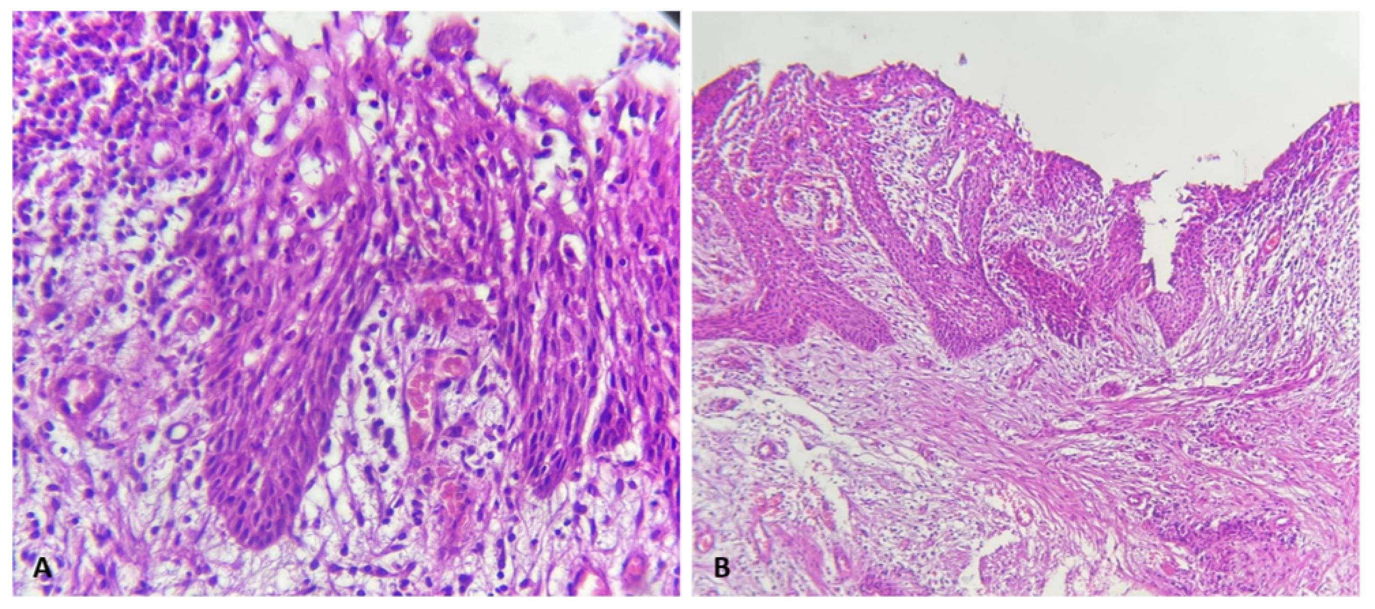
Figure 2.
Histopathologic view. (A) inflamed dentigerous cyst (H&E Stain × 100) and (B) Inflamed dentigerous cyst showing a thicker epithelial lining with hyperplastic rete ridges. Note. The cyst wall displays diffuse mixed inflammatory cells infiltration (H&E stain × 40)
.
Histopathologic view. (A) inflamed dentigerous cyst (H&E Stain × 100) and (B) Inflamed dentigerous cyst showing a thicker epithelial lining with hyperplastic rete ridges. Note. The cyst wall displays diffuse mixed inflammatory cells infiltration (H&E stain × 40)
Afterward, the marsupialization treatment was performed for the patient who has been undergoing this treatment for 16 months. After marsupialization, the patient was referred to the orthodontic department for further treatment.
No extraoral and intraoral swelling was observed on the initial orthodontic examination. Furthermore, there was no pain or discharge. The tilt of the anterior teeth and the first premolar was evident toward the impacted canine site, and the mandibular midline represented a deviation. Moreover, the cusp tip of the canine was visible in the buccal vestibule.
In the radiography images, the size of the lesion was dramatically reduced compared to the serial panoramic prepared from the beginning of the treatment. In addition, the cyst cavity was ossified, and the tooth was almost in a vertical position (Figure 3).
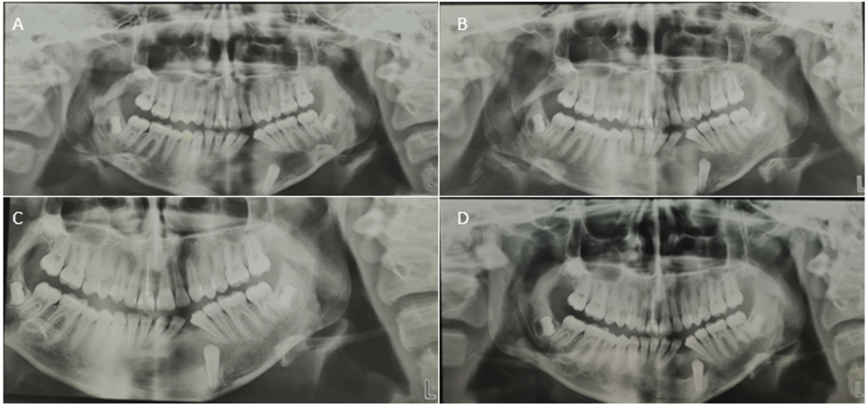
Figure 3.
Serial Panoramic Radiographs. (A) Four months after surgery, (B) Four months later, (C) Eight months later, and (D) Twelve months later
.
Serial Panoramic Radiographs. (A) Four months after surgery, (B) Four months later, (C) Eight months later, and (D) Twelve months later
The initial diagnostic records were obtained, including photographs, cast molds, and radiographs (Figure 4). Thereafter, adjustable edgewise 0.022 inch slots were bonded with metal brackets on the labial surface of mandibular teeth. Then, the initial alignment was performed with nickel-titanium and stainless-steel arch wires. Next, the open coil spring was used on a 0.017 × 0.025-inch arch-wire to open up enough space for the canine tooth. After regaining enough space, 0.012-inch nickel-titanium arch-wire, as an overlay, was employed to help the tooth to continue its eruption, and the tooth was simultaneously directed to its desired position in the dental arch.
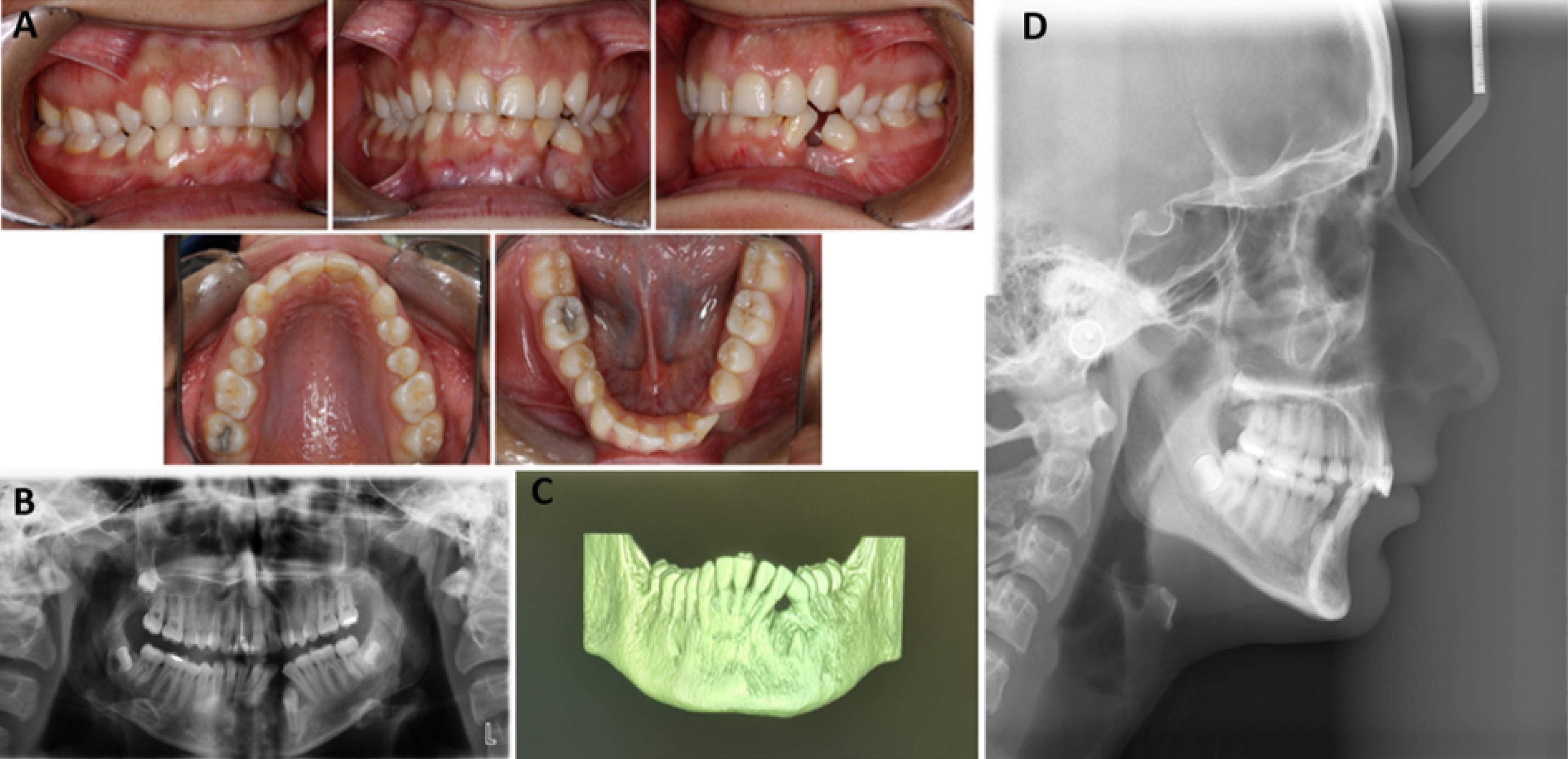
Figure 4.
Initial Diagnostic Records Before Starting Orthodontic Treatment. (A) Intra oral photographs, (B) Panoramic view, (C) 3D reconstruction view, and (D) Lateral cephalometric
.
Initial Diagnostic Records Before Starting Orthodontic Treatment. (A) Intra oral photographs, (B) Panoramic view, (C) 3D reconstruction view, and (D) Lateral cephalometric
The orthodontic treatment lasted for 20 months. Intraoral photographs showed acceptable canine and molar relations, adequate overbite and overjet, improved midline deviation, and well-aligned teeth. The panoramic view demonstrated complete healing of the lesion area (Figure 5).
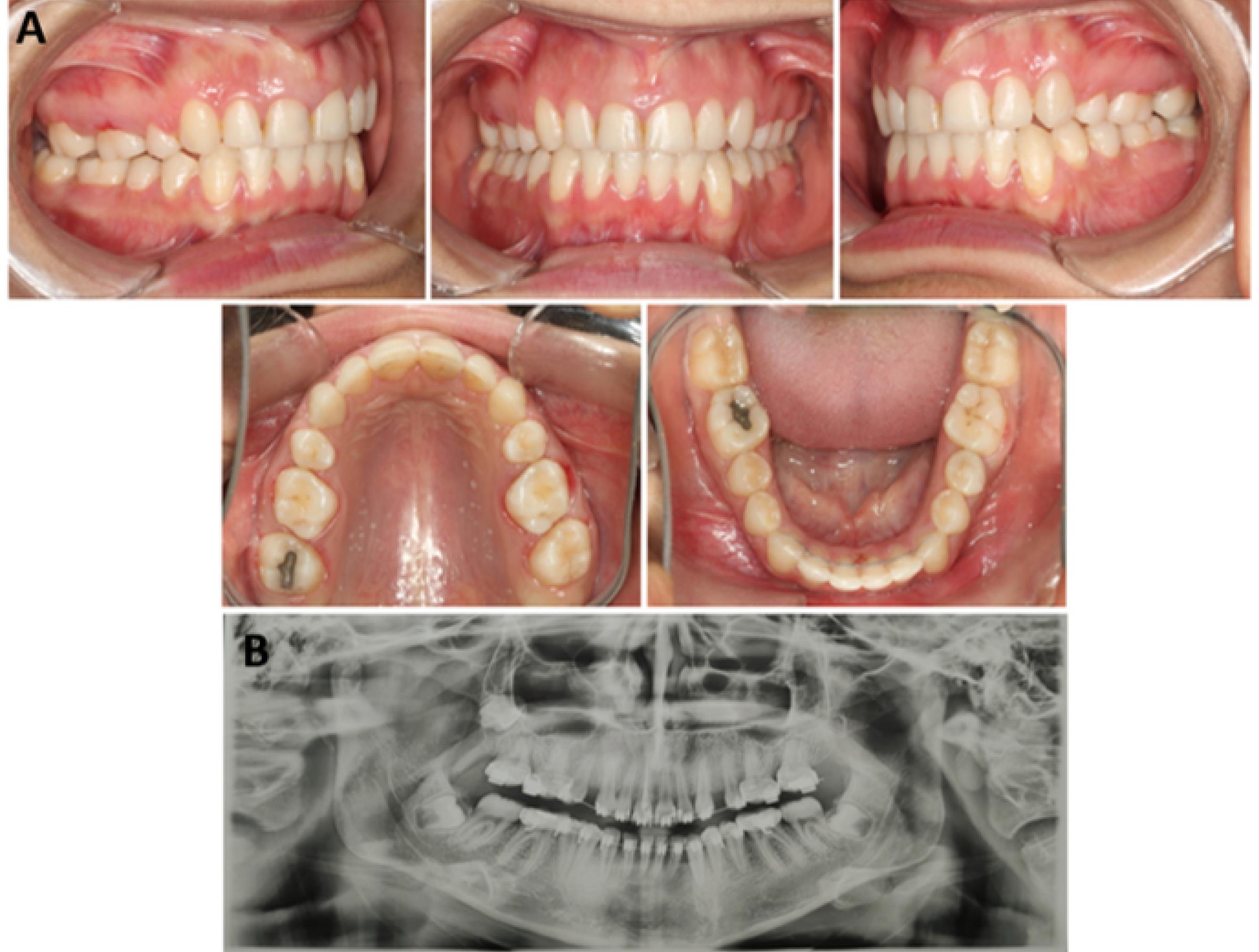
Figure 5.
Final Diagnostic Records. (A) Intraoral photographs showing an acceptable canine and molar relations, adequate overbite and overjet, leading to an improvement in midline deviation and well-aligned teeth and (B) The panoramic view depicting complete healing of the lesion area
.
Final Diagnostic Records. (A) Intraoral photographs showing an acceptable canine and molar relations, adequate overbite and overjet, leading to an improvement in midline deviation and well-aligned teeth and (B) The panoramic view depicting complete healing of the lesion area
Discussion
Marsupialization is a relatively conservative treatment modality used for dentigerous cysts because it reduces the risk of developing jaw fractures and nerve damage. Additionally, spontaneous eruption of the tooth associated with the cyst may occur in marsupialization (4). In most patients aged over 10 years old, spontaneous tooth eruption does not occur, thus in cases where the tooth has not been removed with a cyst, performing the orthodontic treatment is almost always necessary to guide the affected tooth to its final position in the dental arch (5).
Tsironi et al (6) used marsupialization and then fixed orthodontic treatment to guide the unerupted mandibular first molar in association with a dentigerous cyst to the dental arch. In another study, Murakami et al (7) performed the marsupialization of a dentigerous cyst-associated mandibular second premolar. They maintained the space needed for the spontaneous eruption of the tooth by using a space maintainer. Chung et al (8) guided the mandibular first molar associated with a dentigerous cyst to a dental arch using a T-type C-tube placed in the inter radicular space of the posterior maxilla. In a study by Aoki1 et al (9), the orthodontic traction was not successful in guiding maxillary canine after the marsupialization of dentigerous cyst, thus alternative treatments, including tooth extraction and implant placement, were performed in this regard.
The selection of the best treatment to guide the impacted tooth to the dental arch depends on various factors such as the position of the impacted tooth, the type of tooth involved, the patient’s age, and the developmental stage of the root (3). In our case, due to the patient’s age, the high importance of the canine teeth for a favorable occlusion, and the large size of the lesion, a combination of both marsupialization and orthodontic treatment was performed as the best treatment method for the patient.
It was indicated that some other treatment modalities such as surgical repositioning and extraction of the impacted tooth could induce a higher risk of developing related complications. Surgical repositioning or transplantation may cause root resorption, ankylosis, or even pulp necrosis. Therefore, only in cases for whom the orthodontic treatment is contraindicated, these could be the treatment of choice (10).
In cases where marsupialization is the treatment of choice, patient cooperation is essential; because oral hygiene and long-term collaboration are critical to the success of this kind of treatment. Additionally, orthodontic appliances can irritate the adjacent mucosa and consequently cause pain in the patient. Therefore, the patient and the parents should be aware of the necessity of oral hygiene and the use of appliances during treatment for obtaining successful results. Fortunately, our patient had highly good cooperation.
Following the treatment, the patient was delighted and satisfied in terms of occlusion, function, and aesthetics.
Conclusion
The findings of this case study showed that the combination of marsupialization and orthodontic treatment is an effective treatment for large jaw cysts with impacted teeth.
Authors’ Contribution
Conceptualization: Vahid Mollabashi.
Data curation: Hana Salehisaheb, Hamid Naderi.
Investigation: Nafiseh Shamloo, Faezeh Yousefi.
Methodology: Vahid Mollabashi, Hana Salehisaheb, Hamid Naderi.
Project administration: Vahid Mollabashi, Hana Salehisaheb, Hamid Naderi.
Supervision: Vahid Mollabashi.
Validation: Vahid Mollabashi, Nafiseh Shamloo.
Writing–original draft: Hamid Naderi.
Writing–review & editing: Hamid Naderi.
Competing Interests
The authors declare that they have no conflict of interests.
Ethical Approval
None to be declared.
Informed Consent
Verbal and written consent was obtained from the patient.
References
- Aguiló L, Gandía JL. Dentigerous cyst of mandibular second premolar in a five-year-old girl, related to a non-vital primary molar removed one year earlier: a case report. J Clin Pediatr Dent 1998; 22(2):155-8. [ Google Scholar]
- Clauser C, Zuccati G, Barone R, Villano A. Simplified surgical-orthodontic treatment of a dentigerous cyst. J Clin Orthod 1994; 28(2):103-6. [ Google Scholar]
- Serra e Silva FM, Sawazaki R, de Moraes M. Eruption of teeth associated with a dentigerous cyst by only marsupialization treatment: a case report. J Dent Child (Chic) 2007; 74(3):228-30. [ Google Scholar]
- Hyomoto M, Kawakami M, Inoue M, Kirita T. Clinical conditions for eruption of maxillary canines and mandibular premolars associated with dentigerous cysts. Am J Orthod Dentofacial Orthop 2003; 124(5):515-20. doi: 10.1016/j.ajodo.2003.04.001 [Crossref] [ Google Scholar]
- Fujii R, Kawakami M, Hyomoto M, Ishida J, Kirita T. Panoramic findings for predicting eruption of mandibular premolars associated with dentigerous cyst after marsupialization. J Oral Maxillofac Surg 2008; 66(2):272-6. doi: 10.1016/j.joms.2007.06.652 [Crossref] [ Google Scholar]
- Tsironi K, Inglezos E, Vardas E, Mitsea A. Uprighting an impacted permanent mandibular first molar associated with a dentigerous cyst and a missing second mandibular molar-a case report. Dent J (Basel) 2019; 7(3):63. doi: 10.3390/dj7030063 [Crossref] [ Google Scholar]
- Murakami A, Kawabata K, Suzuki A, Murakami S, Ooshima T. Eruption of an impacted second premolar after marsupialization of a large dentigerous cyst: case report. Pediatr Dent 1995; 17(5):372-4. [ Google Scholar]
- Chung KR, Noh MK, Oh SH, Jeong DM, Kim SH, Nelson G. Treatment of 2 impacted molars in a large dentigerous cyst (expansile cystic lesion) with combined orthodontic and surgical therapy. Am J Orthod Dentofacial Orthop 2020; 158(5):752-8. doi: 10.1016/j.ajodo.2020.06.029 [Crossref] [ Google Scholar]
- Aoki N, Ise K, Inoue A, Kosugi Y, Koyama C, Iida M. Multidisciplinary approach for treatment of a dentigerous cyst - marsupialization, orthodontic treatment, and implant placement: a case report. J Med Case Rep 2018; 12(1):305. doi: 10.1186/s13256-018-1829-2 [Crossref] [ Google Scholar]
- McAboy CP, Grumet JT, Siegel EB, Iacopino AM. Surgical uprighting and repositioning of severely impacted mandibular second molars. J Am Dent Assoc 2003; 134(11):1459-62. doi: 10.14219/jada.archive.2003.0074 [Crossref] [ Google Scholar]